Ureteral anomalies frequently occur with kidney anomalies but may occur independently. Complications include:
Obstruction, vesicoureteral reflux, urinary tract infections, and urinary calculus formation (due to urinary stasis)
Urinary incontinence (due to abnormal termination of the ureter in the urethra, perineum, or vagina)
The presence of ureteral anomalies may be suggested in utero by urinary tract abnormalities noted on routine prenatal ultrasound (eg, hydronephrosis). In a neonate or child, anomalies are occasionally detected by physical examination (eg, finding an external ectopic ureteral orifice or a palpable mass).
Ureteral anomalies should be considered in children who have had an episode of pyelonephritis or recurrent urinary tract infections, in girls with persistent urinary incontinence, or in boys with recurrent episodes of orchitis.
Testing typically involves ultrasonography of the kidneys, ureters, and bladder before and after voiding, and then fluoroscopic voiding cystourethrography. Magnetic resonance urography may be used to identify an ectopic ureter associated with a nonfunctioning renal segment (1).
Ureteral anomalies are treated surgically.
Reference
1. Balawender K, Wawrzyniak A, Pliszka A, et al. Ectopic ureter: A concise narrative review with anatomical and clinical commentaries. Trans Res Anat. 2022;29:100220. doi:10.1016/j.tria.2022.100220
Ectopic Ureteral Orifices
Openings of single or duplicated ureters may be malpositioned on the lateral bladder wall, distally along the trigone, in the bladder neck, in the female urethra distal to the sphincter (leading to continuous incontinence despite a normal voiding pattern), in the genital system (prostate and seminal vesicle in the male, uterus or vagina in the female), or externally. Lateral ectopic orifices frequently lead to vesicoureteral reflux, whereas distal ectopic orifices more often cause obstruction and incontinence.
Surgery is needed for obstruction and incontinence and sometimes for vesicoureteral reflux.
Retrocaval Ureter
Anomalous development of the vena cava (pre-ureteric vena cava) allows the infrarenal vena cava to form anterior to the ureter (usually the right); a retrocaval ureter on the left occurs only with persistence of the left cardinal vein system or with complete situs inversus.
Retrocaval ureter can cause ureteral obstruction. For significant ureteral obstruction, the ureter is surgically divided with uretero-ureteral anastomosis anterior to the vena cava or iliac vessel.
Ureter Duplication Anomalies
Incomplete (partial) or complete duplication of one or both ureters may occur with duplication of the ipsilateral renal pelvis.

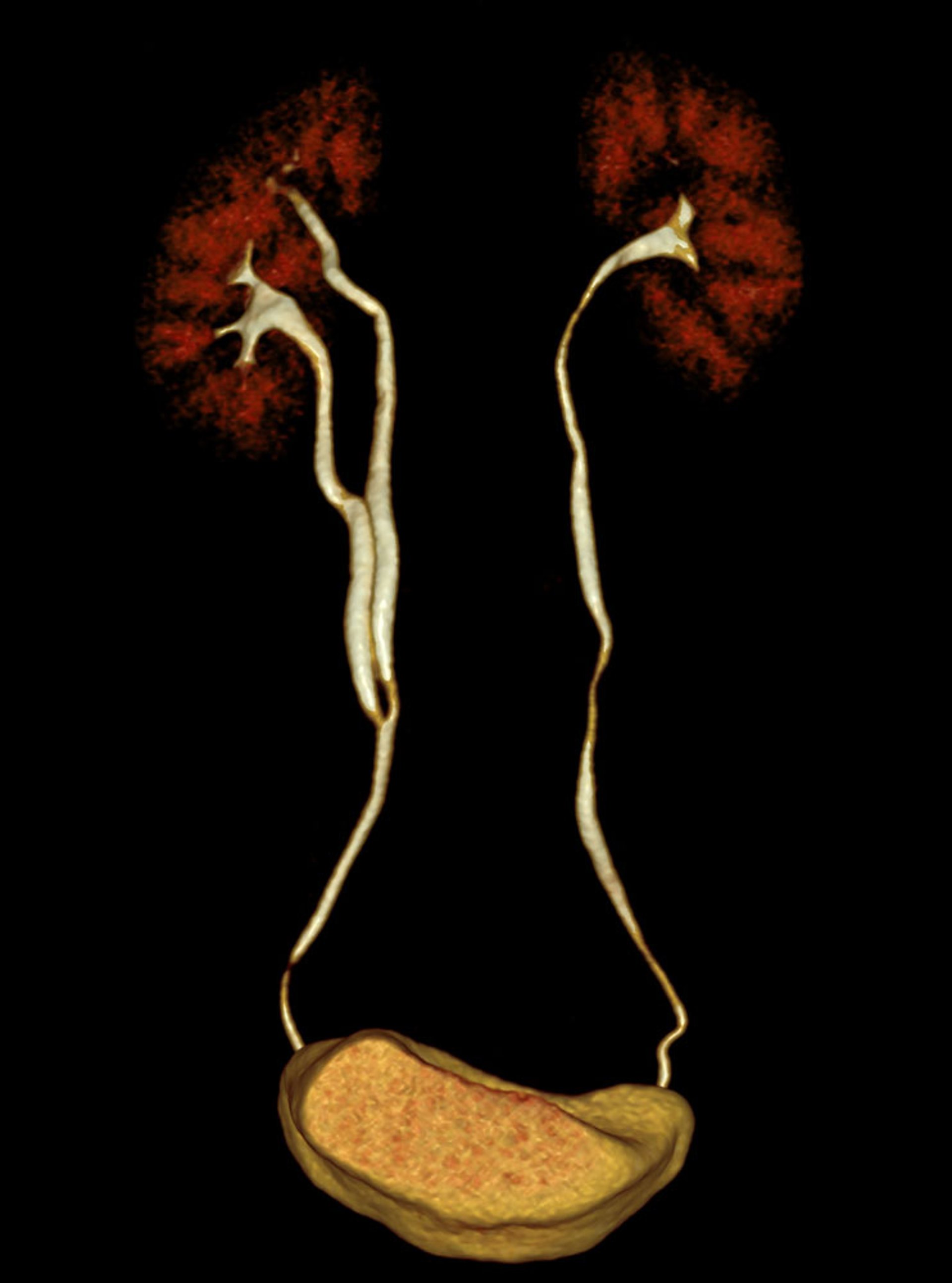
This colored 3D CT scan shows a duplex collecting system of the kidney. This anatomical variant features 2 separate pelvicalyceal systems within 1 kidney (left).
HABIBUR RAHMAN/SCIENCE PHOTO LIBRARY
In complete duplication, the ureter from the upper pole of the kidney opens at a more caudal location than the orifice of the lower pole ureter. As a result, the lower pole tends to reflux and the upper pole tends to become obstructed when pathology is present. Ectopia or stenosis of one or both orifices, vesicoureteral reflux into the lower ureter or both ureters, and ureterocele may occur.
Surgery may be necessary if there is obstruction, vesicoureteral reflux, or urinary incontinence.
Incomplete duplication is rarely of clinical significance.
Ureteral Stenosis
Stenosis may occur at any location in the ureter, most frequently at the ureteropelvic junction and less commonly at the ureterovesical junction (primary megaureter). Consequences include infection, hematuria, and obstruction. Stenoses often diminish as the child grows.
In primary megaureter, ureteral tapering and reimplantation may be needed when dilation increases or infection or obstruction occurs. In ureteropelvic junction obstruction, pyeloplasty (excision of the obstructed segment and reanastomosis) may be done by open, laparoscopic, or robotic techniques.
Ureterocele
Prolapse of the lower end of the ureter into the bladder with pinpoint obstruction may cause progressive ureterectasis, hydronephrosis, infection, occasional calculus formation, and impaired renal function. Ureterocele treatment options include endoscopic transurethral incision and open repair.


This image of a left ureter shows a submucosal cystic dilation, which is a congenital anomaly known as ureterocele. In ureterocele, the distal ureter balloons at its opening into the bladder and forms a sac-like pouch, as seen in the right half of this image.
When a ureterocele involves the upper of 2 duplex ureters, treatment depends on function in that renal segment, which is frequently dysplastic. Removal of the affected renal segment and ureter may be preferable to obstruction repair if that segment is nonfunctional or if significant renal dysplasia is suspected. Alternatively, ipsilateral ureteroureterostomy may be done to bypass the obstruction.
In rare instances, the ureterocele may prolapse beyond the bladder neck, causing a bladder outlet obstruction. In girls, this prolapse may manifest as an interlabial mass.