


Malrotation of the bowel is failure of the bowel to assume its normal place in the abdomen during intrauterine development. Many patients present with midgut volvulus, a surgical emergency. Diagnosis is by abdominal radiograph. Treatment is surgical repair with a Ladd procedure.
(See also Overview of Congenital Gastrointestinal Anomalies.)
Malrotation may be asymptomatic or symptomatic. Prevalence of asymptomatic rotational anomaly is approximately 1 in 500 (1); however, symptomatic malrotation occurs less frequently (1 in 2500 to 6000 live births) (2, 3).
Midgut volvulus is the sudden twisting of the abnormally positioned and anchored intestines around their mesenteric stalk, cutting off blood supply. This is a surgical emergency. Volvulus occurs most often in neonates (4).
During embryonic development, the primitive bowel protrudes from the abdominal cavity. As it returns to the abdomen, the large bowel normally rotates counterclockwise, with the cecum coming to rest in the right lower quadrant. Incomplete rotation, in which the cecum ends up elsewhere (usually in the right upper quadrant or midepigastrium), may cause bowel obstruction due to fibrous retroperitoneal bands (Ladd bands) that stretch across the duodenum or due to a volvulus of the small bowel, which, lacking its normal peritoneal attachment, twists on its narrow, stalk-like mesentery (5).
Other anomalies occur in the majority of patients, most commonly other gastrointestinal (GI) malformations (eg, gastroschisis, omphalocele, diaphragmatic hernia, intestinal atresia, Meckel diverticulum) (2, 5). Congenital heart disease, heterotaxy syndrome (a condition in which the internal organs are abnormally arranged in the chest and abdomen), and genetic syndromes also may occur.
Patients with malrotation can present in infancy or in adulthood; however, approximately 40% present in the first month of life and > 80% in the first 5 years of life (3). Patients can present with acute abdominal pain and bilious emesis, with an acute volvulus, with typical reflux symptoms, with chronic abdominal pain, or with faltering growth. In some patients, malrotation is found incidentally as part of an evaluation for another problem.
Bilious emesis in an infant is an emergency and should be evaluated immediately to make sure the infant does not have malrotation and a midgut volvulus; untreated, the risk of bowel infarction and subsequent short bowel syndrome or death is high.
General references
1. Perez AA, Pickhardt PJ. Intestinal malrotation in adults: prevalence and findings based on CT colonography. Abdom Radiol (NY). 2021;46(7):3002-3010. doi:10.1007/s00261-021-02959-3
2. Forrester MB, Merz RD. Epidemiology of intestinal malrotation, Hawaii, 1986-99. Paediatr Perinat Epidemiol. 2003;17(2):195-200. doi:10.1046/j.1365-3016.2003.00480.x
3. Salehi Karlslätt K, Husberg B, Ullberg U, et al. Intestinal Malrotation in Children: Clinical Presentation and Outcomes. Eur J Pediatr Surg. 2024;;34(3):228-235. doi:10.1055/s-0043-1764239
4. Svetanoff WJ, Srivatsa S, Diefenbach K, Nwomeh BC. Diagnosis and management of intestinal rotational abnormalities with or without volvulus in the pediatric population. Semin Pediatr Surg. 2022;31(1):151141. doi:10.1016/j.sempedsurg.2022.151141
5. Langer JC. Intestinal rotation abnormalities and midgut volvulus. Surg Clin N Am. 2017;97(1):147–159. doi:10.1016/j.suc.2016.08.011
Diagnosis of Malrotation of the Bowel
Abdominal radiographs
Upper GI series
Sometimes barium enema
Sometimes ultrasound
In infants with bilious emesis, plain radiographs of the abdomen should be taken immediately. If they suggest obstruction, for example showing a dilated stomach and proximal small bowel (double-bubble sign), a paucity of bowel gas distal to the duodenum, or both (suggesting a midgut volvulus), further diagnosis and treatment must be performed emergently.
A cautiously performed upper gastrointestinal (GI) series is the generally preferred method for diagnosing malrotation with or without volvulus (1). Barium enema can be helpful in certain cases and is generally considered an adjunct to the upper GI series. Barium enema typically identifies malrotation by showing the cecum outside the right lower quadrant.

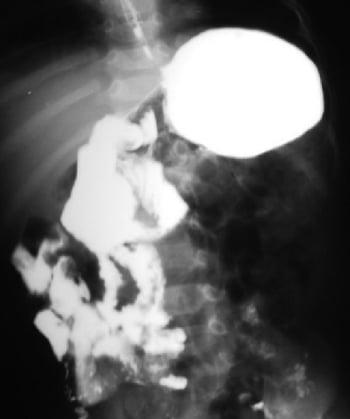
This barium study shows malrotation of the bowel. The duodenojejunal junction is on the right side of the spine, and most of the small intestine is on the right side.
If plain radiographs are nonspecific and no obstruction is present, clinicians sometimes begin with an upper GI series because this may detect other conditions that cause similar symptoms.

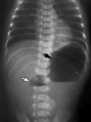
This radiograph shows the typical double-bubble sign seen with complete duodenal obstruction. The smaller bubble represents the proximal, dilated duodenum (white arrow); the larger bubble represents the stomach (black arrow). This sign can be seen with duodenal atresia, duodenal web, annular pancreas, and preduodenal portal vein. Rarely, it can also be seen with complete duodenal obstruction resulting from Ladd bands in a patient with malrotation.
In nonemergent situations, the definitive imaging for malrotation is an upper GI series. Ultrasound is used to diagnose malrotation by looking for retromesenteric localization of the third portion of the duodenum, or reversed mesenteric vessel position, and to diagnose volvulus by looking for the whirlpool sign (2). The use of ultrasound depends on the availability of an experienced radiologist and a radiology technician (3).
Diagnosis references
1. Graziano K, Islam S, Dasgupta R, et al. Asymptomatic malrotation: Diagnosis and surgical management: An American Pediatric Surgical Association outcomes and evidence based practice committee systematic review. J Pediatr Surg. 2015;50:1783–1790. doi:10.1016/j.jpedsurg.2015.06.019
2. Nguyen HN, Kulkarni M, Jose J, et al. Ultrasound for the diagnosis of malrotation and volvulus in children and adolescents: a systematic review and meta-analysis. Arch Dis Child. 2021;106(12):1171-1178. doi:10.1136/archdischild-2020-321082
3. Zhou LY, Li SR, Wang W, et al. Usefulness of sonography in evaluating children suspected of malrotation: Comparison with an upper gastrointestinal contrast study. J Ultrasound Med. 2015;34:1825–1832. doi:10.7863/ultra.14.10017
Treatment of Malrotation of the Bowel
Surgical repair with Ladd procedure
The surgical treatment for malrotation is a Ladd procedure, which consists of detorsion of volvulus (if present), division of Ladd bands (fibrous retroperitoneal bands that can cause duodenal obstruction), widening of the small bowel mesentery, repositioning of the gut (small bowel to right and colon to left), and appendectomy. The Ladd procedure can be performed laparoscopically or as an open procedure (1).
The presence of midgut volvulus with malrotation is an emergency requiring immediate surgery. Performing the Ladd procedure laparoscopically for malrotation without volvulus may decrease the time until enteral nutrition is reintroduced and reduce the length of hospital stay compared to an open procedure (2).
A Ladd procedure may be performed electively in symptomatic patients who have not yet developed a volvulus (3). It may also be performed prophylactically in asymptomatic patients in whom malrotation is found incidentally and in patients with heterotaxy syndrome; however, this practice is controversial (4–6).
Treatment references
1. Ooms N, Matthyssens LE, Draaisma JM, et al. Laparoscopic treatment of intestinal malrotation in children. Eur J Pediatr Surg. 2016;26:376–381. doi:10.1055/s-0035-1554914
2. Xie W, Li Z, Wang Q, Wang L, Pan Y, Lu C. Laparoscopic vs open Ladd's procedure for malrotation in neonates and infants: a propensity score matching analysis. BMC Surg. 2022;22(1):25. Published 2022 Jan 26. doi:10.1186/s12893-022-01487-1
3. Covey SE, Putnam LR, Anderson KT, Tsao K. Prophylactic versus symptomatic Ladd procedures for pediatric malrotation. J Surg Res. 2016;205(2):327-330. doi:10.1016/j.jss.2016.06.097
4. Yu DC, Thiagarajan RR, Laussen PC, Laussen JP, Jaksic T, Weldon CB. Outcomes after the Ladd procedure in patients with heterotaxy syndrome, congenital heart disease, and intestinal malrotation. J Pediatr Surg. 2009;44(6):1089-1095. doi:10.1016/j.jpedsurg.2009.02.015
5. Malek MM, Burd RS. The optimal management of malrotation diagnosed after infancy: a decision analysis. Am J Surg. 2006;191(1):45-51. doi:10.1016/j.amjsurg.2005.10.002
6. Landisch R, Abdel-Hafeez AH, Massoumi R, Christensen M, Shillingford A, Wagner AJ. Observation versus prophylactic Ladd procedure for asymptomatic intestinal rotational abnormalities in heterotaxy syndrome: A systematic review. J Pediatr Surg. 2015;50(11):1971-1974. doi:10.1016/j.jpedsurg.2015.08.002
Key Points
During embryonic development, the bowel begins outside the abdominal cavity and then returns to the abdomen and rotates; incomplete rotation may cause bowel obstruction.
Patients are often asymptomatic, but some have mild, nonspecific symptoms (eg, reflux) or present with life-threatening bowel obstruction (eg, bilious emesis) due to volvulus.
Other malformations, typically gastrointestinal (GI), are present in 30 to 60% of patients.
Do GI radiographs and upper GI series and/or barium enema.
Do surgical repair for symptomatic infants.


