

Transposition of the great arteries is a reversal of the normal connections of the aorta and the pulmonary artery with the heart.
The aorta and pulmonary artery are reversed, which causes oxygen-poor blood to be circulated to the body and oxygen-rich blood to be circulated between the lungs and the heart and not to the body.
Symptoms are apparent at birth and include severe cyanosis (a bluish discoloration of the lips and skin) and difficulty breathing.
The diagnosis is based on echocardiography.
Medication or urgent cardiac catheterization may be needed shortly after birth.
Surgery is done during the first few days of life.
(See also Overview of Heart Defects.)
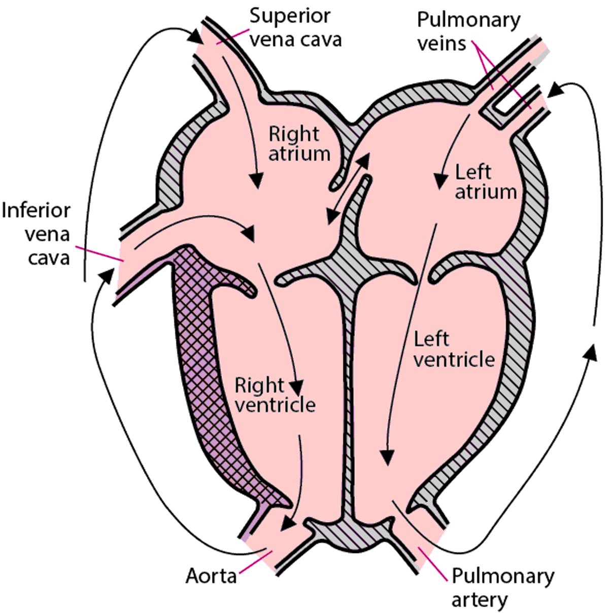
Oxygen-poor blood returning from the body enters the right atrium and then flows to the right ventricle as usual, but then flows to the aorta and the body, bypassing the lungs. Oxygen-rich blood travels back and forth between the heart and lungs (from the lungs to the pulmonary vein, then left atrium and ventricle, then the pulmonary artery) but is not transported to the body.
The body cannot survive without oxygen. However, infants with this heart defect may survive briefly after birth because the foramen ovale (a hole between the right and left atria) and the ductus arteriosus (a blood vessel connecting the pulmonary artery with the aorta—see Normal Fetal Circulation) are still open at birth. These openings allow oxygen-rich blood to mix with oxygen-poor blood, usually supplying enough oxygen to the body to keep the infant alive. Once the ductus closes, as it normally does after birth, if the atrial opening is very small, very little oxygen-rich blood is able to get to the body. The infant will die if the atrial defect is not enlarged immediately.
Transposition of the great arteries can also be accompanied by another heart defect, ventricular septal defect (a hole in the wall separating the two lower heart chambers), which may actually be helpful by allowing more mixing of oxygen-rich and oxygen-poor blood.
Transposition of the Great Arteries
The normal connections of the aorta and the pulmonary artery with the heart are reversed. |

Symptoms of Transposition of the Great Arteries
Transposition of the great arteries usually results in severe cyanosis (bluish discoloration of the lips and skin) and difficulty breathing beginning at birth.
Diagnosis of Transposition of the Great Arteries
Echocardiography
Doctors suspect the diagnosis when they detect very low levels of oxygen in the blood of a newborn. The newborn also has rapid but not labored breathing.
Echocardiography (ultrasonography of the heart) confirms the diagnosis.
Electrocardiography (ECG) and chest x-rays are typically done. The ECG is usually normal. The chest x-ray may show an enlarged or abnormally shaped heart.
Treatment of Transposition of the Great Arteries
Sometimes a medication, such as a prostaglandin, to keep the ductus arteriosus open
Balloon atrial septostomy
Surgery
Usually, surgery is done within the first few days of life. However, other procedures may be done first to keep the infant alive until surgery can be done.
In some infants, doctors give a prostaglandin by vein to keep the ductus arteriosus open. Keeping the ductus arteriosus open may help by increasing blood flow in the lungs and raising oxygen levels in the blood. Doctors closely monitor infants receiving this medication because they occasionally become less stable when receiving a prostaglandin.
Infants who have severe symptoms and do not respond to the prostaglandin may have an atrial opening that is too small to allow adequate mixing of oxygen-rich blood from the left atrium with oxygen-poor blood in the right atrium. In this situation, doctors may do a balloon atrial septostomy procedure. A thin tube (catheter) with a balloon at its tip is passed through a blood vessel in the belly button (umbilical vein) or groin (femoral vein) into the heart. The tip is placed in the opening between the atria (foramen ovale), and the balloon is inflated and pulled through the opening. This procedure widens the foramen and thus enables oxygen-rich blood to go from the left atrium to the right atrium and then out into the body.
Surgery consists of de-attaching the aorta and pulmonary artery and "switching" them to the appropriate ventricles. The heart's coronary arteries are then reattached to the aorta after the aorta is repositioned (this operation is known as the arterial switch procedure).
After surgery, only some children with certain residual defects need to take antibiotics before visits to the dentist and before certain surgeries (such as on the respiratory tract). These antibiotics are used to prevent a serious heart infection called endocarditis.
More Information
The following English-language resources may be useful. Please note that THE MANUAL is not responsible for the content of these resources.
American Heart Association: Common Heart Defects: Provides an overview of common birth defects of the heart for parents and caregivers
American Heart Association: Infective Endocarditis: Provides an overview of infective endocarditis, including summarizing antibiotic use, for parents and caregivers


