

Hemangiomas are abnormal overgrowths of blood vessels that can appear as red or purple lumps in the skin and on other parts of the body.
(See also Overview of Skin Growths and Overview of Growths and Malformations of the Vessels.)
Hemangiomas of infancy are very common. Hemangiomas also develop during middle age and later, especially on the trunk.
Hemangiomas of infancy (also called strawberry or infantile hemangiomas)
These angiomas are the most common tumor of infancy, affecting 5 to 10% of infants by 1 year of age. Hemangiomas of infancy develop soon after birth and tend to enlarge rapidly during the first year of life. After 12 to 18 months, they begin to shrink. Many infantile hemangiomas spontaneously disappear within the first 3 to 5 years of life.
Hemangiomas of infancy grow within and under the skin. They cause the skin to bulge and become bright red or, if they are very deep, bluish. Most deep hemangiomas grow between ¼ and 2 inches (0.5 and 5 centimeters) across, although sometimes they grow much larger. More than half occur on the head and neck. Sometimes, hemangiomas develop in organs, such as the liver (see Hemangiomas of the Liver).

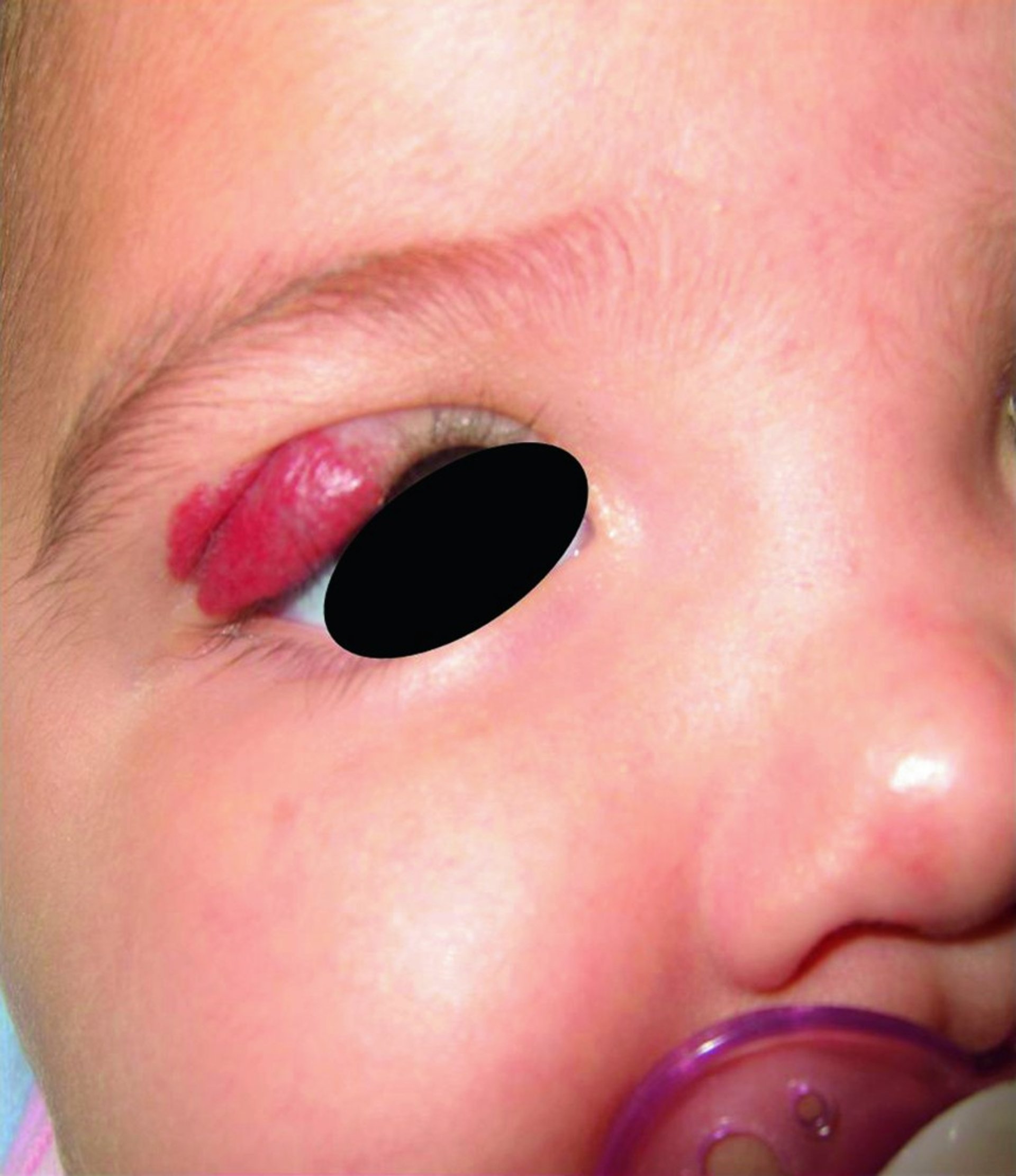
Infantile hemangiomas, which are noncancerous (benign), can appear near the eye or eyelid and can lead to a lazy eye (amblyopia).
© Springer Science+Business Media
Hemangiomas of infancy do not cause pain but occasionally break open (ulcerate) and bleed. Hemangiomas around the eye may grow large enough to block vision, which can lead to permanent vision loss if uncorrected. Hemangiomas may also block the nose or throat, which can block breathing.
Doctors may do magnetic resonance imaging (MRI) if the hemangioma seems to be blocking the nose or throat or another important structure.
Because hemangiomas of infancy usually go away on their own, doctors may not treat them when they first appear unless they grow rapidly, block vision or breathing, ulcerate, or are cosmetically distressing.
When treatment of hemangiomas of infancy is required, doctors may prescribe steroids (also sometimes referred to as glucocorticoids or corticosteroids) that are applied to the skin, injected, or taken by mouth; laser treatments; or propranolol applied to the skin or taken by mouth. Hemangiomas that have ulcerated are treated with compresses, medications that are applied to the wound, and dressings. Surgical removal is usually not recommended because the vast majority of hemangiomas go away on their own and there is less scarring when they are left alone. For older children in whom the hemangioma has shrunk to the greatest degree, surgery may improve the appearance of the skin.; or propranolol applied to the skin or taken by mouth. Hemangiomas that have ulcerated are treated with compresses, medications that are applied to the wound, and dressings. Surgical removal is usually not recommended because the vast majority of hemangiomas go away on their own and there is less scarring when they are left alone. For older children in whom the hemangioma has shrunk to the greatest degree, surgery may improve the appearance of the skin.
Drug Information for the Topic


