

Viral conjunctivitis is a highly contagious, acute conjunctival infection usually caused by an adenovirus. Symptoms include irritation, photophobia, and watery discharge. Diagnosis is clinical; sometimes viral cultures or immunodiagnostic testing is indicated. Infection is self-limited, but severe cases sometimes require topical corticosteroids.
Etiology of Viral Conjunctivitis
Conjunctivitis may accompany the common cold and other systemic viral infections (especially measles, but also chickenpox, rubella, and mumps). Localized viral conjunctivitis without systemic manifestations usually results from adenoviruses (up to 90% of viral conjunctivitis) and sometimes enteroviruses or herpes simplex virus (1.3 to 4.8% of viral conjunctivitis) (1).
Epidemic keratoconjunctivitis is a severe form of viral conjunctivitis that is usually caused by adenovirus serotypes Ad 5, 8, 11, 13, 19, and 37. Adenoviruses can also be identified by genotypes. Genotype HAdV-D is associated with conjunctivitis and HAdV-D53 and HAdV-D54 have been associated with epidemic keratoconjunctivitis. Pharyngoconjunctival fever usually results from serotypes Ad 3, 4, and 7.
Outbreaks of acute hemorrhagic conjunctivitis, a rare conjunctivitis associated with infection by enterovirus type 70, have occurred in Africa and Asia. Ebola virus (which is associated with the highly contagious and potentially fatal Ebola hemorrhagic fever) and SARS-CoV-2 infections (which are associated with the highly contagious and potentially fatal COVID-19 infection) can manifest with bilateral conjunctival hyperemia, tearing, and systemic symptoms (2). Caution and appropriate personal protective equipment should be used when examining patients with conjunctivitis, systemic symptoms, and travel from high-risk regions.
Etiology references
1. Azari AA, Barney NP. Conjunctivitis: a systematic review of diagnosis and treatment [published correction appears in JAMA. 2014 Jan 1;311(1):95. Dosage error in article text]. JAMA. 2013;310(16):1721-1729. doi:10.1001/jama.2013.280318
2. Navel V, Chiambaretta F, Dutheil F. Haemorrhagic conjunctivitis with pseudomembranous related to SARS-CoV-2. Am J Ophthalmol Case Rep. 2020;19:100735. Published 2020 May 6. doi:10.1016/j.ajoc.2020.100735
Symptoms and Signs of Viral Conjunctivitis
After an incubation period of about 5 to 12 days, conjunctival hyperemia, watery discharge, and ocular irritation usually begin in one eye and spread rapidly to the other. Follicles may be present on the palpebral conjunctiva. A preauricular lymph node is often enlarged and painful.
Many patients have had contact with someone with conjunctivitis, a recent upper respiratory infection, or both.

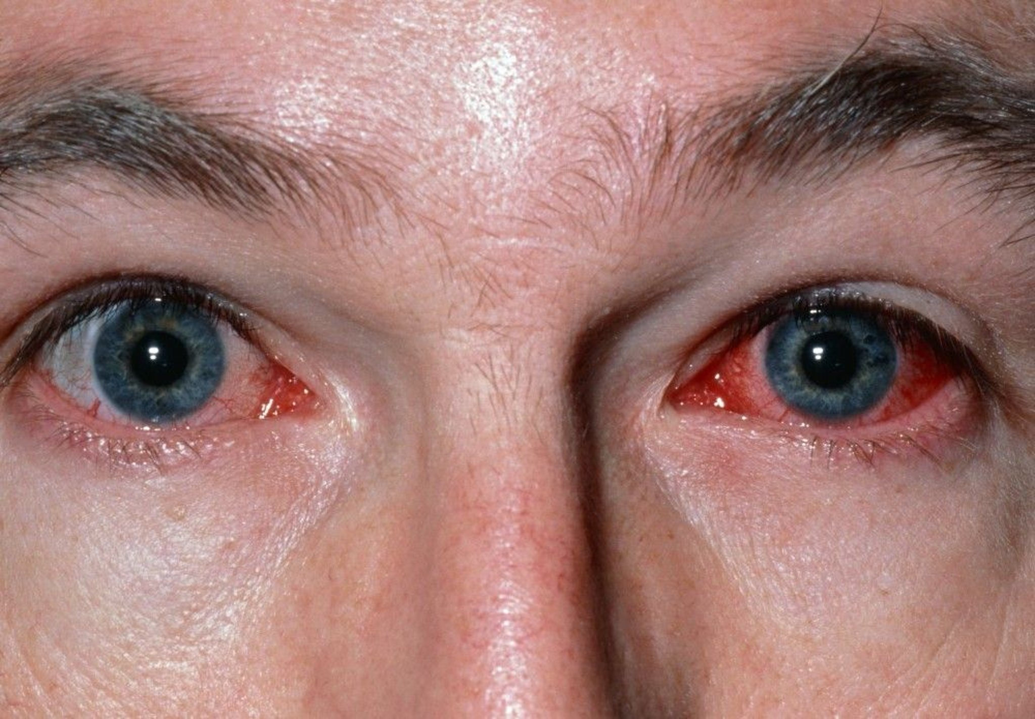
This photo shows bilateral conjunctival erythema (more in the left eye) in a patient with viral conjunctivitis. A discharge may also be present, typically clear. Itching is not usually a feature.
DR P. MARAZZI/SCIENCE PHOTO LIBRARY
In epidemic keratoconjunctivitis, patients may have photophobia and foreign body sensation due to corneal involvement. Chemosis may be present. Pseudomembranes of fibrin and inflammatory cells on the tarsal conjunctiva, focal corneal inflammation, or both may blur vision. Even after conjunctivitis has resolved, residual corneal subepithelial opacities (multiple, coin-shaped, 0.5 to 1.0 mm in diameter) may be visible with a slit lamp for up to 2 years (1). Corneal opacities occasionally result in decreased vision and significant halos and starbursts.
Symptoms and signs reference
1. Jonas RA, Ung L, Rajaiya J, Chodosh J. Mystery eye: Human adenovirus and the enigma of epidemic keratoconjunctivitis. Prog Retin Eye Res. 2020;76:100826. doi:10.1016/j.preteyeres.2019.100826
Diagnosis of Viral Conjunctivitis
Ocular examination
Diagnosis of conjunctivitis and differentiation between bacterial, viral, and noninfectious conjunctivitis (see table ) are usually clinical; however, differentiation between viral and bacterial conjunctivitis can be imprecise because symptoms may overlap. Special tissue cultures are necessary for growth of the virus but are rarely indicated. Nucleic acid amplification tests (NAAT) and other rapid, office-based immunodiagnostic tests, can be useful especially when the inflammation is severe and other diagnoses (eg, orbital cellulitis) must be ruled out (1).
Features that may help differentiate between viral and bacterial conjunctivitis can include purulence of ocular discharge, presence of preauricular lymphadenopathy, and, in epidemic keratoconjunctivitis, chemosis.
Patients with photophobia are stained with fluorescein and examined with a slit lamp. Epidemic keratoconjunctivitis may cause punctate corneal staining. Herpes simplex keratitis may also be seen by fluorescein staining and Patients with photophobia are stained with fluorescein and examined with a slit lamp. Epidemic keratoconjunctivitis may cause punctate corneal staining. Herpes simplex keratitis may also be seen by fluorescein staining andslit-lamp examination.
Secondary bacterial infection of viral conjunctivitis is very rare. However, if any signs suggest bacterial conjunctivitis (eg, purulent discharge), cultures or other studies may be useful.
Differentiating Features in Acute Conjunctivitis
Etiology | Discharge/Cell Type | Eyelid Edema | Node Involvement | Itching |
|---|---|---|---|---|
Purulent/polymorphonuclear leukocytes | Moderate | Usually none | None | |
Clear/mononuclear cells | Minimal | Often present | Usually None | |
Clear, mucoid, ropy/eosinophils | Moderate to severe | None | Mild to intense |
Diagnosis reference
1. Azari AA, Barney NP. Conjunctivitis: a systematic review of diagnosis and treatment [published correction appears in JAMA. 2014 Jan 1;311(1):95. Dosage error in article text]. JAMA. 2013;310(16):1721-1729. doi:10.1001/jama.2013.280318
Treatment of Viral Conjunctivitis
Supportive measures
Viral conjunctivitis is highly contagious, and transmission precautions must be followed.
Physicians take the following precautions to avoid transmitting infection:
Use hand sanitizer or wash their hands properly (fully lather hands, scrub hands for at least 20 seconds, rinse well, and turn off the water using a paper towel)
Disinfect equipment after examining patients
Patients should do the following:
Use hand sanitizer and/or wash their hands thoroughly after touching their eyes or nasal secretions
Avoid touching the noninfected eye after touching the infected eye
Avoid sharing towels or pillows
Avoid swimming in pools
Eyes should be kept free of discharge and should not be patched. Small children with conjunctivitis should be kept home from school to avoid spreading the infection.
Viral conjunctivitis is self-limiting, lasting 1 week in mild cases to up to 3 weeks in severe cases. It requires only cool compresses for symptomatic relief. However, patients who have severe photophobia or whose vision is affected may benefit from topical corticosteroids (eg, prednisolone acetate 1% drops 4 times a day). Corticosteroids, if prescribed, are usually prescribed by an ophthalmologist. Viral conjunctivitis is self-limiting, lasting 1 week in mild cases to up to 3 weeks in severe cases. It requires only cool compresses for symptomatic relief. However, patients who have severe photophobia or whose vision is affected may benefit from topical corticosteroids (eg, prednisolone acetate 1% drops 4 times a day). Corticosteroids, if prescribed, are usually prescribed by an ophthalmologist.Herpes simplex keratitis must be ruled out first (by fluorescein staining and slit-lamp examination) because corticosteroids can exacerbate it. Topical cyclosporin A drops are overall less effective but are helpful if corticosteroid drop use is limited by adverse effects. must be ruled out first (by fluorescein staining and slit-lamp examination) because corticosteroids can exacerbate it. Topical cyclosporin A drops are overall less effective but are helpful if corticosteroid drop use is limited by adverse effects.
In severe cases, any conjunctival pseudomembranes should be removed at the slit-lamp examination to reduce the chances of conjunctival scarring and symblepharon formation.
Key Points
Most viral conjunctivitis is a highly contagious infection caused by adenoviruses or enteroviruses.
Features that may help differentiate between viral and bacterial conjunctivitis can include purulence of ocular discharge, presence of preauricular lymphadenopathy, and, in epidemic keratoconjunctivitis, chemosis.
Diagnosis is usually clinical.
Treatment is usually cool compresses and measures to prevent spread.
Drug Information for the Topic


