

Gallbladder and bile duct tumors can cause extrahepatic biliary obstruction. Symptoms may be absent but often are constitutional or reflect biliary obstruction. Diagnosis is based on ultrasound plus magnetic resonance cholangiopancreatography (MRCP), CT angiography, or endoscopic retrograde cholangiopancreatography (ERCP) depending upon cancer type. Prognosis is generally poor. When feasible, surgical resection with adjuvant chemotherapy can be offered for cholangiocarcinoma; mechanical bile duct drainage can often relieve pruritus, recurrent sepsis, and pain due to biliary obstruction.
(See also Overview of Biliary Function.)
Cholangiocarcinomas and other bile duct tumors are rare (worldwide incidence < 1 to 3/100,000 people and much higher where liver flukes are endemic) but usually malignant (1, 2, 3). Cholangiocarcinomas occur predominantly in the extrahepatic bile ducts: 60 to 70% in the perihilar region (Klatskin tumors), approximately 25% in the distal extrahepatic ducts, and the remainder intrahepatically (4). Established risk factors include primary sclerosing cholangitis, alcohol use, infestation with liver flukes, choledochal cyst, cirrhosis, and viral hepatitis.
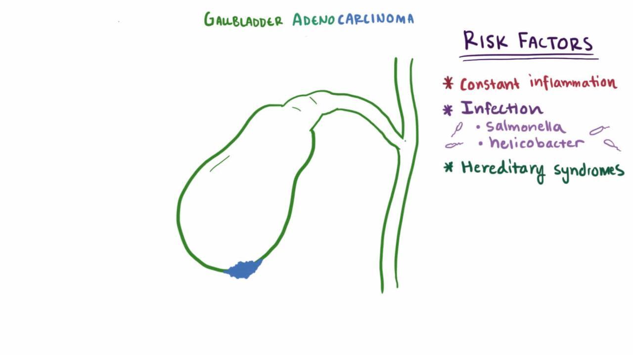
Gallbladder carcinoma is uncommon (worldwide incidence 1.4/100,000 ) (5). It is more common among older patients, women, patients with gallstones, and American Indians. Extensive gallbladder calcification due to chronic cholecystitis (porcelain gallbladder) is weakly associated with gallbladder carcinoma (6). Most (69 to > 95%) patients also have gallstones (7, 8). Prognosis is poor with overall median survival of approximately 6 months with 5-year survival just 5% (9). Cure is possible when cancer is found early (eg, incidentally at cholecystectomy).

Gallbladder polyps are usually asymptomatic benign mucosal projections that develop in the lumen of the gallbladder. They are found in approximately 5% of adults (10). Most are < 10 mm in diameter and composed of cholesterol ester and triglycerides; the presence of such polyps is called cholesterolosis. Patients with gallbladder polyps < 10 mm should undergo surveillance ultrasound at a frequency based on their risk factors and polyp size. Cholecystectomy is sometimes recommended for polyps >10 mm.
Other, much less common benign polyps include adenomas (causing adenomyomatosis) and inflammatory polyps.
General references
1. Banales JM, Cardinale V, Carpino G, et al. Expert consensus document: Cholangiocarcinoma: current knowledge and future perspectives consensus statement from the European Network for the Study of Cholangiocarcinoma (ENS-CCA). Nat Rev Gastroenterol Hepatol. 2016;13(5):261-280. doi: 10.1038/nrgastro.2016.51
2. Bowlus CL, Arrivé L, Bergquist A, et al. AASLD practice guidance on primary sclerosing cholangitis and cholangiocarcinoma. Hepatology. 2023;77(2):659-702. doi:10.1002/hep.32771
3. Lauterio A, De Carlis R, Centonze L, et al. Current Surgical Management of Peri-Hilar and Intra-Hepatic Cholangiocarcinoma. Cancers (Basel). 2021;13(15):3657. doi:10.3390/cancers13153657
4. Buckholz AP, Brown RS Jr. Cholangiocarcinoma: Diagnosis and Management. Clin Liver Dis. 2020;24(3):421-436. doi:10.1016/j.cld.2020.04.005
5. Vuthaluru S, Sharma P, Chowdhury S, Are C. Global epidemiological trends and variations in the burden of gallbladder cancer. J Surg Oncol. 2023;128(6):980-988. doi:10.1002/jso.27450
6. Schnelldorfer T. Porcelain gallbladder: a benign process or concern for malignancy?. J Gastrointest Surg. 2013;17(6):1161-1168. doi:10.1007/s11605-013-2170-0
7. Hsing AW, Gao YT, Han TQ, et al. Gallstones and the risk of biliary tract cancer: a population-based study in China. Br J Cancer. 2007;97(11):1577-1582. doi:10.1038/sj.bjc.6604047
8. Cariati A, Piromalli E, Cetta F. Gallbladder cancers: associated conditions, histological types, prognosis, and prevention. Eur J Gastroenterol Hepatol. 2014;26(5):562-569. doi:10.1097/MEG.0000000000000074
9. Hundal R, Shaffer EA. Gallbladder cancer: epidemiology and outcome. Clin Epidemiol. 2014;6:99-109. doi:10.2147/CLEP.S37357
10. Babu BI, Dennison AR, Garcea G. Management and diagnosis of gallbladder polyps: a systematic review. Langenbecks Arch Surg. 2015;400(4):455-462. doi:10.1007/s00423-015-1302-2
Symptoms and Signs of Gallbladder and Bile Duct Tumors
Most patients with cholangiocarcinomas present with pruritus and painless obstructive jaundice. Early perihilar tumors may cause only vague abdominal pain, anorexia, and weight loss. Other features include fatigue, acholic stool, a palpable mass, hepatomegaly, or a distended gallbladder (Courvoisier sign, with distal cholangiocarcinoma). Pain may resemble that of biliary colic (reflecting biliary obstruction) or may be constant and progressive. Sepsis (secondary to acute cholangitis), although unusual, may be induced by endoscopic retrograde cholangiopancreatography.
Metastatic disease and noncancerous biliary tract tumors may also cause biliary obstruction and, therefore, symptoms.
Manifestations of gallbladder carcinoma may range from incidental findings at cholecystectomy performed to relieve biliary pain to cholelithiasis to advanced disease with constant pain, weight loss, and an abdominal mass or obstructive jaundice.
Most gallbladder polyps cause no symptoms.
Diagnosis of Gallbladder and Bile Duct Tumors
Sometimes laboratory tests
Ultrasound (sometimes endoscopic), followed by magnetic resonance cholangiopancreatography (MRCP) or CT cholangiography
Sometimes endoscopic retrograde cholangiopancreatography (ERCP) or tissue sampling
Cholangiocarcinomas are suspected when extrahepatic biliary obstruction is unexplained. Cholangiocarcinoma and gallbladder cancer is particularly suspected in patients with primary sclerosing cholangitis (PSC) or ulcerative colitis. In patients with primary sclerosing cholangitis, serum carcinoembryonic antigen (CEA) and carbohydrate antigen (CA) 19-9 levels are measured periodically to check for cholangiocarcinoma (1). Laboratory test results reflect the degree of cholestasis.
Diagnosis of cholangiocarcinoma ultimately requires a tissue biopsy, but can be suspected based on ultrasound (or endoscopic ultrasound). CT or MRI is sometimes performed and may provide additional information, particularly for gallbladder carcinomas Typically MRCP or CT cholangiography follows (see Imaging Tests of the Liver and Gallbladder).
For cholangiocarcinoma of the common bile duct, or sometimes the right or left hepatic ducts, ERCP can be used to detect the tumor but also, with cytology brushings, can provide a tissue diagnosis and sometimes make ultrasound- or CT-guided needle biopsy unnecessary. Contrast-enhanced CT assists in staging.
Laparotomy is often necessary to determine disease extent in cholangiocarcinoma, which guides treatment.
For symptoms suggesting gallstone disease, ultrasound may lead to the initial suspicion or diagnosis of gallbladder cancer, with subsequent staging by CT or MRI (2). Biopsy or fine needle aspiration is performed to identify the presence of malignancy. If metastatic disease is the initial clinical presentation, biopsy or fine needle aspiration of the metastatic disease may yield the initial diagnosis.
Diagnosis references
1. Buckholz AP, Brown RS Jr. Cholangiocarcinoma: Diagnosis and Management. Clin Liver Dis. 2020;24(3):421-436. doi:10.1016/j.cld.2020.04.005
2. Roa JC, García P, Kapoor VK, et al. Gallbladder cancer. Nat Rev Dis Primers. 2022;8(1):69. doi:10.1038/s41572-022-00398-y
Treatment of Gallbladder and Bile Duct Tumors
For cholangiocarcinomas, sometimes surgical resection, sometimes with chemotherapy
For unresectable cholangiocarcinomas, stenting (or another bypass procedure) or occasionally partial resection
For gallbladder carcinoma, usually symptomatic treatment
For gallbladder polyps > 10 mm in size, cholecystectomy
For cholangiocarcinoma, surgical resection is the only potentially curative therapy for localized disease, but many patients are not surgical candidates. Prognosis is poor even with resection (post-resection 5-year survival rate is 40 to 60% for intrahepatic disease, 30 to 50% for perihilar disease, and 27% for distal cholangiocarcinoma) (1, 2).
Surgical resection for intrahepatic disease involves partial hepatic resection. Surgery for perihilar cholangiocarcinoma involves lobectomy, bile duct resection, and hepaticojejunostomy.
Surgery for distal cholangiocarcinoma involves a pancreaticoduodenectomy (Whipple procedure). Neoadjuvant chemotherapy may be used to improve resectability. Preoperative biliary drainage for extrahepatic strictures due to resectable cholangiocarcinoma is indicated for obstructive jaundice and cholangitis (3). Adjuvant chemotherapy is recommended for all types of cholangiocarcinoma.
For patients who are not surgical candidates (eg, those with hilar cholangiocarcinomas with CT evidence of spread), stent placement or surgically bypass of the obstruction relieves pruritus, jaundice, and fatigue (2). Stenting is performed via percutaneous transhepatic cholangiography or endoscopic retrograde cholangiopancreatography (ERCP). Distal duct cholangiocarcinomas are stented endoscopically with ERCP.
If cholangiocarcinoma appears localized, surgical exploration determines resectability by hilar resection or pancreaticoduodenectomy.
Liver transplantation for localized perihilar cholangiocarcinoma, after neoadjuvant chemotherapy, is offered at some transplant centers (1).
Treatment of gallbladder carcinomas is individualized, with options including resection (often with extended cholecystectomy involving removal of gallbladder, part of the liver, and regional lymph nodes), chemotherapy, or targeted immunotherapy (4).
Gallbladder polyps are treated with cholecystectomy when > 10 mm (5).
Treatment references
1. Buckholz AP, Brown RS Jr. Cholangiocarcinoma: Diagnosis and Management. Clin Liver Dis. 2020;24(3):421-436. doi:10.1016/j.cld.2020.04.005
2. Bowlus CL, Arrivé L, Bergquist A, et al. AASLD practice guidance on primary sclerosing cholangitis and cholangiocarcinoma. Hepatology. 2023;77(2):659-702. doi:10.1002/hep.32771
3. Elmunzer BJ, Maranki JL, Gómez V, et al. ACG Clinical Guideline: Diagnosis and Management of Biliary Strictures. Am J Gastroenterol. 2023;118(3):405-426. doi:10.14309/ajg.0000000000002190
4. Roa JC, García P, Kapoor VK, et al. Gallbladder cancer. Nat Rev Dis Primers. 2022;8(1):69. doi:10.1038/s41572-022-00398-y
5. Babu BI, Dennison AR, Garcea G. Management and diagnosis of gallbladder polyps: a systematic review. Langenbecks Arch Surg. 2015;400(4):455-462. doi:10.1007/s00423-015-1302-2
Key Points
Biliary tract cancer (usually cholangiocarcinoma or gallbladder carcinoma) is uncommon.
Suspect cancer if patients have an unexplained extrahepatic biliary obstruction or abdominal mass.
Diagnose cancers by imaging, beginning with ultrasound.
For cholangiocarcinoma, offer resection and adjuvant chemotherapy when disease is localized and stenting or bypassing obstructions in nonresectable cholangiocarcinoma.
Consider liver transplantation for selected patients with hilar cholangiocarcinoma.


