

Neuropathic arthropathy is a rapidly destructive arthropathy due to impaired pain perception and position sense, which can result from various underlying disorders, most commonly diabetes and stroke. Common manifestations include joint effusion, deformity, and instability. Pain may be disproportionately mild due to the underlying neuropathy. Diagnosis requires radiographic confirmation. Treatment consists of external or sometimes surgical joint stabilization to slow disease progression and reduce pain.
Pathophysiology of Neuropathic Arthropathy
Many conditions predispose to neuropathic arthropathy (see table ). Impaired deep pain sensation or proprioception affects the joint’s normal protective reflexes, often allowing trauma (especially repeated minor episodes) and small periarticular fractures to go unrecognized. Increased blood flow to bone from reflex vasodilation results in active bone resorption, which can contribute to bone and joint damage.
Each new injury sustained by the joint causes more distortion as it heals. Hemorrhagic joint effusions and multiple small fractures can occur, accelerating disease progression. Ligamentous laxity, muscular hypotonia, and rapid destruction of joint cartilage are common, predisposing to joint dislocations, which also accelerate disease progression. Advanced neuropathic arthropathy can cause hypertrophic changes, destructive changes, or both.
Conditions Underlying Neuropathic Arthropathy
Amyloid neuropathy (secondary amyloidosis) |
Arnold-Chiari malformation |
Congenital insensitivity to pain |
Degenerative spinal disease with nerve root compression |
Familial-hereditary neuropathies:
|
Gigantism with hypertrophic neuropathy |
Spina bifida with meningomyelocele (in children) |
Subacute combined degeneration of the spinal cord |
Tabes dorsalis |
Tumors and injuries of the peripheral nerves (see Peripheral Neuropathy) and spinal cord (see Spinal Trauma and Spinal Cord Tumors) |
Symptoms and Signs of Neuropathic Arthropathy
Arthropathy does not usually develop until years after onset of the neurologic condition but can then progress rapidly and lead to complete joint disorganization in a few months. Pain is a common early symptom. However, because the ability to sense pain is commonly impaired, the degree of pain is often unexpectedly mild for the degree of joint damage. A prominent, often hemorrhagic, effusion and subluxation and instability of the joint are usually present during early stages. Acute joint dislocation sometimes also occurs.
During later stages, pain may be more severe if the disease has caused rapid joint destruction (eg, periarticular fractures or tense hematomas). During advanced stages, the joint is swollen from bony overgrowth and massive synovial effusion. Deformity results from dislocations and displaced fractures. Fractures and bony healing may produce loose pieces of cartilage or bone that can slough into the joint. A coarse, grating, often audible crepitus, usually more unpleasant for the observer than for the patient, may develop.
Although many joints can be involved, the knee and the ankle are most often affected. Distribution depends largely on the underlying disease. Thus, tabes dorsalis affects the knee and hip, and diabetes mellitus affects the foot and ankle. Syringomyelia commonly affects the spine and upper limb joints, especially the elbow and shoulder. Frequently, only 1 joint is affected and usually no more than 2 or 3 (except for the small joints of the feet), in an asymmetric distribution.
Infectious arthritis may develop with or without systemic symptoms (eg, fever, malaise), particularly with diabetes, and requires a high index of suspicion.
Diagnosis of Neuropathic Arthropathy
Radiographs
The diagnosis of neuropathic arthropathy should be considered in a patient with a predisposing neurologic disorder who develops a destructive but relatively painless arthropathy, usually several years after the onset of the underlying neurologic condition. Diagnosis is established by characteristic abnormalities on a radiograph in a patient with a predisposing condition and typical symptoms and signs.
Abnormalities on a radiograph in early-stage neuropathic arthropathy are often similar to those in osteoarthritis (see diagnosis of osteoarthritis). The cardinal signs later in disease progression are
Bone fragmentation
Bone destruction
New bone growth
Loss of joint space
There may also be synovial effusion and joint subluxation. The bones may become deformed, and new bone forms adjacent to the cortex, starting within the joint capsule and often extending up the shaft, particularly in long bones. Calcification and ossification may occur in the soft tissues. Large, bizarrely shaped osteophytes may be present at the joint margins or within joints. Large curved (parrot’s beak) osteophytes frequently develop in the spine in the absence of clinical spinal disease.
Neuropathic arthropathy progresses more rapidly than osteoarthritis.

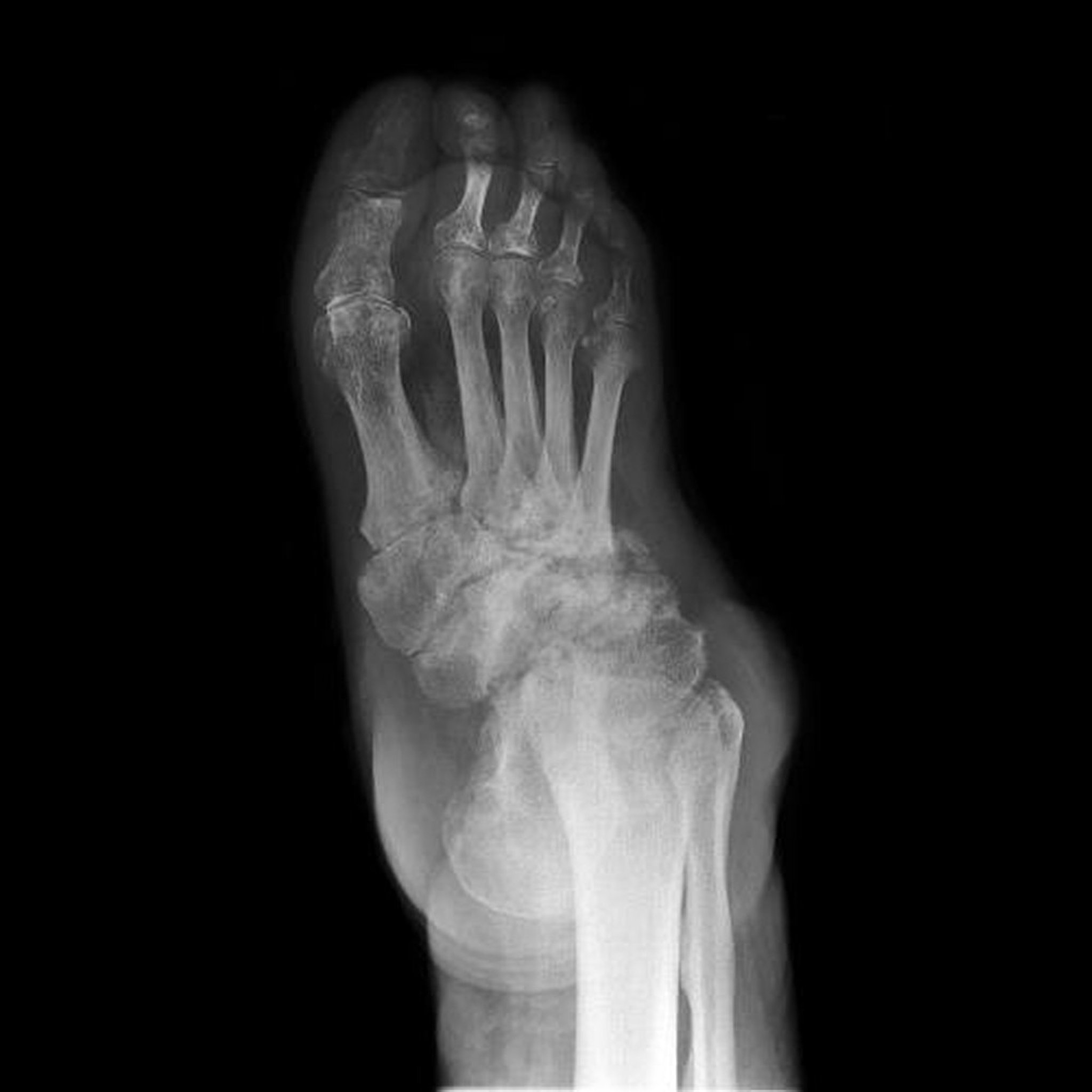
This radiograph shows neuropathic arthropathy of the foot (also known as Charcot foot). Destruction, deformities, and loss of joint spaces of the tarsal bones is extensive.
SCIENCE PHOTO LIBRARY
Treatment of Neuropathic Arthropathy
Treatment of cause
Sometimes surgery
Treatment of the underlying neurologic condition may slow progression of the arthropathy and, if joint destruction is still in the early stages, partially reverse the process.
Early diagnosis of asymptomatic or minimally symptomatic fractures facilitates early treatment; stabilization (with splints, special boots, or calipers) protects the joint from further injury, possibly stopping disease evolution. Stabilization may even prevent neuropathic arthropathy in a patient at risk.
For a grossly disorganized joint, arthrodesis using internal fixation, compression, and an adequate bone graft may be successful. For grossly disorganized hip and knee joints, if neuropathic arthropathy is not expected to be progressive, successful outcomes can be obtained with total hip and knee replacements. However, patients are at higher risk for postoperative complications such as loosening and dislocation of the prosthesis (1).
Treatment reference
1. Zhang Z, Chi J, Raso J, Xu H, Cui Q: Outcomes following total hip arthroplasty in patients who have Charcot neuroarthropathy of the hip [published online ahead of print, 2023 Jun 8]. J Arthroplasty S0883-5403(23)00630-7, 2023. doi:10.1016/j.arth.2023.05.088
Key Points
Neuropathic arthropathy is a rapidly destructive arthropathy that occurs when perception of pain and position sense are impaired (eg, due to diabetes or stroke).
Joint destruction out of proportion to pain is typical, often with rapid progression to joint disorganization in advanced stages.
Confirm the diagnosis with radiographic evidence of joint destruction (initially similar to changes seen in osteoarthritis) out of proportion to pain in patients with a predisposing neurologic disorder.
Be alert to the possibility of infection.
Treat the cause when possible and protect the joint from further injury by physical means (eg, by stabilization).
Refer patients for surgery when appropriate.


