


Horner syndrome affects one side of the face, causing the eyelid to droop, the pupil to become small (constricted), and sweating to decrease. The cause is disruption of the nerve fibers that connect the brain to the eye.
Horner syndrome may occur on its own or result from a disorder that disrupts nerve fibers connecting the brain with the eye.
The upper eyelid droops, the pupil remains small, and the affected side of the face may sweat less.
Doctors test the pupil to see whether it can widen and may do imaging tests to look for a cause.
The cause, if identified, is treated.
(See also Overview of the Autonomic Nervous System.)
Horner syndrome can develop in people of any age.
Causes of Horner Syndrome
Some of the nerve fibers that connect the eyes and brain take a circuitous route. From the brain, they go down the spinal cord. They exit the spinal cord in the chest, then go back up the neck beside the carotid artery, through the skull, and into the eye. If these nerve fibers are disrupted anywhere along their pathway, Horner syndrome results.
Horner syndrome may occur on its own or be caused by another disorder. For example, it can be caused by disorders of the head, brain, neck, chest, or spinal cord, such as the following:
Other tumors
Swollen lymph glands in the neck (cervical adenopathy)
Dissection of the aorta or carotid artery (a tear in the lining of the artery's wall)
A thoracic aortic aneurysm (a bulge in the aorta's wall)
Injuries
Horner syndrome may be present at birth (congenital).
Symptoms of Horner Syndrome
Horner syndrome affects the eye on the same side as the disrupted nerve fibers.
Symptoms of Horner syndrome include a drooping upper eyelid (ptosis) and a constricted pupil (miosis). In some people, the constricted pupil interferes with being able to see in the dark. However, most people do not notice a difference in vision.
The affected side of the face may sweat less than normal or not at all, and rarely, it appears flushed.

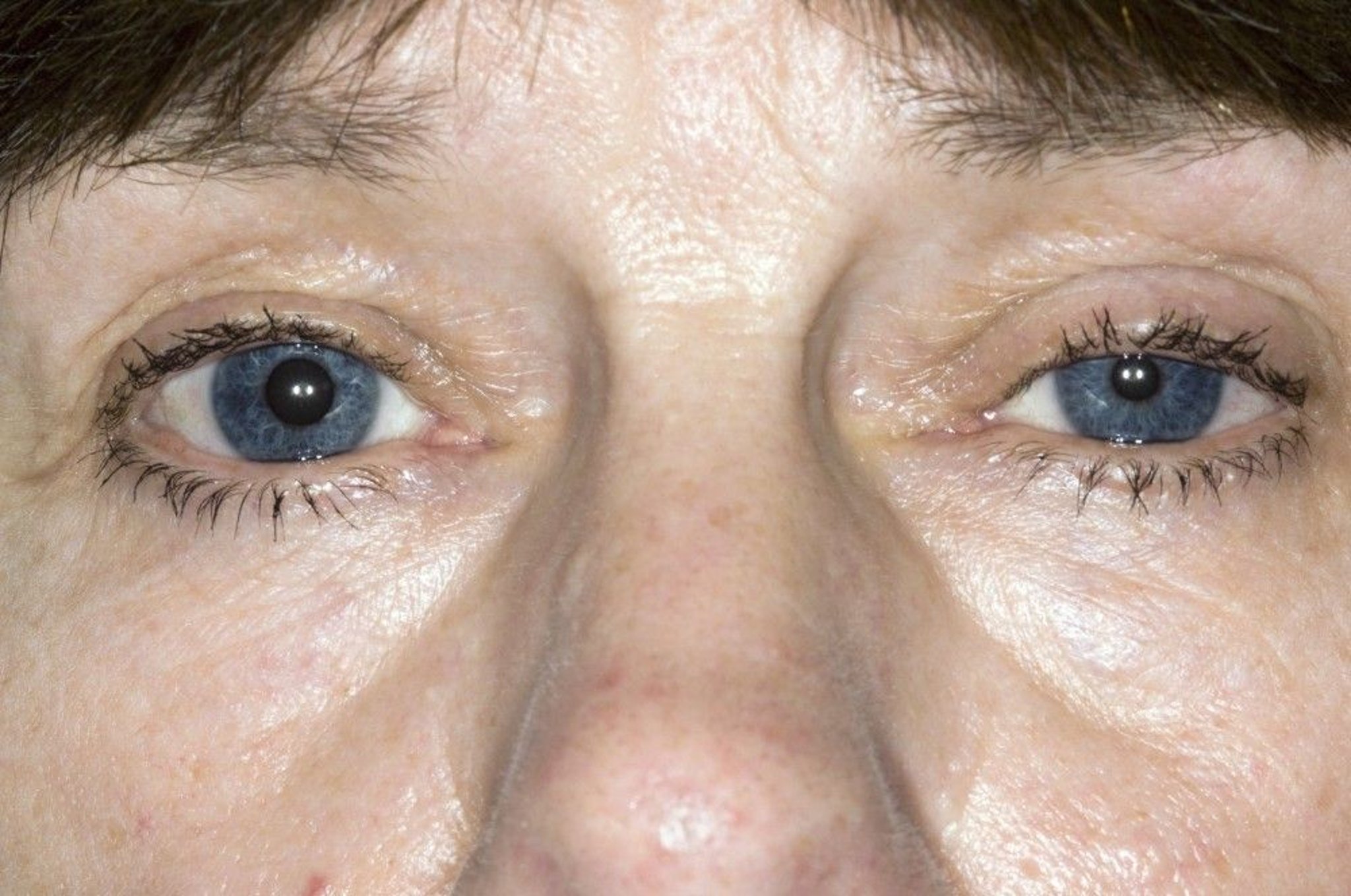
This photo shows a person with Horner syndrome that affects the left eye. The left eyelid droops (called ptosis), and the left pupil is constricted (called miosis).
DR P. MARAZZI/SCIENCE PHOTO LIBRARY
In the congenital form, the iris of the affected eye remains blue-gray as it is at birth.
Diagnosis of Horner Syndrome
Eye drop tests
Magnetic resonance imaging or computed tomography to identify the cause
Horner syndrome is suspected based on symptoms.
To confirm the diagnosis of Horner syndrome and determine where the problem is, doctors do a 2-part test applying specific medication drops to the eye to see how the pupils react to the medications.
If Horner syndrome is likely, magnetic resonance imaging (MRI) or computed tomography (CT) of the brain, spinal cord, chest, and/or neck is done to look for tumors and other serious disorders that may be disrupting nerve fibers connecting the brain and the eye.
Treatment of Horner Syndrome
Treatment of the cause if identified
The cause of Horner syndrome, if identified, is treated. However, there is no specific treatment for Horner syndrome. Often, no treatment is necessary because typically, the eyelid only droops very slightly.


