


Twelve pairs of nerves—the cranial nerves—lead directly from the brain to various parts of the head, neck, and trunk. Some of the cranial nerves are involved in the special senses (such as seeing, hearing, and taste), and others control muscles in the face or regulate glands. The nerves are named and numbered (according to their location, from the front of the brain to the back).
Viewing the Cranial Nerves
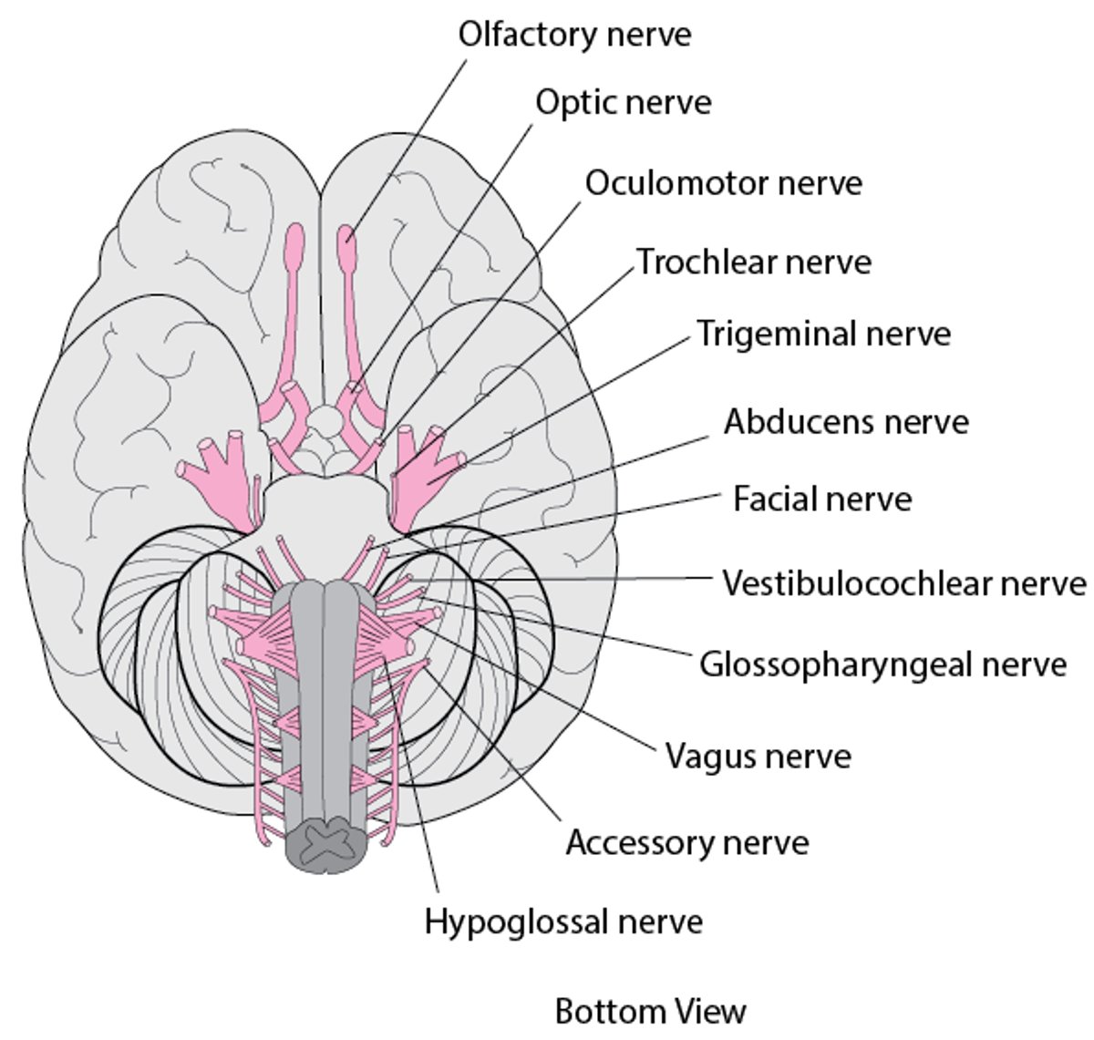
Twelve pairs of cranial nerves emerge from the underside of the brain, pass through openings in the skull, and lead to parts of the head, neck, and trunk. |

A cranial nerve disorder may result when the following are damaged or malfunction:
Areas of the brain that control cranial nerves (called centers, or nuclei), as may occur when a stroke damages the area that controls the facial nerve
The nerve fibers that connect cranial nerve centers within the brain, as occurs in internuclear ophthalmoplegia
Only 1 cranial nerve, as occurs in oculomotor palsy, trigeminal neuralgia, Bell palsy, and hemifacial spasm
Some cranial nerve disorders interfere with eye movement. Eye movement is controlled by 3 pairs of muscles. These muscles move the eye up and down, right and left, and diagonally. The muscles are controlled by the following cranial nerves:
If 1 of these nerves or the area in the brain that controls these muscles is damaged, the muscles may become paralyzed to varying degrees (called a palsy), and people may not be able to move their eyes normally. How eye movement is affected depends on which nerve is affected. People with 1 of these palsies may have double vision when they look in certain directions.
Did You Know...
|
Causes of Cranial Nerve Disorders
Cranial nerve disorders have many causes, including the following:
Head injuries
Tumors
Infections, such as COVID-19, Lyme disease, and shingles
An inadequate blood supply (as occurs in diabetes or stroke)
Pressure on a nerve due to abnormalities in a blood vessel, such as a bulge (aneurysm) in an artery or an abnormal connection between an artery and a vein (arteriovenous malformation)
Disorders that cause nerve cells to degenerate, as occurs in amyotrophic lateral sclerosis (ALS) or multiple sclerosis
Disorders that cause inflammation of blood vessels (vasculitis), such as giant cell arteritis
Certain medications, particularly antibiotics such as aminoglycosides and streptomycin
Some toxins, such as mercury
Symptoms of Cranial Nerve Disorders
Symptoms of cranial nerve disorders depend on which nerves are damaged and how they were damaged. Cranial nerve disorders can affect smell, taste, vision, sensation in the face, facial expression, hearing, balance, speech, swallowing, and muscles of the neck.
For example, vision may be affected in various ways:
If one of the second cranial nerves (optic nerve) is damaged, vision in the affected eye may be partially or completely lost.
If any of the 3 cranial nerves that control eye movement (third, fourth, or sixth cranial nerve) is damaged, people cannot move their eyes normally. Symptoms include double vision when looking in certain directions.
If the third cranial nerve (oculomotor nerve) is paralyzed, people cannot raise their upper eyelid. It droops down over the eye and interferes with vision.
If the eighth cranial nerve (auditory or vestibulocochlear nerve) is damaged or malfunctions, people may have problems hearing and/or have vertigo—a feeling that they, their environment, or both are spinning.
Cranial nerve disorders can also cause various kinds of facial or head pain.
Diagnosis of Cranial Nerve Disorders
A doctor's evaluation
Tests of cranial nerve function
Usually magnetic resonance imaging
When doctors suspect a cranial nerve disorder, they ask the person detailed questions about the symptoms. They also test the function of the cranial nerves by asking the person to do simple tasks, such as to follow a moving target with the eyes.
Imaging of the brain with magnetic resonance imaging (MRI) is often needed.
Testing Cranial Nerves
Cranial Nerve Number | Name | Function | Test |
I | Olfactory | Smell | The ability to smell is tested by asking the person to identify items with very specific odors (such as soap, coffee, and cloves) placed under the nose. Each nostril is tested separately. |
II | Optic | Vision | The ability to see is tested by asking the person to read an eye chart. Peripheral (side) vision is tested by asking the person to look straight ahead while the doctor gradually moves a finger toward the person's center of vision from above, below, left, and right. The person is then asked to say when the finger is first seen. |
Detection of light | The ability to detect light is tested by shining a bright light (as from a flashlight) into each pupil in a darkened room. | ||
III | Oculomotor | Eye movement upward, downward, and inward | The ability to move each eye up, down, and inward is tested by asking the person to follow a target moved by the examiner. |
Narrowing (constriction) or widening (dilation) of the pupil in response to changes in light | The pupils’ response to light is checked by shining a bright light (as from a flashlight) into each pupil in a darkened room. | ||
Raising the eyelids | The upper eyelid is checked for drooping (ptosis). | ||
IV | Trochlear | Eye movement downward and inward | The ability to move each eye down and inward is tested by asking the person to follow a target moved by the examiner. |
V | Trigeminal | Facial sensation | Sensation in areas of the face is tested using a pin and a wisp of cotton. The blink reflex is tested by touching the cornea of the eye with a cotton wisp. |
Chewing | Strength and movement of muscles that control the jaw are tested by asking the person to clench the teeth and open the jaw against resistance. | ||
VI | Abducens | Eye movement outward | The ability to move each eye outward beyond the midline is tested by asking the person to look to the side. |
VII | Facial | The ability to move muscles in the face (for example, in facial expressions), taste in the front two-thirds of the tongue, production of saliva and tears, and control of a muscle involved in hearing | The ability to move the face is tested by asking the person to smile, to open the mouth and show the teeth, and to close the eyes tightly. Taste is tested using substances that are sweet (sugar), sour (lemon juice), salty (salt), and bitter (aspirin, quinine, or aloes). |
VIII | Auditory (vestibulocochlear) | Hearing | Hearing is tested with a tuning fork or with headphones that play tones of different frequencies (pitches) and loudness (audiometry). |
Balance | Balance is tested by asking the person to walk a straight line. | ||
IX | Glossopharyngeal | Swallowing, the gag reflex, and speech | Because both the ninth and tenth cranial nerves control swallowing and the gag reflex, they are tested together. The person is asked to swallow. The person is asked to say “ah-h-h” to check movement of the palate (roof of the mouth) and uvula (the small, soft projection that hangs down at the back of throat). The back of the throat may be touched with a tongue blade, which evokes the gag reflex in most people. The person is asked to speak to determine whether the voice sounds nasal (another test of palate movement). |
X | Vagus | Swallowing, the gag reflex, and speech | |
Control of muscle in some internal organs and the heart rate | This function is not tested as part of the cranial nerve examination. | ||
XI | Accessory | Neck turning and shoulder shrugging | The person is asked to turn the head and to shrug the shoulders against resistance provided by the examiner. |
XII | Hypoglossal | Tongue movement | The person is asked to stick out the tongue, which is observed for deviation to one side or the other. |
Treatment of Cranial Nerve Disorders
Treatment of the cause
Treatment of specific cranial nerve disorders depends on the cause.
Drug Information for the Topic


