

Hyperpigmentation has multiple causes and may be focal or diffuse. Most cases are due to an increase in melanin production and deposition.
(See also Overview of Pigmentation Disorders.)
Focal hyperpigmentation is most often postinflammatory in nature, occurring after injury (eg, cuts and burns) or other causes of inflammation (eg, acne, lupus). Focal linear hyperpigmentation is commonly due to phytophotodermatitis, which is a phototoxic reaction that results from ultraviolet (UV) light combined with psoralens (specifically furocoumarins) in plants (eg, limes, parsley, celery—see Chemical photosensitivity). Focal hyperpigmentation can also result from neoplastic processes (eg, lentigines, melanoma), melasma, freckles, or café-au-lait macules. causes focal hyperpigmentation and a velvety plaque most often on the axillae and posterior neck.
Diffuse hyperpigmentation can result from and also has systemic and neoplastic causes (especially lung carcinomas and melanoma with systemic involvement). After eliminating medications as a cause of diffuse hyperpigmentation, patients should be tested for the most common systemic causes. These causes include Addison disease, hemochromatosis, and primary biliary cholangitis. Skin findings are nondiagnostic; therefore, a skin biopsy is not necessary or helpful. The search for underlying cancer should be based on a review of systems.
Melasma (Chloasma)
Melasma consists of dark brown, roughly symmetric patches of hyperpigmentation with irregular borders on the face (usually on the forehead, temples, cheeks, cutaneous upper lip, or nose). It occurs primarily in pregnant women (melasma gravidarum, also called the mask of pregnancy) and in women taking oral contraceptives. Melasma is more prevalent among and lasts longer in people with dark skin.
Melasma can affect the epidermis and dermis.

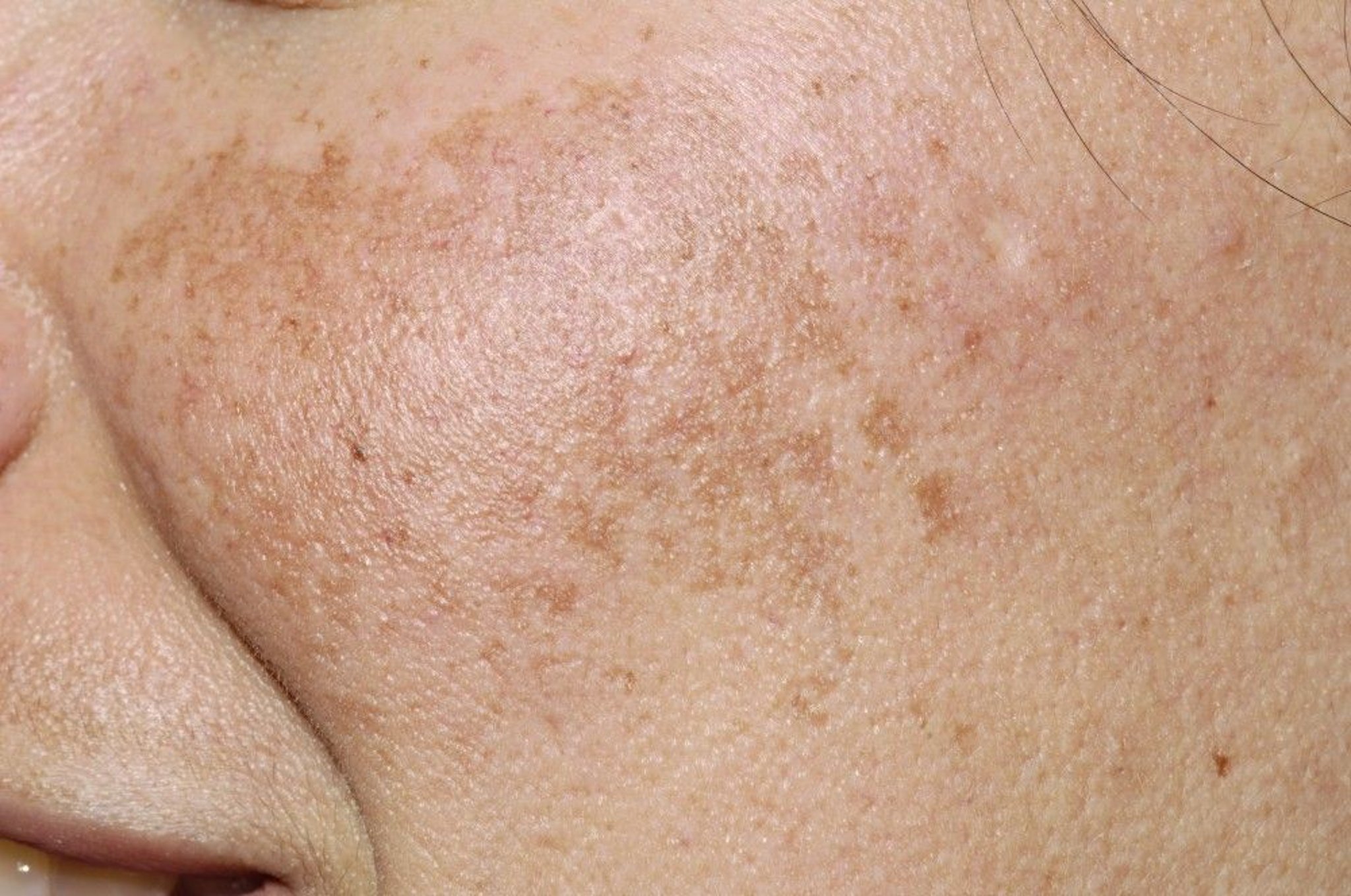
This photo shows brown patches on the cheek of a patient with melasma.
DR P. MARAZZI/SCIENCE PHOTO LIBRARY
Because melasma risk increases with increasing sun exposure, the mechanism probably involves overproduction of melanin by hyperfunctional melanocytes. Other than sun exposure, aggravating factors include
Autoimmune thyroid disorders
Photosensitizing medications
Antiseizure medications
Sex hormones, including oral contraceptives
In women, melasma fades slowly and incompletely after childbirth or cessation of hormone use. In men, melasma rarely fades.
The mainstay of melasma management is strict photoprotection agents. Patients should use sunscreen with a broad-spectrum (UVA and UVB) sun protection factor (SPF) of 50 or higher, wear sun-protective clothing and hats (UV protection factor 50 or higher), and avoid direct sun exposure. During and after therapy, strict sun protection must be maintained. Because visible light is not blocked by most sunscreens, patients should use a tinted sunscreen (eg, that contains zinc oxide or titanium dioxide). The addition of antioxidants to the sunscreen and oral adjunctive photoprotection agents such as The mainstay of melasma management is strict photoprotection agents. Patients should use sunscreen with a broad-spectrum (UVA and UVB) sun protection factor (SPF) of 50 or higher, wear sun-protective clothing and hats (UV protection factor 50 or higher), and avoid direct sun exposure. During and after therapy, strict sun protection must be maintained. Because visible light is not blocked by most sunscreens, patients should use a tinted sunscreen (eg, that contains zinc oxide or titanium dioxide). The addition of antioxidants to the sunscreen and oral adjunctive photoprotection agents such asPolypodium leucotomas can enhance protection (1, 2). Because of potential health and environmental toxicities, use of oxybenzone/benzophenone-3 has often been discouraged (3); however, its health effects are unknown because data on clinically meaningful outcomes are limited (4, 5).
Other treatment depends on whether the pigmentation is epidermal or dermal; epidermal pigmentation becomes accentuated with a Wood light (365 nm) or can be diagnosed with biopsy. Only epidermal pigmentation responds to treatment. Most topical melasma treatments are used in combination rather than individually.
Triple topical therapy is first-line treatment that is often effective and consists of a combination of
Hydroquinone 2 to 4% Hydroquinone 2 to 4%
Tretinoin 0.05 to 1% Tretinoin 0.05 to 1%
A class V to VII topical corticosteroid (see table )
Hydroquinone depigments the skin by blocking the enzymatic oxidation of tyrosine 3,4-dihydroxyphenylalanine (DOPA) and inhibiting melanocyte metabolic processes. Hydroquinone depigments the skin by blocking the enzymatic oxidation of tyrosine 3,4-dihydroxyphenylalanine (DOPA) and inhibiting melanocyte metabolic processes.Hydroquinone should be tested behind one ear or on a small patch on the forearm for 1 week before use on the face because it may cause irritation or an allergic reaction.
Tretinoin promotes keratinocyte turnover and can exfoliate skin that contains epidermal pigment. Tretinoin promotes keratinocyte turnover and can exfoliate skin that contains epidermal pigment.
Corticosteroids help block synthesis and secretion of melanin.
Two promising technologies being tried in conjunction with triple topical therapy are the Q-switched Nd:YAG (1064 nm) laser modalities (6).
If triple topical therapy is not available, hydroquinone 2 to 4% alone can be used; however, chronic continuous use can theoretically increase the risk of exogenous ochronosis, which is a permanent form of hyperpigmentation. If triple topical therapy is not available, hydroquinone 2 to 4% alone can be used; however, chronic continuous use can theoretically increase the risk of exogenous ochronosis, which is a permanent form of hyperpigmentation.Hydroquinone 2% is useful as maintenance.
Azelaic acid 15 to 20% cream can be used in place of or with Azelaic acid 15 to 20% cream can be used in place of or withhydroquinone and/or tretinoin. Azelaic acid is a tyrosinase inhibitor that reduces melanin production. In addition, topical kojic acid has been increasingly used; it is a chelating agent that blocks tyrosine conversion to melanin.
During pregnancy, azelaic acid 15 to 20% cream and chemical peeling with glycolic acid are safe to use. NOTE: Hydroquinone, tranexamic acid, trichloroacetic acid peels, and , trichloroacetic acid peels, andtretinoin are not safe to use during pregnancy.
Second-line treatment options for patients with severe melasma unresponsive to topical bleaching agents include chemical peeling with glycolic acid or 30 to 50% trichloroacetic acid. Laser treatments also have been used but are not standard therapy. Second-line treatment options for patients with severe melasma unresponsive to topical bleaching agents include chemical peeling with glycolic acid or 30 to 50% trichloroacetic acid. Laser treatments also have been used but are not standard therapy.
A randomized study has shown improvement with oral tranexamic acid in patients with moderate-to-severe melasma (A randomized study has shown improvement with oral tranexamic acid in patients with moderate-to-severe melasma (7). However, relapse can occur when treatment is stopped.
Melasma references
1. Goh CL, Chuah SY, Tien S, et al: Double-blind, placebo-controlled trial to evaluate the effectiveness of Polypodium leucotomos extract in the treatment of melasma in Asian skin: A pilot study. J Clin Aesthet Dermatol 11(3):14-19, 2018.
2. Lim HW, Kohli I, Ruvolo E, et al: Impact of visible light on skin health: The role of antioxidants and free radical quenchers in skin protection. J Am Acad Dermatol 86(3S):S27-S37, 2022. doi: 10.1016/j.jaad.2021.12.024
3. DiNardo JC, Downs CA. Dermatological and environmental toxicological impact of the sunscreen ingredient oxybenzone/benzophenone-3. J Cosmet Dermatol. 2018;17(1):15-19. doi:10.1111/jocd.12449
4. American Academy of Dermatology: American Academy of Dermatology comments on follow-up study on absorption of sunscreen ingredients. January 21, 2020.
5. Matta MK, Florian J, Zusterzeel R, et al. Effect of Sunscreen Application on Plasma Concentration of Sunscreen Active Ingredients: A Randomized Clinical Trial [published correction appears in JAMA. 2020 Mar 17;323(11):1098. doi: 10.1001/jama.2020.1950]. JAMA. 2020;323(3):256-267. doi:10.1001/jama.2019.20747
6. Arora P, Sarkar R, Garg VK, Arya L. Lasers for treatment of melasma and post-inflammatory hyperpigmentation. J Cutan Aesthet Surg. 2012;5(2):93-103. doi:10.4103/0974-2077.99436
7. Del Rosario E, Florez-Pollack S, Zapata L Jr, et al: Randomized, placebo-controlled, double-blind study of oral tranexamic acid in the treatment of moderate-to-severe melasma. J Am Acad Dermatol 78(2):363–369, 2018. doi: 10.1016/j.jaad.2017.09.053
Lentigines
Lentigines (singular: lentigo) are flat, tan to brown, oval macules. They are commonly due to chronic sun exposure (solar lentigines; sometimes called liver spots, although they are not related to hepatic dysfunction) and occur most frequently on the face and back of the hands. They typically first appear during middle age and increase in number with age. Although progression from lentigines to melanoma has not been established, lentigines are an independent risk factor for melanoma.
If lentigines are a cosmetic concern, they are treated with cryotherapy or laser; hydroquinone is not effective.
Nonsolar lentigines are sometimes associated with systemic disorders, such as Peutz-Jeghers syndrome (in which profuse lentigines of the lips occur), multiple lentigines syndrome (or LEOPARD syndrome, which stands for multiple Lentigines, Electrocardiogram [ECG] conduction abnormalities, Ocular hypertelorism, Pulmonic stenosis, Abnormal genitals, Retardation of growth, and sensorineural Deafness), or xeroderma pigmentosum.

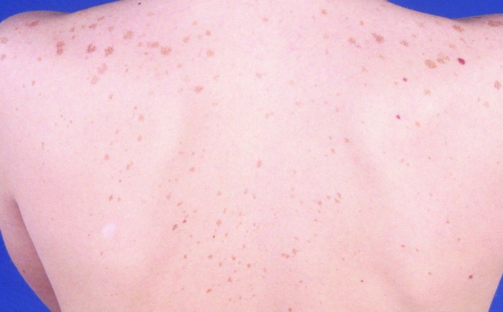
This photo shows lentigo simplex manifesting as multiple, small, hyperpigmented macules on the back.
© Springer Science+Business Media
Drug- or Substance-Induced Hyperpigmentation
Changes are usually diffuse but sometimes have drug- or substance-specific distribution patterns or hues (see table ). Mechanisms include
Increased melanin in the epidermis (tends to be more brown)
Increased melanin in the epidermis and high dermis (mostly brown with hints of gray or blue)
Increased melanin in the dermis (tends to be more grayish or blue)
Dermal deposition of the drug or substance, metabolite, or complex between the drug or substance and melanin (usually slate or bluish gray)
Medications may cause secondary hyperpigmentation. For example, focal hyperpigmentation frequently occurs after medication-induced lichen planus (also known as lichenoid drug eruption).
Hyperpigmentation Effects of Some Medications and Heavy Metals
Substance | Effect |
|---|---|
Medications | |
Amiodarone | Slate-gray to violaceous discoloration of sun-exposed areas; yellowish brown deposits in the dermis |
Antimalarials | Yellow-brown to gray to bluish black discoloration of pretibial areas, face, oral cavity, and nails; medication–melanin complexes in the dermis; hemosiderin around capillaries |
Bleomycin | Flagellate hyperpigmented streaks on the back, often in areas of scratching or minor trauma |
Cancer chemotherapy agents, including busulfan, cyclophosphamide, dactinomycin, daunorubicin, and 5-fluorouracil (5-FU) | Diffuse hyperpigmentation |
Desipramine Imipramine | Grayish blue discoloration on sun-exposed areas; golden-brown granules in upper dermis |
Hydroquinone | Bluish black discoloration of ear cartilage and face after years of use ("rebound hyperpigmentation") |
Phenothiazines, including chlorpromazine | Grayish blue discoloration on sun-exposed areas; golden-brown granules in upper dermis |
Tetracyclines, particularly minocycline | Grayish discoloration of teeth, nails, sclerae, oral mucosa, acne scars, face, forearms, and lower legs |
Heavy metals | |
Arsenic | Can manifest with hyperpigmentation or hypopigmentation in a raindrop-like pattern ("raindrop" hypopigmentation) White transverse lines of the fingernails (Mees lines [leukonychia striata]) |
Bismuth | Blue-gray discoloration of face, neck, and hands |
Gold | Blue-gray deposits around the eyes (chrysiasis) |
Mercury | Slate-gray discoloration of skinfolds |
Silver | Diffuse slate-gray discoloration (argyria), especially in sun-exposed areas |
In fixed drug eruptions, round or oval red plaques or blisters form at the same site each time the causative medication is taken; residual postinflammatory hyperpigmentation usually persists, especially in dark skin types. Typical lesions occur on the face (especially the lips), hands, feet, and genitals. Typical inciting medications include antibiotics (sulfonamides, tetracyclines, trimethoprim, and fluoroquinolones), nonsteroidal anti-inflammatory drugs, and barbiturates.In fixed drug eruptions, round or oval red plaques or blisters form at the same site each time the causative medication is taken; residual postinflammatory hyperpigmentation usually persists, especially in dark skin types. Typical lesions occur on the face (especially the lips), hands, feet, and genitals. Typical inciting medications include antibiotics (sulfonamides, tetracyclines, trimethoprim, and fluoroquinolones), nonsteroidal anti-inflammatory drugs, and barbiturates.
Treatment of drug- or substance-induced hyperpigmentation involves stopping the causative drug or substance; the hyperpigmentation fades very slowly in some if not all cases.
Because many drugs and substances that cause skin pigmentation also cause photosensitivity reactions, patients should avoid sun exposure. People with dark skin tones are more susceptible to postinflammatory hyperpigmentation and should also practice strict sun protective measures to limit exacerbation of postinflammatory hyperpigmentation.
Key Points
Common causes of focal hyperpigmentation include injury, inflammation, phytophotodermatitis, lentigines, melasma, freckles, café-au-lait macules, and acanthosis nigricans.
Common causes of widespread hyperpigmentation include melasma, drugs, substances, cancers, and other systemic disorders.
Test patients who have widespread hyperpigmentation not caused by drugs or substances for disorders such as primary biliary cholangitis, hemochromatosis, and Addison disease.
Treat melasma initially with a combination of hydroquinone 2 to 4%, tretinoin 0.05 to 1%, and a class V to VII topical corticosteroid.Treat melasma initially with a combination of hydroquinone 2 to 4%, tretinoin 0.05 to 1%, and a class V to VII topical corticosteroid.
If lentigines are a cosmetic concern, treat with cryotherapy or laser.
Treat drug- or substance-induced hyperpigmentation by stopping exposure to the offending agent.
Drug Information for the Topic


