


Pityriasis lichenoides is an acquired inflammatory skin condition that may develop in response to foreign antigens (eg, infections or substances) and may be associated with cutaneous T-cell lymphoma. Diagnosis is clinical. Treatment may include various topical and oral medications.
Pityriasis lichenoides is an acquired inflammatory skin disorder characterized by a spectrum of clinical presentations.
The two principal subtypes are (1):
Pityriasis lichenoides et varioliformis acuta (PLEVA), which presents as crops of erythematous papules that may become necrotic or ulcerated
Pityriasis lichenoides chronica (PLC), which manifests as persistent, small, red-brown maculopapules with fine scale
The acute form (PLEVA) typically appears in children and young adults, with crops of asymptomatic chickenpox-like lesions that typically resolve, often with scarring, within weeks to months. Lesions that appear erythematous on light skin may appear more violaceous or brown on dark skin. The chronic form (PLC) initially manifests as flatter, reddish-brown, scaling papules that may take months or longer to resolve. Lesions may also evolve from the acute to the chronic form.
The average age of onset is around 6 years, with a slight male predominance (2).
The etiology remains poorly characterized, but proposed mechanisms include hypersensitivity reactions to infectious agents, medication exposures, or a premycotic lymphoproliferative process (1).

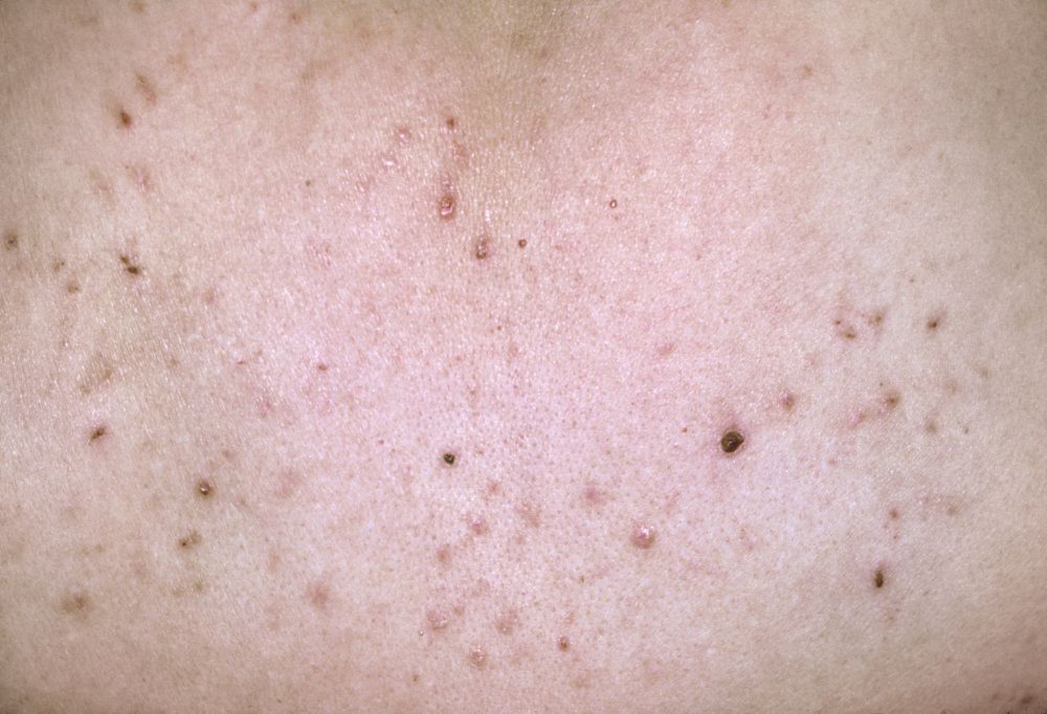
Acute pityriasis lichenoides is characterized by small erythematous or purpuric macules and papules; lesions can be centrally vesiculated or have hemorrhagic crusting.
DR P. MARAZZI/SCIENCE PHOTO LIBRARY

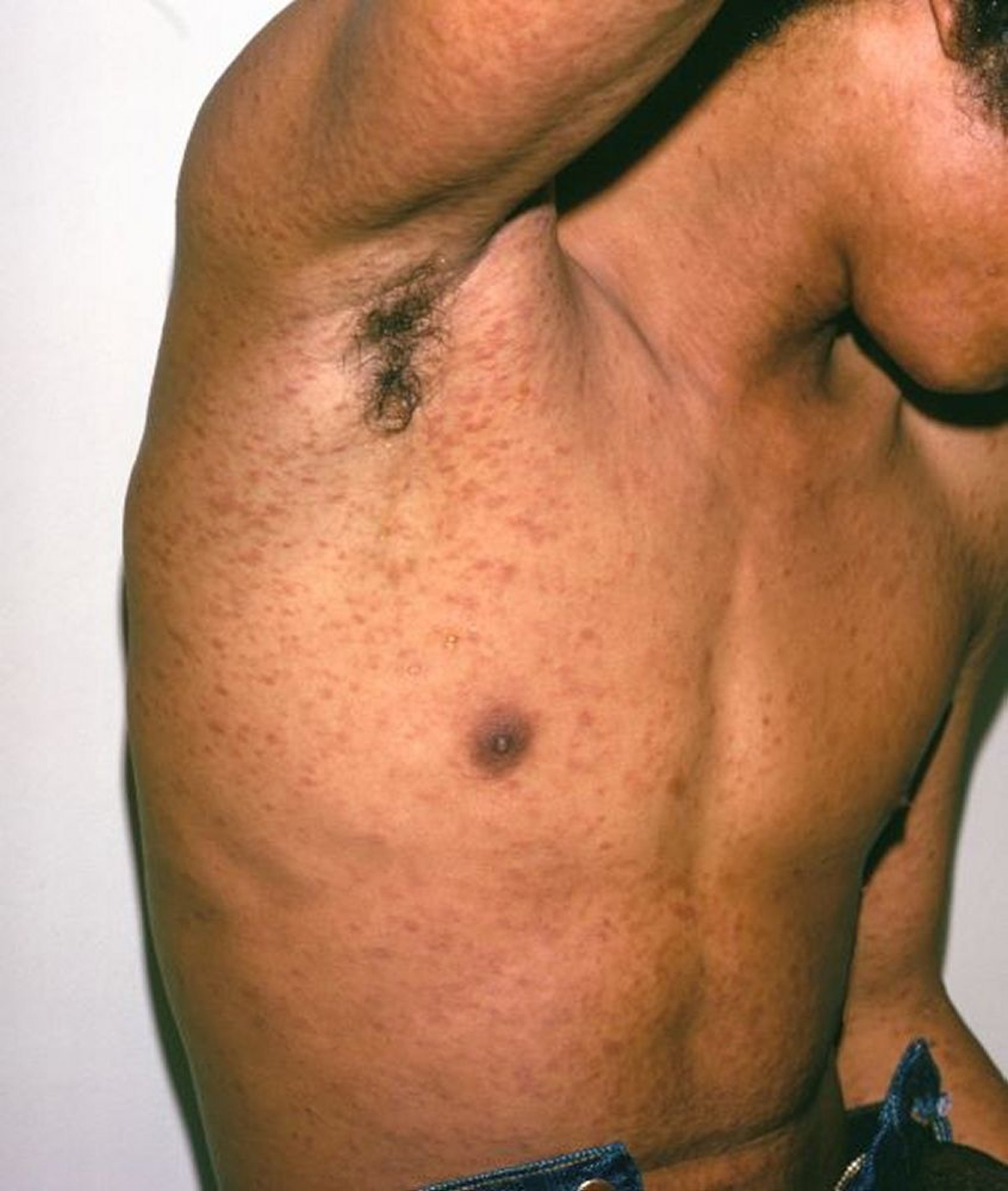
This image shows widely scattered, round, erythematous papules on the trunk and extremities of this patient with pityriasis lichenoides et varioliformis acuta (PLEVA), an acute form of pityriasis lichenoides. Erythema can look more violaceous or brown on dark skin tones.
Image courtesy of Karen McKoy, MD.
General references
1. Khachemoune A, Blyumin ML. Pityriasis lichenoides: pathophysiology, classification, and treatment. Am J Clin Dermatol. 2007;8(1):29-36. doi:10.2165/00128071-200708010-00004
2. Geller L, Antonov NK, Lauren CT, et al. Pityriasis Lichenoides in Childhood: Review of Clinical Presentation and Treatment Options. Pediatr Dermatol. 2015;32(5):579-592. doi:10.1111/pde.12581
Diagnosis of Pityriasis Lichenoides
Primarily history and physical examination
Sometimes biopsy
The diagnosis of pityriasis lichenoides is based on clinical appearance and distribution.
Biopsy is performed when clinical findings are inconclusive.
Differential diagnoses of pityriasis lichenoides include the following:
Hypopigmented mycosis fungoides (a form of cutaneous T-cell lymphoma)
Treatment of Pityriasis Lichenoides
Various topical and oral treatments
Treatment of pityriasis lichenoides involves topical glucocorticoids, topical tacrolimus, oral antibiotics, phototherapy (especially narrow-band UVB), and immunosuppressants, all of which have been used with varying success (1). In one prospective cohort evaluating longitudinal outcomes, the most effective treatments were phototherapy (47% response rate), followed by sunlight (33%), topical glucocorticoids (27%), and antibiotics (25%) (2).
Treatment references
1. Bowers S, Warshaw EM: Pityriasis lichenoides and its subtypes. J Am Acad Dermatol 55:557–572, 2006. doi: 10.1016/j.jaad.2005.07.058
2. Zang JB, Coates SJ, Huang J, et al. Pityriasis lichenoides: Long-term follow-up study. Pediatr Dermatol. 2018;35(2):213-219. doi:10.1111/pde.13396
Drug Information for the Topic


