

Pityriasis rosea is a self-limited, inflammatory disease characterized by diffuse, scaling papules or plaques. Diagnosis is clinical. Treatment is usually unnecessary.
Pityriasis rosea is an acute, self-limited exanthematous skin disease that most commonly presents with a single large erythematous plaque that is followed by multiple smaller scaly papules and plaques, usually on the trunk.
Pityriasis rosea is a relatively common skin condition. In one seminal population-based epidemiologic study, the average annual incidence rate was 172 per 100,000 person-years (1). It most commonly occurs between ages 10 and 35. There may be a slight female preponderance.
The cause of pityriasis rosea may be a viral infection (some research has implicated human herpesviruses (HHV 6, 7, and 8) (2, 3). Some medications (eg, angiotensin-converting enzyme inhibitors, hydrochlorothiazide, captopril, barbiturates, metronidazole, allopurinol), may cause a pityriasis rosea–like reaction.
General references
1. Chuang TY, Ilstrup DM, Perry HO, Kurland LT. Pityriasis rosea in Rochester, Minnesota, 1969 to 1978. J Am Acad Dermatol. 1982;7(1):80-89. doi:10.1016/s0190-9622(82)80013-3
2. Mubki TF, Bin Dayel SA, Kadry R: A case of pityriasis rosea concurrent with the novel influenza A (H1N1) infection. Pediatr Dermatol 28(3):341-342, 2011. doi: 10.1111/j.1525-1470.2010.01090.x
3. Prantsidis A, Rigopoulos D, Papatheodorou G, et al: Detection of human herpesvirus 8 in the skin of patients with pityriasis rosea. Acta Derm Venereol 89(6):604-606, 2009. doi: 10.2340/00015555-0703
Symptoms and Signs of Pityriasis Rosea
The condition classically begins with a single, primary, 2- to 10-cm herald patch that usually appears on the trunk, or less commonly, the proximal limbs. A general centripetal eruption of 0.5- to 2-cm rose- or fawn-colored oval papules and plaques follows within 7 to 14 days. The lesions have a scaly, slightly raised border (collarette of scale) and resemble the rash of ringworm (tinea corporis).
The rose or fawn color is not as evident in patients with dark skin. Patches that appear more pink in patients with light skin appear violaceous and/or hyperpigmented in patients with dark skin.
Most patients experience pruritus, occasionally severely. Papules may dominate with little or no scaling in children and pregnant women.
Children more commonly have inverse pityriasis rosea (lesions in the axillae or groin that spread centrifugally).

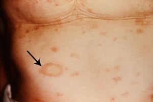
Initially, most people develop one large scaly patch (herald patch; arrow), and within 1 to 2 weeks, smaller pink patches can occur on the trunk, arms, and legs.
Initially, most people develop one large scaly patch (herald patch; arrow), and within 1 to 2 weeks, smaller pink patch
Image courtesy of the Public Health Image Library of the Centers for Disease Control and Prevention.

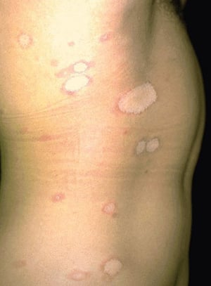
In pityriasis rosea, the initial herald patch is followed by centripetal eruption of oval papules and plaques with a slightly raised border, typically appearing along skin lines.
In pityriasis rosea, the initial herald patch is followed by centripetal eruption of oval papules and plaques with a sl
Image provided by Thomas Habif, MD.

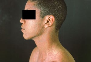
This image shows erythematous and violaceous, ovoid, scaly papules coalescing into patches on the head and torso of a patient with pityriasis rosea.
This image shows erythematous and violaceous, ovoid, scaly papules coalescing into patches on the head and torso of a p
Image courtesy of Karen McKoy, MD.

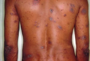
This image shows violaceous, scaly plaques on the back and arms of a patient with pityriasis rosea. Patches that appear more pink in patients with light skin appear violaceous and/or hyperpigmented in patients with dark skin.
This image shows violaceous, scaly plaques on the back and arms of a patient with pityriasis rosea. Patches that appear
Image courtesy of Karen McKoy, MD.

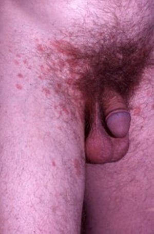
This photo shows atypical (inverse) pityriasis rosea characterized by oval scaly plaques in the inguinal creases.
This photo shows atypical (inverse) pityriasis rosea characterized by oval scaly plaques in the inguinal creases.
Image provided by E. Laurie Tolman, MD.
Initially, most people develop one large scaly patch (herald patch; arrow), and within 1 to 2 weeks, smaller pink patches can occur on the trunk, arms, and legs.
Initially, most people develop one large scaly patch (herald patch; arrow), and within 1 to 2 weeks, smaller pink patch
Image courtesy of the Public Health Image Library of the Centers for Disease Control and Prevention.
In pityriasis rosea, the initial herald patch is followed by centripetal eruption of oval papules and plaques with a slightly raised border, typically appearing along skin lines.
In pityriasis rosea, the initial herald patch is followed by centripetal eruption of oval papules and plaques with a sl
Image provided by Thomas Habif, MD.
This image shows erythematous and violaceous, ovoid, scaly papules coalescing into patches on the head and torso of a patient with pityriasis rosea.
This image shows erythematous and violaceous, ovoid, scaly papules coalescing into patches on the head and torso of a p
Image courtesy of Karen McKoy, MD.
This image shows violaceous, scaly plaques on the back and arms of a patient with pityriasis rosea. Patches that appear more pink in patients with light skin appear violaceous and/or hyperpigmented in patients with dark skin.
This image shows violaceous, scaly plaques on the back and arms of a patient with pityriasis rosea. Patches that appear
Image courtesy of Karen McKoy, MD.
This photo shows atypical (inverse) pityriasis rosea characterized by oval scaly plaques in the inguinal creases.
This photo shows atypical (inverse) pityriasis rosea characterized by oval scaly plaques in the inguinal creases.
Image provided by E. Laurie Tolman, MD.
Classically, lesions orient along skin cleavage lines, giving pityriasis rosea a Christmas tree–like distribution when multiple lesions appear on the back.
A prodrome of malaise, headache, and sometimes arthralgia can precede the lesions in a minority of patients.
Diagnosis of Pityriasis Rosea
Primarily history and physical examination
The diagnosis of pityriasis rosea is based on its characteristic clinical appearance and distribution.
Differential diagnoses include:
Pityriasis lichenoides chronica
Serologic testing for syphilis is indicated when the palms or soles are affected, when a herald patch is not seen, or when lesions occur in an unusual sequence or distribution.
Treatment of Pityriasis Rosea
Supportive management (antipruritic therapy, topical glucocorticoids)
Sometimes, acyclovirSometimes, acyclovir
No specific treatment is necessary because the eruption usually spontaneously remits within 5 weeks and recurrence is rare.
Artificial or natural sunlight may hasten resolution.
Antipruritic therapy such as topical glucocorticoids, oral antihistamines (eg, hydroxyzine), or other topical measures (eg, calamine lotion) may be used as needed. Antipruritic therapy such as topical glucocorticoids, oral antihistamines (eg, hydroxyzine), or other topical measures (eg, calamine lotion) may be used as needed.
Limited data suggest that a short course of acyclovir may be helpful in patients who present early and have widespread disease, or present with flu-like symptoms (Limited data suggest that a short course of acyclovir may be helpful in patients who present early and have widespread disease, or present with flu-like symptoms (1). Of note, pityriasis rosea during pregnancy (especially during the first 15 weeks of gestation) is associated with preterm birth or fetal demise. Pregnant women should be offered acyclovir; however, antiviral therapy has not been proven to reduce obstetric complications (). Of note, pityriasis rosea during pregnancy (especially during the first 15 weeks of gestation) is associated with preterm birth or fetal demise. Pregnant women should be offered acyclovir; however, antiviral therapy has not been proven to reduce obstetric complications (2).
Treatment references
1. Contreras-Ruiz J, Peternel S, Jiménez Gutiérrez C, et al: Interventions for pityriasis rosea. Cochrane Database Syst Rev 2019(10):CD005068, 2019. doi: 10.1002/14651858.CD005068.pub3
2. Stashower J, Bruch K, Mosby A, et al: Pregnancy complications associated with pityriasis rosea: A multicenter retrospective study. J Am Acad Dermatol 85(6):1648-1649, 2021. doi: 10.1016/j.jaad.2020.12.063
Key Points
Pityriasis rosea is a self-limited, inflammatory disorder of the skin possibly caused by human herpesvirus types 6, 7, or 8 or medications.
An initial 2- to 10-cm herald patch is followed by centripetal eruption of oval papules and plaques with a slightly raised and scaly border, typically appearing along skin lines.
Diagnose based on clinical appearance and distribution.
Treat with antipruritic medications as needed and possibly topical glucocorticoids and/or sunlight.
Pityriasis rosea during the first 15 weeks of pregnancy is associated with preterm birth or fetal demise.
Pregnant women should be offered antiviral therapy, even though this has not been proven to reduce obstetric complications.
Drug Information for the Topic


