


Colonic diverticulosis is the presence of 1 or more diverticula in the colon. Most diverticula are asymptomatic, but some become inflamed or bleed. Diagnosis is by ultrasound, colonoscopy, capsule endoscopy, barium enema, CT, or MRI. Asymptomatic diverticulosis requires no treatment. When symptoms develop, treatment varies depending on clinical manifestations.
A colonic diverticulum is a saclike pouch of colonic mucosa and submucosa that protrudes through the muscular layer of the colon; because it does not contain all layers of the bowel, it is considered a pseudodiverticulum (see also Definition of Diverticular Disease).

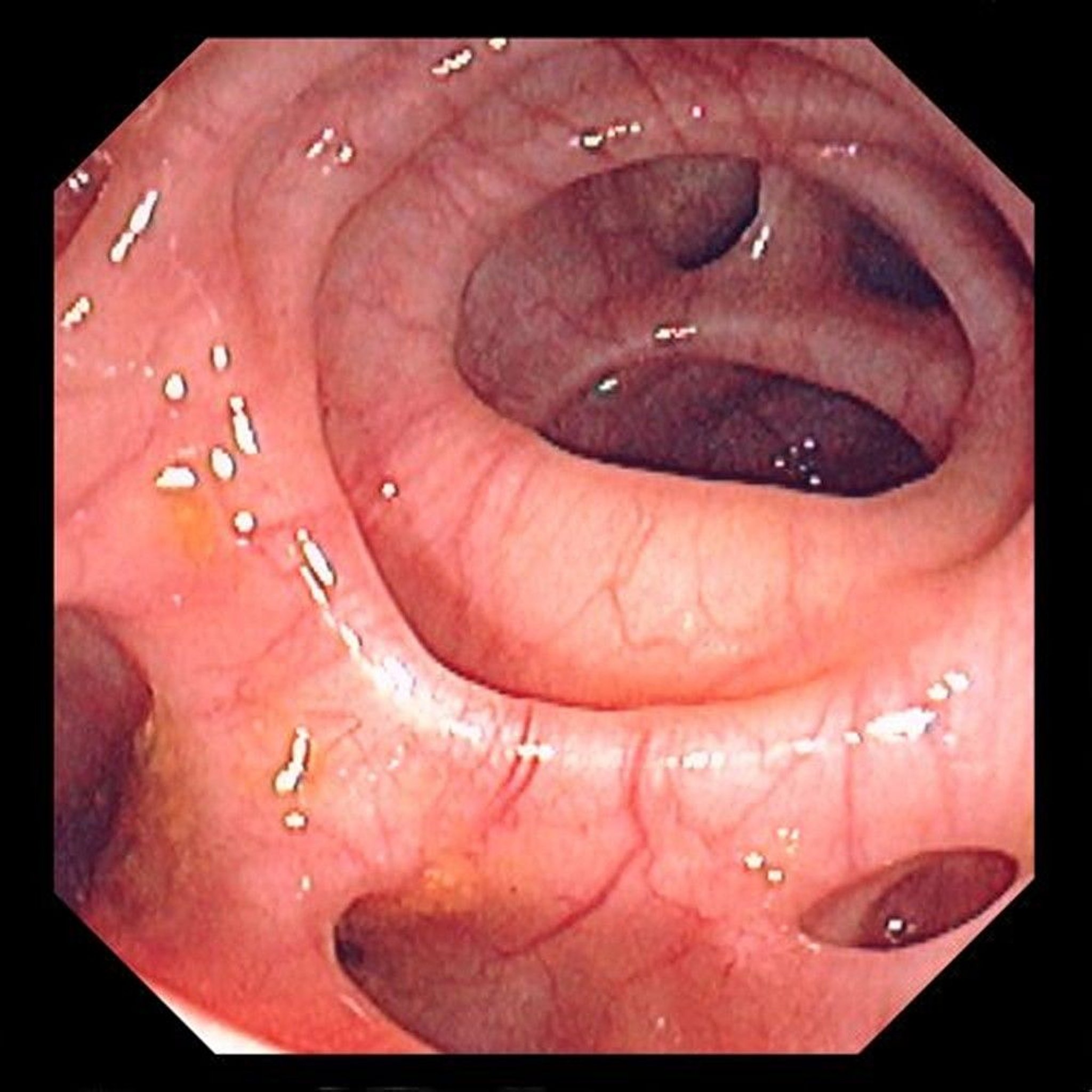
This image shows multiple diverticuli in the colon.
Image provided by David M. Martin, MD.
Although diverticula can occur anywhere in the large bowel, they usually occur in the sigmoid portion of the colon (1). They rarely occur below the peritoneal reflection and involve the rectum. Diverticula vary in diameter but typically are 5 to 10 mm in size (2). Giant diverticula, which are extremely rare, are defined as diverticula > 4 cm in diameter; sizes up to 25 cm have been reported (3). People who have colonic diverticulosis often have several diverticula.
Prevalence generally increases with age and varies widely by geographic region (4). Half of people ≥ 60 years and approximately 70% of people ≥ 80 years have diverticulosis (5).

General references
1.Brown RF, Lopez K, Smith CB, Charles A. Diverticulitis: A Review. JAMA. Published online July 24, 2025. doi:10.1001/jama.2025.10234
2. Stollman N, Raskin JB. Diverticular disease of the colon. Lancet. 2004;363(9409):631-639. doi:10.1016/S0140-6736(04)15597-9
3. Macht R, Sheldon HK, Fisichella PM. Giant Colonic Diverticulum: a Rare Diagnostic and Therapeutic Challenge of Diverticular Disease. J Gastrointest Surg. 2015;19(8):1559-1560. doi:10.1007/s11605-015-2773-8
4. Tursi A, Scarpignato C, Strate LL, et al. Colonic diverticular disease. Nat Rev Dis Primers. 2020;6(1):20. Published 2020 Mar 26. doi:10.1038/s41572-020-0153-5
5. Feuerstein JD, Falchuk KR. Diverticulosis and Diverticulitis. Mayo Clin Proc. 2016;91(8):1094-1104. doi:10.1016/j.mayocp.2016.03.012
Etiology of Colonic Diverticulosis
The etiology of colonic diverticulosis is multifactorial and not entirely known.
There may be a correlation between symptomatic diverticular disease and environmental factors such as a diet low in fiber or high in red meat, sedentary lifestyle, obesity, smoking, and use of nonsteroidal anti-inflammatory drugs (NSAIDs), aspirin, glucocorticoids, and opioids (1, 2, 3). The consumption of nuts, seeds, corn, or popcorn does not appear to be related to the risk or development of diverticular disease (3, 4).
Other risk factors include heritable factors and alterations in the colonic wall structure and motility. Although genetics play a strong role in the development of diverticular disease, clinically useful genetic testing is not yet available (5).
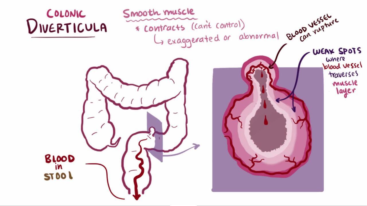
Diverticula are possibly caused by an increase in intraluminal pressure due to spasms of the muscular layer, which leads to mucosal extrusion through the weakest points of the muscular layer of the bowel—areas adjacent to intramural blood vessels.
The etiology of giant diverticula is unclear. One theory is that a narrow neck-opening leads to a ball-valve effect with intermittent obstruction of the opening, causing the diverticulum to enlarge. A very large giant diverticulum is often a true perforation of a smaller diverticulum that was contained and walled off and became lined mostly by granulation tissue.
Etiology references
1. Schultz JK, Azhar N, Binda GA, et al. European Society of Coloproctology: guidelines for the management of diverticular disease of the colon. Colorectal Dis. 2020;22 Suppl 2:5-28. doi:10.1111/codi.15140
2. Tursi A, Scarpignato C, Strate LL, et al. Colonic diverticular disease. Nat Rev Dis Primers. 2020;6(1):20. Published 2020 Mar 26. doi:10.1038/s41572-020-0153-5
3. Brown RF, Lopez K, Smith CB, Charles A. Diverticulitis: A Review. JAMA. Published online July 24, 2025. doi:10.1001/jama.2025.10234
4. Strate LL, Liu YL, Syngal S, et al. Nut, corn, and popcorn consumption and the incidence of diverticular disease. JAMA. 2008;300(8):907-914. doi:10.1001/jama.300.8.907
5. Tursi A, Brandimarte G, Di Mario F, et al. Global guidelines on diverticular disease of the colon: the Fiesole Consensus report. Gut. Published online December 31, 2025. doi:10.1136/gutjnl-2025-336902
Symptoms and Signs of Colonic Diverticulosis
Approximately75% of patients with diverticulosis are asymptomatic or have only intermittent constipation. Approximately 25% become symptomatic with pain or bleeding when inflammatory or hemorrhagic complications develop (1).
Patients with diverticulosis sometimes develop nonspecific gastrointestinal (GI) symptoms, including abdominal pain, bloating, constipation, diarrhea, and passage of mucus from the rectum. This constellation is sometimes referred to as symptomatic uncomplicated diverticular disease (SUDD). However, some specialists believe these symptoms are due to another disorder (eg, irritable bowel syndrome), and the presence of diverticula is coincidental rather than causal (2, 3, 4).
Complications of diverticulosis
Complications of colonic diverticular disease are more common among people who smoke, have obesity, and use nonsteroidal anti-inflammatory drugs (NSAIDs) or immunosuppressed patients such as those with advanced HIV or those undergoing cancer chemotherapy (5). Complications include:
Diverticular bleeding
Segmental colitis associated with diverticular disease (SCAD)
Diverticulitis is painful inflammation of a diverticulum, which develops in approximately 1 to 5% of those with diverticulosis (6, 7). It may be uncomplicated or complicated. Complications of acute diverticulitis are abscess, fistula, obstruction, and perforation (5). Complications of chronic diverticulitis are related to stenotic disease, which can include both obstructions and strictures.
Diverticular bleeding occurs in 10 to 15% of patients with diverticulosis (8, 9).
Segmental colitis associated with diverticular disease (SCAD) refers to manifestations of colitis (eg, hematochezia, abdominal pain, diarrhea) that develop in a few patients with diverticulosis. The degree to which the diverticulosis is causal is unclear.
Diverticular bleeding
Diverticular bleeding is the most common cause (30 to 65%) of acute lower GI bleeding in adults (10). One study of approximately 3700 patients showed that the cumulative incidence of lower GI bleeding from asymptomatic diverticulosis was approximately 2% at 5 years and 10% at 10 years (11).
The pathophysiology of diverticular bleeding is unknown, but several mechanisms are hypothesized, including:
Local trauma from impacted feces in a diverticulum that can erode the adjacent vessel
Enlargement of the diverticulum that can stretch (and ultimately tear) the vessel
NSAIDs have been reported to increase the risk of hemorrhage (9).
Most diverticula are in the distal (left) colon in Western countries and in the proximal (right) colon in Asian countries (12, 13). However, the right colon is the source of diverticular bleeding in > 50% of patients (14, 15, 16). Right-sided diverticula in Western patients tend to have wider necks and thinner domes, exposing the vasa recta (the nutrient blood vessels) to greater mechanical stress and stretching, making them more prone to rupture and massive hemorrhage compared to left-sided diverticula (17). Patients with pancolonic diverticulosis have a higher incidence of bleeding.
Diverticular bleeding manifests as painless hematochezia. Because the bleeding vessel is an arteriole, the amount of blood loss is usually moderate to severe. Fresh blood or maroon-colored stool is the typical manifestation; rarely, right-sided diverticular bleeding can manifest as melena. Diverticular bleeding usually occurs without concomitant diverticulitis.

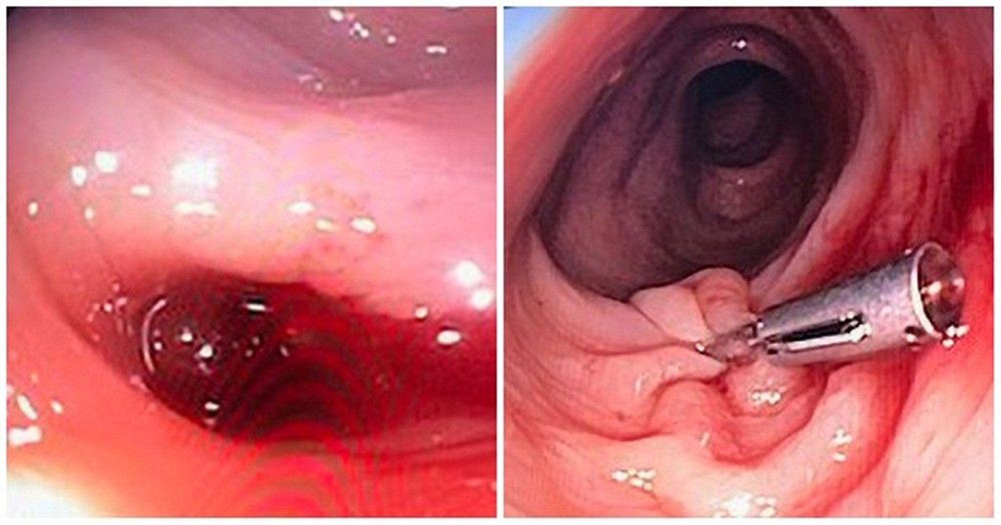
This photo shows a diverticulum with a clotted blood vessel (left) that caused lower gastrointestinal bleeding. Placement of an endoclip (right) during colonoscopy stopped the bleeding.
Photos courtesy of Drs. Joel A. Baum and Rafael A. Ching Companioni.
The majority of episodes of bleeding cease spontaneously (18, 19). The remainder require intervention, typically endoscopic (9).
Patients who have had a diverticular bleeding episode are at risk of rebleeding. After an episode of diverticular bleeding, reported early (≤ 30 days) and late (> 30 days) rebleeding rates are 17 to 33% and 26 to 32%, respectively. (9, 20)
Diagnosis of Colonic Diverticulosis
CT or MRI
Sometimes ultrasound, colonoscopy, capsule endoscopy, or barium enema
Sometimes fecal calprotectin testing
Intestinal ultrasound and point-of-care ultrasound have emerged as highly accurate, frontline diagnostic modalities for evaluating diverticular disease, particularly during episodes of acute diverticulitis (1, 2).
However, CT and sometimes MRI are preferred for definitive diagnosis due to their ability to image the entire bowel wall (3, 4, 5).
Asymptomatic diverticula may be found incidentally during colonoscopy, capsule endoscopy, barium enema, CT, or MRI.
Colonoscopy may also be used to exclude malignancy, which can be misdiagnosed as diverticulitis (3).
Fecal calprotectin is useful in distinguishing symptomatic uncomplicated diverticular disease (SUDD) from irritable bowel syndrome (IBS) (6), a significant clinical challenge due to their overlapping symptom profiles, which include chronic abdominal pain, bloating, and altered bowel habits (7). Fecal calprotectin levels are characteristically elevated in a majority of SUDD patients, with concentrations correlating directly to the severity of abdominal pain, whereas values remain entirely within normal limits for patients suffering from functional IBS (8). Consequently, the integration of fecal calprotectin testing alongside precise symptom evaluation—specifically identifying the prolonged lower left quadrant pain typical of SUDD versus the shorter, defecation-relieved pain of IBS—provides clinicians with a robust, objective framework to differentiate between these disorders. (For additional detail, see Symptomatic Uncomplicated Diverticular Disease).
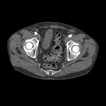
This axial cross-sectional CT image through the pelvis shows the typical appearance of diverticulosis (the dark areas in the wall of the sigmoid colon).
Lower GI bleeding due to diverticulosis is suspected when painless rectal bleeding develops, particularly in an older adult patient or in a patient who has a history of diverticular disease. Evaluation of lower GI bleeding typically includes colonoscopy (9, 10), which can be performed after rapid colonic preparation: 4 to 6 L of polyethylene glycol solution delivered orally, ideally via a nasogastric tube, and given over 3 to 4 hours until the rectal effluent is clear of blood and stool. Endoscopic findings in diverticular bleeding range from active bleeding to a nonbleeding visible vessel, an adherent clot that is resistant to washing off, and dark, flat spots.
If the source cannot be seen with colonoscopy and ongoing bleeding is sufficiently rapid (> 0.5 to 1 mL/minute), CT angiography or radionuclide imaging may localize the source.
Treatment of Colonic Diverticulosis
No treatment for asymptomatic diverticulosis
Management of specific symptoms
Diverticular bleeding treated as a lower GI bleed
Asymptomatic diverticulosis is not necessarily considered a disease per se and requires no treatment or dietary changes (1). There is no association between consumption of nuts, seeds, corn, or popcorn and diverticulitis, diverticular hemorrhage, or uncomplicated diverticulosis, and avoidance of these foods is no longer recommended (2, 3). Nonsteroidal anti-inflammatory drugs (NSAIDs) and opioid analgesics may increase the risk of diverticular perforation and bleeding, therefore these medications should be used only with appropriate caution and after extensive discussion with the patient about the risks (4).
For diverticulosis with nonspecific GI symptoms, For patients with diverticulosis and nonspecific bowel symptoms, evidence that fiber directly improves symptoms is limited; management should be individualized and constipation treated with bulk-forming agents as needed. However, to reduce the risk of future diverticulitis episodes, patients should be counseled on lifestyle measures including a high-quality diet (often fiber-rich) (5), maintaining a normal BMI, routine physical activity, smoking cessation, and avoidance of non-aspirin NSAIDs when possible. Nonetheless, higher dietary fiber intake is consistently associated with favorable cardiometabolic and mortality outcomes and is reasonable as part of overall lifestyle counseling. Antispasmodics (eg, belladonna) are not of benefit and may cause adverse effects. Surgery is unwarranted for uncomplicated disease with the exception of giant diverticula.
Pearls & Pitfalls
|
Treatment of diverticular bleeding
Diverticular bleeding stops spontaneously in most patients (6, 7). Initial management is as for lower GI bleeding. Treatment of diverticular bleeding is often given during the diagnostic procedure.
A colonoscopy should be the first test performed after adequate colon preparation in patients with minor lower GI bleeding and in patients in whom bleeding has clinically ceased. In these patients, observation without colonoscopy should be considered when lower GI bleeding has subsided and when the patient has had, within the year, a well-prepared colonoscopy that showed diverticulosis but no colorectal cancer or other possible etiologies of GI bleeding (eg, angioectasias, postpolypectomy bleeding).
Colonoscopic identification of stigmata of recent hemorrhage (active bleeding, adherent clot, dark spot, and a visible vessel) allows for endoscopic options to control bleeding, including epinephrine injection, application of endoclips or fibrin sealant, heater probe or bipolar coagulation, and band ligation. Both early and late recurring bleeding rates are lower in patients with definitive colonic diverticular bleeding that is treated endoscopically than in those with presumptive colonic diverticular bleeding that is treated conservatively (8). Additional techniques include endoscopic detachable snare ligation and over-the-scope clip. Comparative data suggest differences in rebleeding outcomes between modalities, and choice should be guided by stigmata of recent hemorrhage, location, endoscopist expertise, and local resources (9).
CT angiography should be performed initially in patients with ongoing hemodynamically significant lower GI bleeding; however, this test has low diagnostic yield in those with minor bleeding or no clinical evidence of continuing bleeding (10). If CT angiography is abnormal, direct transcatheter angiography can be performed, permitting a number of techniques to be used to control the bleeding, particularly embolization. Embolization is technically successful approximately > 95% of the time. Angiographic complications of bowel ischemia or infarction are less common (< 5%) with current super-selective catheterization techniques.
Surgery is rarely needed but is recommended for patients who have had multiple or persistent episodes of diverticular bleeding refractory to therapy or who have hemodynamic instability despite aggressive resuscitation.
If angiography or surgery is being considered, identifying the specific bleeding diverticulum endoscopically or using a nuclear medicine study during active bleeding gives direction to the interventional radiologist and may limit the size of a potential surgical resection. When the bleeding site is known, the need for subtotal colectomy (with its associated higher morbidity and mortality) is markedly reduced, and a hemicolectomy or segmental colectomy may be performed instead. However, patients who have continued to have a life-threatening hemorrhage and no identifiable bleeding diverticulum may require a subtotal colectomy. Patients with diverticular bleeding who have an unclear localization of the bleeding site have a mortality rate of 43% after colonic surgery, whereas patients who have a defined localization of the bleeding site have a mortality rate of 7% after surgery (1).
Key Points
Colonic diverticula are saclike mucosal pouches that protrude from the colon.
Diverticulosis is increasingly common with age; it is present in half of people over 60 years and 70% of people over 80 years old.
Diverticulosis is usually asymptomatic, but approximately 25% of patients develop symptoms and/or complications, including inflammation (diverticulitis) and lower gastrointestinal bleeding.
Asymptomatic diverticulosis requires no treatment.
Diverticular bleeding stops spontaneously in most patients; control the remainder during colonoscopy or angiography, or rarely with surgery.
Symptomatic Uncomplicated Diverticular Disease (SUDD)
Symptomatic uncomplicated diverticular disease refers to the presence of persistent and recurrent nonspecific abdominal symptoms in patients with diverticulosis in the absence of overt colitis or diverticulitis.
Some medical authorities consider symptomatic uncomplicated diverticular disease (SUDD) to be a form of irritable bowel syndrome that occurs coincidentally in patients with diverticulosis (1, 2, 3).
Patients with SUDD have left lower quadrant abdominal pain with bloating, constipation, diarrhea, or passage of mucus from the rectum. In general, patients have a very low incidence of complications.
Diagnosing and differentiating symptomatic uncomplicated diverticular disease (SUDD) from irritable bowel syndrome (IBS) remains a significant clinical challenge due to their overlapping symptom profiles, which include chronic abdominal pain, bloating, and altered bowel habits (4). While both conditions lack the overt systemic inflammation, leukocytosis, or high-grade C-reactive protein (CRP) elevations characteristic of acute diverticulitis, they diverge at the mucosal level (4). IBS operates primarily as a functional disorder of gut-brain interaction with normal endoscopic and mucosal architecture, whereas SUDD is increasingly recognized as an entity driven by localized, low-grade mucosal inflammation (5. Fecal calprotectin—a noninvasive calcium-binding protein complex derived from migrating neutrophils—serves as a pivotal surrogate biomarker to expose this underlying microscopic pathology (6).
Colonoscopy, CT, and ultrasound may be also useful in evaluating SUDD and the degree of associated diverticulosis (7).
Evidence-based treatment for SUDD remains limited and heterogeneous. Symptom-directed management (eg, constipation management and dietary optimization) is reasonable. The roles of probiotics, rifaximin, and mesalamine remain uncertain and vary by guideline/region, and they should be individualized rather than routinely recommended (1).
Drug Information for the Topic


