Pelvic examination is performed if indicated based on symptoms, such as pelvic pain, abnormal vaginal bleeding, vaginal discharge, or vulvovaginal symptoms (eg, pruritus) or the need to perform a gynecologic procedure.
The question of whether to perform pelvic examinations for detection of ovarian cancer, bacterial vaginosis, trichomoniasis, and genital herpes in asymptomatic women has been studied, but data are inadequate to recommend for or against this practice (1). Thus, screening pelvic examinations are not required but may be performed if this decision is made based on shared decision-making between the patient and clinician. Also, pelvic examination is not indicated for initiation or renewal of contraception, except for an intrauterine device.
If a patient is asymptomatic and does not wish to have a screening pelvic examination, certain screening tests can be performed with urine testing or vaginal swabs. Screening for gonorrhea and chlamydia can be performed with urine testing or vaginal swab collected by the patient or a clinician (2); self-collection kits are also available for syphilis and trichomoniasis in some countries (3). Self-collected samples can be taken at home or in the clinic.
For cervical cancer screening, self-collected human papillomavirus (HPV) testing kits are available. The advantages of HPV testing via speculum examination is that it allows cervical cytology sample collection and examination of the cervix for lesions. The American Cancer Society guidelines state that primary HPV screening using clinician-collected cervical specimens is preferred; self-collected vaginal specimens are acceptable for average-risk individuals ages 25 to 65 years, with repeat testing in 3 years after a negative result (4). These recommendations apply only to combinations of collection devices and HPV assays approved by the U.S. Food and Drug Administration (FDA) for HPV testing in a clinical setting or at home. If HPV testing self-collection is used, abnormal results typically require a pelvic examination for follow-up evaluation or treatment.
Patients may prefer self-collection for STI and HPV testing because this allows privacy and is less uncomfortable than a speculum examination. These methods also remove barriers to health care access based on cost, time, transportation, physical limitations, or trauma history (5, 6). Patients should be counseled to use only tests and laboratories approved by appropriate health authorities and in conjunction with care by a qualified health care professional.
The components of the pelvic examination—including external genital examination, speculum examination, and bimanual examination—should be explained to the patient; it should also be explained to the patient that while she may consent to be examined, she reserves the right to refuse certain components of that examination (eg, a patient may consent to external genital examination but decline speculum examination). The clinician should explain what will happen before each step of the examination.
The American College of Obstetricians and Gynecologists (ACOG) recommends that a chaperone be present for all breast, genital, and rectal examinations (1). Best practices include having the chaperone directly observe the clinician's direct physical contact with the patient (7, 8).

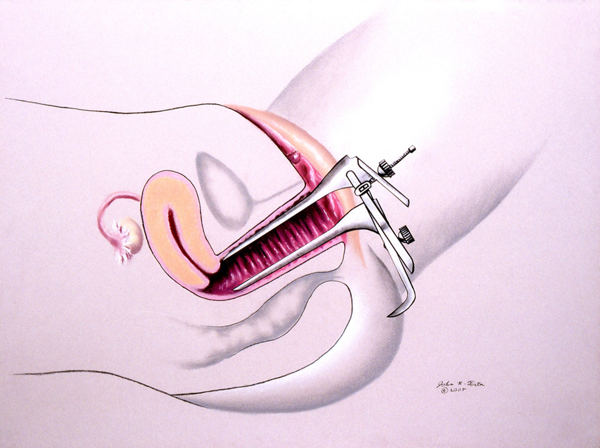
This illustration shows a pelvic examination with a speculum.
JOHN FOSTER/SCIENCE PHOTO LIBRARY
References
1. ACOG Committee Opinion No. 754. The Utility of and Indications for Routine Pelvic Examination. Obstet Gynecol. 2018;132(4):e174-e180. doi:10.1097/AOG.0000000000002895
2. Cantor A, Dana T, Griffin JC, et al. Screening for Chlamydial and Gonococcal Infections: Updated Evidence Report and Systematic Review for the US Preventive Services Task Force. JAMA. 2021;326(10):957-966. doi:10.1001/jama.2021.105771
3. World Health Organization. WHO recommendations on self-care interventions: Self-collection of samples for sexually transmitted infections (STIs). December 30, 2022 update. Accessed June 16, 2026.
4. Perkins RB, Wolf AMD, Church TR, et al. Self-collected vaginal specimens for human papillomavirus testing and guidance on screening exit: An update to the American Cancer Society cervical cancer screening guideline. CA Cancer J Clin. 2026;76(1):e70041. doi:10.3322/caac.70041
5. Vialard F, Anand A, Leung Soo C, et al. Self-sampling strategies (with/without digital innovations) in populations at risk of Chlamydia trachomatis and Neisseria gonorrhoeae: a systematic review and meta-analyses. Sex Transm Infect. 2023;99(6):420-428. Published 2023 Aug 17. doi:10.1136/sextrans-2022-055557
6. Qin J, Martinez G, Holt HK, et al. Preferences Among U.S. Women for Cervical Cancer Screening with Self-Collected Specimens for Human Papillomavirus Testing. Obstet Gynecol. 2026;147(3):306-312. doi:10.1097/AOG.0000000000006147
7. An official position statement of the Association of Women’s Health, Obstetric and Neonatal Nurses. The Use of Chaperones during Sensitive Examinations and Treatments. J Obstet Gynecol Neonatal Nurs. 2022;51(2):e1-e2. doi:10.1016/j.jogn.2021.12.0027.
8. Bignell CJ. Chaperones for genital examination. BMJ. 1999;319(7203):137-138. doi:10.1136/bmj.319.7203.137
Pelvic Examination Procedure
The examiner should have adequate light and the speculum and any other instruments or supplies readily available.
For the female pelvic examination, the patient lies supine on an examination table with her feet in stirrups (dorsal lithotomy position) and is usually draped.
The pelvic examination includes the following:
External examination of mons pubis and vulva
Speculum examination
Bimanual examination
Rectovaginal examination (sometimes)
A pelvic examination is indicated for
Symptomatic patients (eg, those with pelvic pain or abnormal uterine bleeding)
Asymptomatic patients with specific indications (eg, need for cervical cancer screening)
Before beginning the pelvic examination, the patient should be asked to void to empty the bladder. An empty bladder is important for the accurate assessment of uterovaginal prolapse, because increased bladder volume may reduce the extent of prolapse. Also, a full bladder may add further discomfort to the pelvic examination in some patients. Prior to the examination, many clinicians collect a urine sample if needed for analysis, culture, or STI testing.
A pelvic examination has not been found to be an effective screening test for ovarian cancer (see ACOG Committee Opinion No. 754: The Utility of and Indications for Routine Pelvic Examination).
External examination
During an external pelvic examination, there is inspection and palpation of the pubic and groin areas and the external genitalia. Prior to each examination, the clinician must take several important steps (1– 4):
Obtain the patient's informed consent for each component of the examination.
Offer a chaperone as per institutional policy and patient preference.
Use a trauma-informed approach throughout the examination (1).
The mons pubis and the hair-bearing areas of the labia majora are inspected for lesions or other findings based on symptoms (eg, folliculitis, lice). Hair distribution and skin changes may vary with age, hormonal status, ethnicity, gender-affirming therapy, and personal grooming practices and should not be considered abnormal in isolation.
The skin of the groin and the perineum is inspected for erythema, abnormal pigmentation, and skin lesions (eg, warts, excoriations, erosions, ulcers, fissures, pustules, nodules, tumors). Other findings, such as edema, scarring, lichenification, hypopigmentation or depigmentation, ecchymosis, signs of trauma, and lichen sclerosus or lichen planus, should be noted. The complete external examination includes examination of the entire vulva from the clitoris to the area below the anus to confirm any architectural changes in the skin. Examination of the perianal region is important because inflammatory, infectious, dermatologic, and neoplastic conditions frequently involve both the vulva and perianal skin (2, 3).
Clinicians should be familiar with normal vulvar anatomy and variation. Asymmetry of the labia, variation in pigmentation, size of the clitoris, and prominence of vestibular glands are common normal findings (2).
The clitoral hood is inspected for mobility, adhesions, scarring, inflammation, or lesions. Limited mobility may occur with lichen sclerosus, prior surgery, trauma, or chronic inflammatory disease.
The urethral meatus is inspected for lesions (eg, urethral carbuncle). Assessment should also include evidence of urethral prolapse, urethral diverticulum, or periurethral masses when clinically indicated.
The labia minora and majora are examined. Vulval skin disorders can cause changes in color, texture, and architecture of the vulval anatomy.
Gentle separation of the labia permits visualization of the vestibule, hymenal remnants, urethral meatus, vaginal introitus, and openings of the Bartholin glands while limiting the patient's discomfort. Structural abnormalities due to prior procedures (eg, episiotomy, female genital mutilation/cutting [FGM/C], gender-affirming surgery, or obstetric trauma) should be documented.
A cotton-swab test is performed on patients with vulvar pain or suspected vulvodynia to localize tenderness and differentiate localized vs generalized pain. Inspection should also assess for signs of vulvar dermatoses, trauma, neoplasia, and STIs when clinically indicated.
Vulvar Anatomy
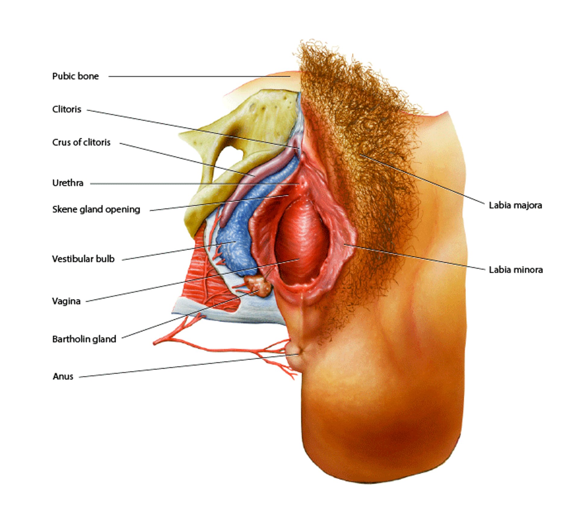
The vagina is a fibromuscular canal lined by nonkeratinized stratified squamous epithelium and contains no glands. Vaginal lubrication is mainly from transudation through the vaginal epithelium and secretions from the cervix and Bartholin glands. The urethra is located in the anterior portion of the vaginal introitus. The paraurethral (Skene) glands open adjacent to the urethral meatus and are homologous to the male prostate. The tissue surrounding the vagina is the vulva, which includes the labia minora, labia majora, and the glans and hood of the clitoris (most anteriorly and in the midline, anterior to the urethra). The vestibule is the region bounded by the labia minora and contains the urethral meatus, vaginal introitus, and the openings of the Bartholin and paraurethral glands. Anterior to the clitoris is the mons pubis (tissue that covers the pubic bone). Posterior to the vagina is the perineum, an area of skin between the vagina and anus. The crura of the clitoris (purple) extend subcutaneously and are composed of erectile tissue. The bulb of the vestibule (blue) also consists of erectile tissue. On each side, posterolateral to the vaginal introitus (at approximately the 4- and 8-o'clock positions), the Bartholin (greater vestibular) glands are located. They secrete mucus that contributes to the lubrication of the vulvar vestibule, especially during sexual arousal. BO VEISLAND/SCIENCE PHOTO LIBRARY |

Patients who report experiencing vulvovaginal pain during sexual activity or daily activities should be evaluated for vulvodynia, vestibular pain (localized provoked vestibulodynia), pelvic floor myalgia, interstitial cystitis/bladder pain syndrome, or genito-pelvic pain/penetration disorder (formerly known as vaginismus). A cotton-swab test is performed by gently applying light pressure to standardized locations of the vulva and vestibule to identify the location, severity, and distribution of pain. The patient should be asked to rate pain intensity (eg, on a 0 to 10 numeric rating scale, with 10 being the most intense pain) at each site. The cotton-swab test is used to diagnose allodynia and localized vestibular tenderness, which are characteristic of vulvodynia. A moistened cotton swab is gently applied from the labia majora toward the vestibule in a systematic clockwise fashion (commonly using the clock-face method) to evaluate sensory abnormalities and localized pain. Localized pain elicited by light pressure over the vulvar vestibule is characteristic of localized provoked vestibulodynia (5, 6).
A digital palpation of the pelvic floor muscles during the internal examination is done to assess pelvic floor muscle spasm or hypertonicity. If pelvic floor dysfunction is suspected, a single lubricated finger is used to palpate the levator ani, obturator internus, and other pelvic floor muscles for tenderness, increased resting tone, trigger points, and the patient's ability to voluntarily contract and relax the pelvic floor musculature (5, 6).
Neurologic assessment, which includes evaluation of the anocutaneous (anal wink) reflex and perineal sensation, may be performed when neurologic injury, pelvic floor neuropathy, obstetric sphincter injury, or significant bowel or bladder dysfunction is suspected. Routine testing is not required in asymptomatic patients. This is done by using a cotton swab to stroke the perianal skin, which should cause the external anal sphincter to contract (5, 6).
Next, the introitus is inspected. The Bartholin glands are palpated between the thumb and index finger only when enlargement, tenderness, or a cyst or abscess is suspected. In women older than 40 years with a new Bartholin gland mass, biopsy or excision should be considered to exclude Bartholin gland carcinoma.
When prolapse is suspected, the patient is examined during the Valsalva maneuver and, when appropriate, in both the lithotomy and standing positions. The degree of prolapse may be documented using the Pelvic Organ Prolapse Quantification (POP-Q) system (6). The examiner checks the vaginal opening for signs of pelvic organ prolapse: an anterior bulge (suggesting cystocele), a posterior bulge (suggesting rectocele), and displacement of the cervix toward the introitus (suggesting prolapsed uterus).
Speculum examination
The speculum is either a metal or plastic instrument used to retract the vaginal walls to enable the clinician to examine the vagina and cervix. Speculum size and type should be individualized according to patient anatomy, age, parity, menopausal status, gender-affirming surgery, and patient comfort (4, 7).
There are 2 main types of speculum: Graves (curved blades) and Pederson (straight blades). Both come in different sizes; the Pederson has a narrow and a pediatric size. A smaller speculum should be used if needed based on the patient's anatomy or discomfort.
Before the speculum examination, the patient is asked to relax the legs and hips. Throughout the examination, clinicians should explain each step, obtain ongoing verbal consent, and stop immediately if requested by the patient (4, 7).
The speculum is sometimes warmed prior to the examination and lubricated with water-based lubricant before insertion. A small amount of water-based lubricant may be applied to the posterior blade or external surface of the speculum to improve patient comfort and does not significantly affect cervical cytology or HPV testing when applied appropriately. Excess lubricant should be avoided because it may interfere with some microbiologic specimens. If water-based lubricant is not available, the speculum can be rinsed with warm water prior to insertion.
In many patients, the cervical position is uncertain or patient discomfort is anticipated; a gentle digital examination before speculum insertion helps with identification of the cervical position and appropriate speculum placement (1, 6, 7).
This illustration shows a pelvic examination with a speculum.
JOHN FOSTER/SCIENCE PHOTO LIBRARY
The speculum is inserted gently while separating the labia minora, while maintaining communication with the patient. The speculum is inserted slowly with the blades nearly vertical (in approximately the 1- and 7-o'clock positions) to avoid pressure on the urethra. Once fully inserted, the speculum is rotated to the horizontal position, then the blades are gently opened to visualize the cervix (4, 7).
If the clinician is unable to visualize the entire cervix, the speculum should be slightly withdrawn while the blades are open until the cervix is fully visible. If the patient experiences significant pain, the examination should be paused and the cause of discomfort evaluated before proceeding. If visualization of the cervix is still difficult, several maneuvers may improve visualization, including posterior repositioning of the speculum, slight withdrawal and reinsertion, asking the patient to place her hands beneath her buttocks to elevate the pelvis, hyperflexion of the hips, or changing the speculum size or type. A longer or wider speculum helps with better visualization in cases with elevated BMI.
Documentation should include all of the following: color, size, position, ectropion, lesions, polyps, ulceration, masses, discharge, friability, and bleeding. Whenever possible, the transformation zone should be identified, particularly during cervical cancer screening or colposcopic evaluation. The character of cervical mucus or discharge (color, consistency, quantity, and odor) should be noted because it may provide clues to infectious or inflammatory conditions. In patients with trichomoniasis, the cervix may demonstrate punctate hemorrhages (the classic "strawberry cervix"), although this finding is present in only a minority of cases.
If cervical cancer screening is indicated, cervical cytology and/or HPV testing should be obtained according to current screening guidelines before application of acetic acid or biopsy. If any abnormality of the cervix is identified, evaluation should not be delayed because of a recent normal cervical cytology or HPV test. Any visible cervical abnormalities require appropriate further diagnostic evaluation, including biopsy when indicated and referral for further evaluation. Biopsy is typically performed using cervical punch biopsy forceps. Cervical polyps may be removed when appropriate (8, 9).

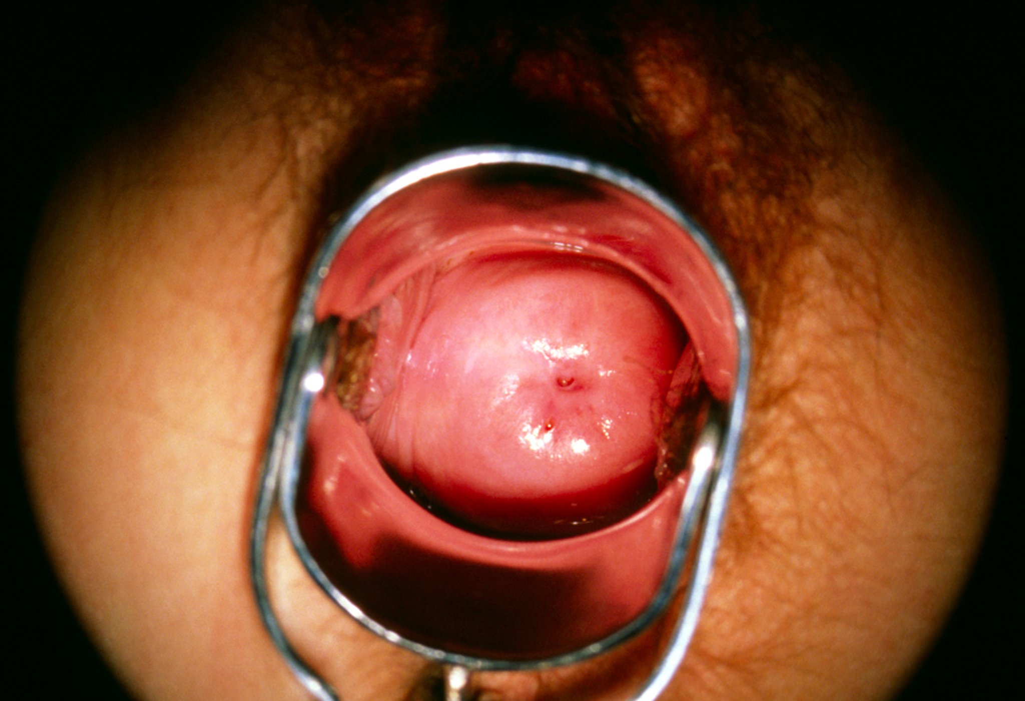
This image of the cervix seen through a colposcope shows the ectocervix (pale pink, shiny, round surface), which is covered with squamous epithelium. At the center of the ectocervix is the external cervical os, an opening that continues as the endocervix (also called the cervical canal), which is lined with glandular epithelium.
SCIENCE PHOTO LIBRARY
Normal vaginal discharge is usually clear to white and without a strong or unpleasant odor. The amount and consistency vary with age, hormonal status, pregnancy, sexual arousal, and use of hormonal contraception. Abnormal vaginal discharge may be associated with an unpleasant odor, abnormal color or consistency, vulvovaginal irritation, pruritus, dysuria, dyspareunia, or pelvic discomfort. Thin, gray discharge with a fishy odor suggests bacterial vaginosis, frothy yellow-green discharge may occur with trichomoniasis, and thick white, adherent discharge is characteristic of vulvovaginal candidiasis (4, 7).
The common diagnostic tests may include vaginal pH, a whiff test, saline and potassium hydroxide wet-mount microscopy, and gonorrhea and chlamydia nucleic acid amplification testing (NAAT). FDA-cleared molecular vaginitis panels or fungal culture are indicated when recurrent or complicated vulvovaginal candidiasis is suspected. For cervical cancer screening, a specimen is collected using a cervical broom, a spatula plus an endocervical brush, or another FDA-approved sampling device, depending on the type of screening being performed. The specimen is placed in a liquid-based solution for cervical cytology, primary HPV testing, cotesting, and, in selected settings, NAAT testing for chlamydia trachomatis, Neisseria gonorrhoeae, and Trichomonas vaginalis, depending on indications. Excess mucus, blood, or discharge may be gently removed with a large cotton swab before specimen collection. Avoid vigorous cleaning or repeated swabbing. A small amount of water-based lubricant applied to the speculum does not significantly interfere with liquid-based cytology or HPV testing when used appropriately (4, 10).
Current cervical cancer screening recommendations vary according to age, screening history, and risk factors. Screening options include primary HPV testing, cervical cytology alone, or cotesting with cervical cytology and HPV testing at guideline-recommended intervals (8, 9, 10).
Bimanual examination
The bimanual examination is done with 2 fingers in the vagina and the other hand on the lower abdomen to palpate the vagina, cervix, uterus, and ovaries. The fallopian tubes are only palpable if there is a tubal mass.
The index and middle fingers of the dominant hand are inserted into the vagina to just below the cervix. The other hand is placed just above the pubic symphysis and gently presses down to determine the size, position, consistency, and mobility of the uterus and, if possible, the ovaries. Normally, the uterus is about 7 cm by 5 cm by 3 cm and tilts anteriorly (anteversion), but it may tilt posteriorly (retroversion) to various degrees. The uterus may also be bent at an angle anteriorly (anteflexion) or posteriorly (retroflexion).

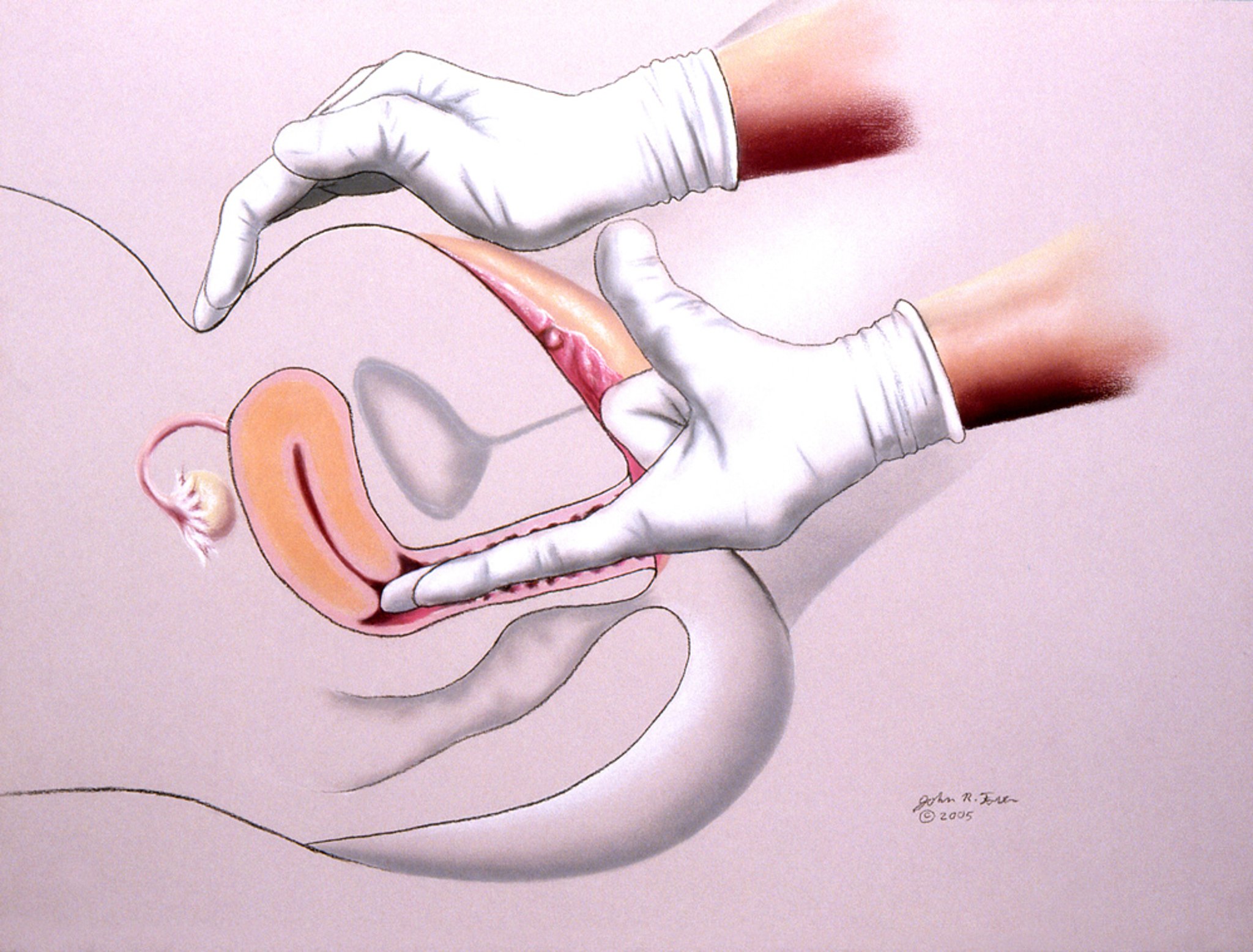
This illustration shows a pelvic examination being performed by a health care professional.
JOHN FOSTER/SCIENCE PHOTO LIBRARY

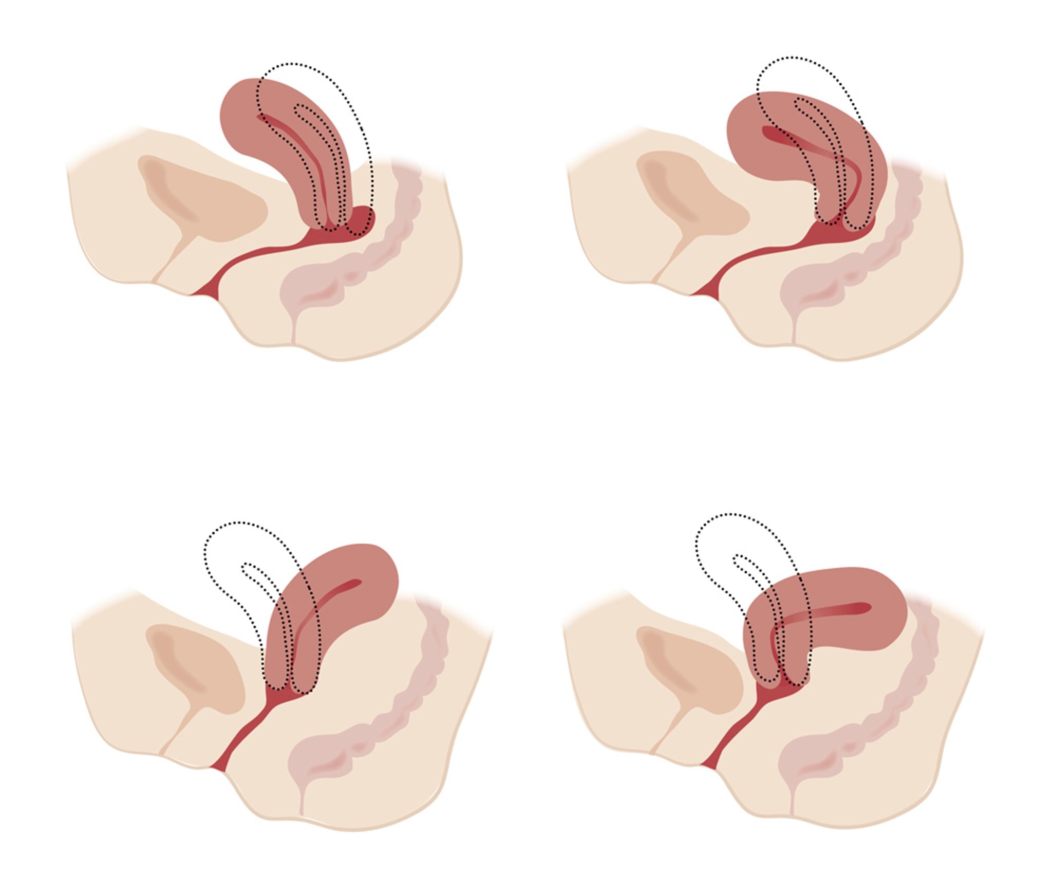
This illustration shows anatomical variations of the uterus. The uterus of a mature, nonpregnant female is between 7 to 9 cm long and 6.5 cm wide, and the muscular walls are about 3.5 cm thick. It is held loosely in position by ligaments, peritoneal tissue folds, and the pressure of adjacent organs like the urinary bladder, colon, and rectum. In most women, the uterus is anteverted, tipped forward so that it rests on the urinary bladder. However, it may be retroverted, or tipped backward. Various degrees of flexion are normal. Women who have given birth often have a retroflexed uterus.
JEANETTE ENGQVIST/SCIENCE PHOTO LIBRARY
The uterus is normally mobile, firm, and smooth; irregularity suggests uterine fibroids (leiomyomas). A fixed uterus may suggest adhesions from endometriosis, prior surgeries, or infections. Any palpable nodules along the cul-de-sac or uterosacral ligament posteriorly may suggest endometriosis.
Normally, the ovaries are about 2 cm by 3 cm in young women and are not palpable in postmenopausal women. Any enlargement of ovaries or adnexal masses may be palpated. Adnexal tenderness may be present in patients with a ruptured hemorrhagic ovarian cyst, ectopic pregnancy, ovarian or adnexal torsion, tubo-ovarian abscess, acute or chronic pelvic inflammatory disease, and rare cases of retrocecal appendicitis on the right side. An ovarian cyst or tumors or pedunculated fibroids may be tender if there are hemorrhagic changes.
Significant pain when the cervix is gently moved from side to side (cervical motion tenderness) suggests pelvic inflammation and is one of the diagnostic criteria for pelvic inflammatory disease.
Rectovaginal examination
After bimanual palpation, if indicated, the examiner palpates the rectovaginal septum by inserting the index finger in the vagina and the middle finger in the rectum. The rectovaginal examination may detect an ovarian mass.

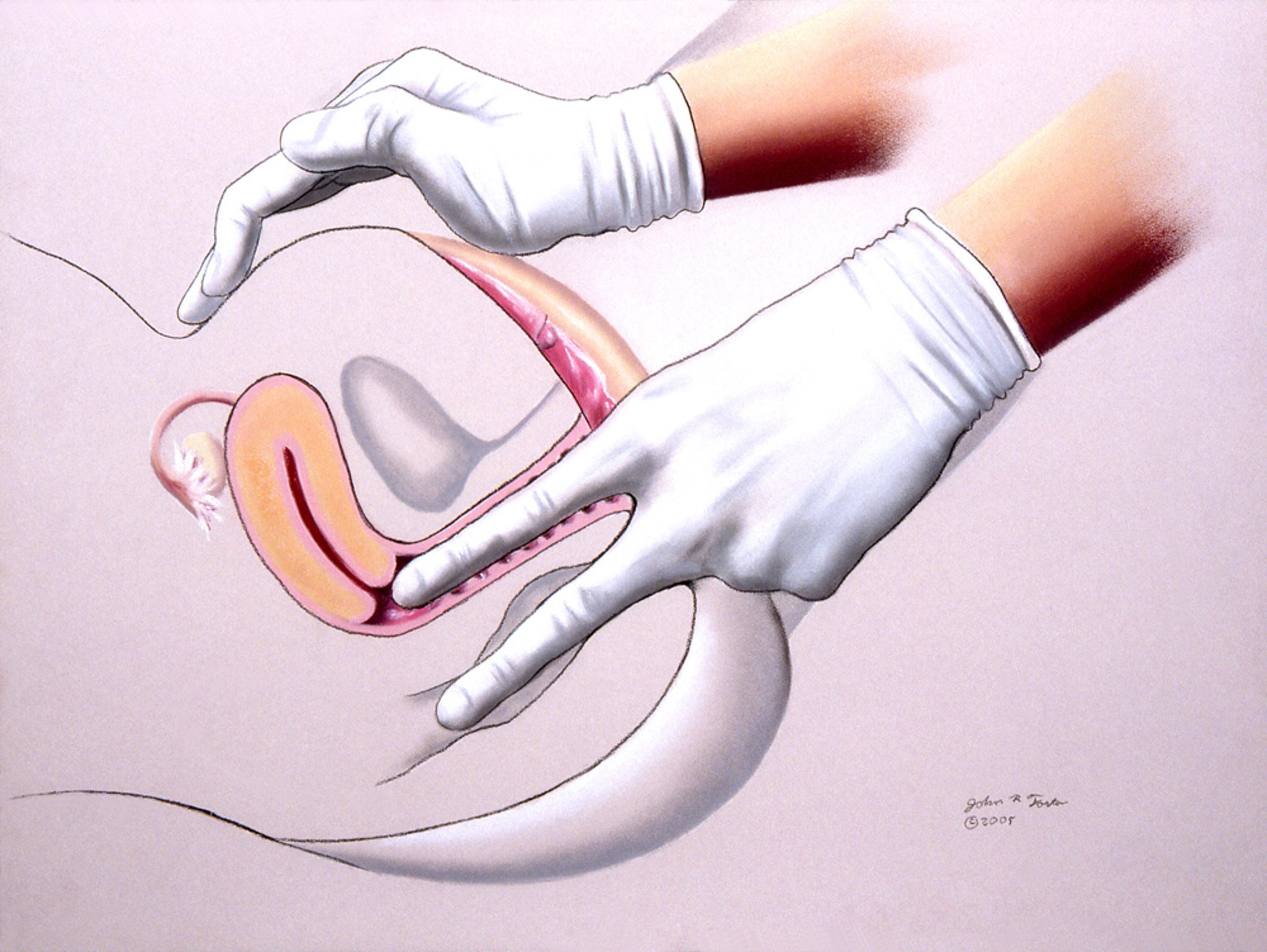
This illustration shows a pelvic rectovaginal examination being performed by a health care professional.
JOHN FOSTER/SCIENCE PHOTO LIBRARY
Pelvic examination references
1. Caring for Patients Who Have Experienced Trauma. ACOG Committee Opinion Summary, Number 825. Obstet Gynecol. 2021;137(4):757-758. doi:10.1097/AOG.0000000000004328
2. American College of Obstetricians and Gynecologists (ACOG) Committee on Gynecologic Practice Bulletin 224: Diagnosis and Management of Vulvar Skin Disorders. July 2020 (reaffirmed 2026).
3. Bornstein J, Goldstein AT, Stockdale CK, et al. 2015 ISSVD, ISSWSH and IPPS Consensus Terminology and Classification of Persistent Vulvar Pain and Vulvodynia. Obstet Gynecol. 2016;127(4):745-751. doi:10.1097/AOG.0000000000001359
4. Jonathan S. Berek. Berek & Novak's Gynecology. 17th edition Lippincott Williams & Wilkins (LWW)
5. American College of Obstetricians and Gynecologists (ACOG). ACOG Persistent Vulvar Pain. Committee Opinion Number 673. September 2016 (reaffirmed 2024).
6. AUGS/IUA. Pelvic Organ Prolapse: Interactive Assessment Tool. Accessed July 7, 2026.
7. Handa VL, Van Le L. Te Linde's Operative Gynecology. Wolters Kluwer.
8. ACOG PUBLICATIONS: ACOG COMMITTEE STATEMENT. Screening for Cervical Cancer. Obstetrics & Gynecology. 148(1):p e63-e67, July 2026. DOI: 10.1097/AOG.0000000000006257
9. Vadaparampil ST, Fuzzell LN, Brownstein NC, et al. A cross-sectional survey examining clinician characteristics, practices, and attitudes associated with adoption of the 2019 American Society for Colposcopy and Cervical Pathology risk-based management consensus guidelines. Cancer. 2023;129(17):2671-2684. doi:10.1002/cncr.34838
10. Centers for Disease Control and Prevention. STI Treatment Guidelines, 2021. Last reviewed: June 13, 2013. Accessed July 7, 2026.
Drug Information for the Topic