


Hereditary hemorrhagic telangiectasia is a hereditary disorder of vascular malformation transmitted as an autosomal dominant trait affecting men and women.
(See also Overview of Vascular Bleeding Disorders.)
More than 80% of patients have mutations in one of the following genes (1):
Endoglin (ENG) gene, which encodes a receptor for transforming growth factor beta-1 (TGF-β1) and transforming growth factor beta-3
Activin A receptor-like type 1 (ACVRL1) gene, which encodes the activin receptor-like kinase (ALK1)
SMADH4 (MADH4) gene, which encodes SMAD4, a protein active in the TGF beta signalling pathway
General reference
1. Kritharis A, Al-Samkari H, Kuter D. Hereditary hemorrhagic telangiectasia: Diagnosis and management from the hematologist’s perspective. Haematologica. 103:1433–1443, 2018. doi: 10.3324/haematol.2018.193003
Symptoms and Signs of Hereditary Hemorrhagic Telangiectasia
The most characteristic lesions of hereditary hemorrhagic telangiectasia are small red-to-violet telangiectatic lesions on the face, lips, oral and nasal mucosa, and tips of the digits. Similar lesions may be present throughout the mucosa of the gastrointestinal (GI) tract, resulting in recurrent GI bleeding. Patients may experience recurrent, profuse nosebleeds.
Some patients have pulmonary arteriovenous malformations (AVMs). These AVMs may cause significant right-to-left shunts, which can result in dyspnea, fatigue, cyanosis, or erythrocytosis. However, the first sign of the presence of AVMs may be a brain abscess, transient ischemic attack, or stroke as a result of infected or noninfected emboli. Cerebral or spinal AVMs occur in some families and may cause subarachnoid hemorrhage, seizures, or paraplegia. Hepatic AVMs may lead to liver failure and high output heart failure.
Chronic iron deficiency anemia is commonly present.

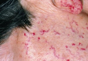
This photo shows a close-up of the face of a patient with multiple telangiectasias secondary to hereditary hemorrhagic telangiectasia.
This photo shows a close-up of the face of a patient with multiple telangiectasias secondary to hereditary hemorrhagic
DR P. MARAZZI/SCIENCE PHOTO LIBRARY

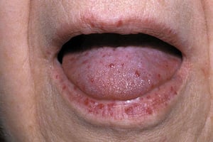
Papular, punctate, and linear telangiectases occur predominantly on the tongue, lips, digit tips, perioral region, and trunk.
Papular, punctate, and linear telangiectases occur predominantly on the tongue, lips, digit tips, perioral region, and
By permission of the publisher. From Deitcher S. In Atlas of Clinical Hematology. Edited by JO Armitage. Philadelphia, Current Medicine, 2004.


DR P. MARAZZI/SCIENCE PHOTO LIBRARY
This photo shows a close-up of the face of a patient with multiple telangiectasias secondary to hereditary hemorrhagic telangiectasia.
This photo shows a close-up of the face of a patient with multiple telangiectasias secondary to hereditary hemorrhagic
DR P. MARAZZI/SCIENCE PHOTO LIBRARY
Papular, punctate, and linear telangiectases occur predominantly on the tongue, lips, digit tips, perioral region, and trunk.
Papular, punctate, and linear telangiectases occur predominantly on the tongue, lips, digit tips, perioral region, and
By permission of the publisher. From Deitcher S. In Atlas of Clinical Hematology. Edited by JO Armitage. Philadelphia, Current Medicine, 2004.
DR P. MARAZZI/SCIENCE PHOTO LIBRARY
Diagnosis of Hereditary Hemorrhagic Telangiectasia
Clinical evaluation
Sometimes endoscopy or angiography
Sometimes genetic testing
Diagnosis of hereditary hemorrhagic telangiectasia is based on the finding of characteristic arteriovenous malformations on the face, mouth, nose, palms, digits, and/or internal organs in the context of epistaxis and family history. The Curaçao criteria include the following:
Spontaneous recurrent epistaxis
Multiple telangiectasias in typical locations
Documented visceral arteriovenous malformations (eg, in the lung, liver, brain, and spine)
First-degree family member with hereditary hemorrhagic telangiectasia
Hereditary hemorrhagic telangiectasia is definite if 3 of these criteria are met and possible if 2 are met (1, 2).
Sometimes endoscopy or angiography or both are needed. Laboratory findings are usually normal except for iron deficiency anemia in many patients.
Testing for the ENG,ACVRL1, and SMADH4 (MADH4) mutations may be helpful in some patients with atypical features or for screening asymptomatic family members.
Diagnosis references
1. Shovlin CL, Guttmacher AE, Buscarini E, et al. Diagnostic criteria for hereditary hemorrhagic telangiectasia (Rendu-Osler-Weber syndrome). Am J Med Genet. 91(1):66–67, 2000. doi: 10.1002/(sici)1096-8628(20000306)91:1<66::aid-ajmg12>3.0.co;2-p
2. Faughnan ME, Mager JJ, Hetts SW, et al. Second international guidelines for the diagnosis and management of hereditary hemorrhagic telangiectasia. Ann Intern Med. 173(12):989–1001, 2020. doi: 10.7326/M20-1443
Screening for Hereditary Hemorrhagic Telangiectasia
If a family history of pulmonary, hepatic, or cerebral arteriovenous malformations exists, screening at puberty and at the end of adolescence with pulmonary CT, hepatic CT, and cerebral MRI is recommended. Screening also involves checking for iron deficiency anemia due to the chronic blood loss. Screening may also include blood tests for the gene mutations mentioned above.
Treatment of Hereditary Hemorrhagic Telangiectasia
Sometimes laser ablation, surgical resection, or embolization of symptomatic arteriovenous malformations
Supplemental oral or intravenous iron therapy
Possibly blood transfusions
Sometimes antifibrinolytic medications (eg, aminocaproic acid, tranexamic acid)
Sometimes angiogenesis inhibitors (eg, bevacizumab, pomalidomide, thalidomide)
Treatment for most patients involves a combination of interventions, including local treatment for epistaxis, laser and surgical interventions for arteriovenous malformations, blood transfusions, iron supplementation, and systemic therapy with antifibrinolytic agents and angiogenesis inhibitors. For example, accessible telangiectasias (eg, in the nose or gastrointestinal tract via endoscopy) may be treated with laser ablation and arteriovenous malformations in the lungs can be treated by surgical resection or coil embolization.
Liver lesions may not be amenable to surgery or embolization. Measurement of serum ammonia level may help assess the degree of hepatic shunting of blood (1).
Repeated blood transfusions may be needed.
Many patients require continuous oral iron therapy to replace iron lost due to repeated mucosal bleeding (see treatment of Iron Deficiency Anemia). Many patients also require parenteral (IV) iron, and sometimes recombinant erythropoietin is necessary.
Medical therapy is increasingly being used to treat hereditary hemorrhagic telangiectasia and can reduce the need for red blood cell transfusion, iron administration, and surgical intervention (2, 3). Options include medications that inhibit fibrinolysis (eg, aminocaproic acid, tranexamic acid) and medications that inhibit angiogenesis (eg, bevacizumab, pomalidomide, thalidomide, pazopanib) (4). Bevacizumab, in particular, has been shown to decrease the incidence of nasal and gastrointestinal bleeding in patients with hereditary hemorrhagic telangiectasia (5, 6, 2). Also, a trial that compared pomalidomide with placebo demonstrated statistically significant improvements in epistaxis severity and disease specific quality of life scores (7).
To avoid paradoxical embolization of particulate matter to the brain through pulmonary arteriovenous malformations, all IV fluids need to be given through a filter.
Treatment references
1. Bloom PP, Rodriguez-Lopez J, Witkin AS, et al. Ammonia predicts hepatic involvement and pulmonary hypertension in patients with hereditary hemorrhagic telangiectasia. Clin Transl Gastroenterol. 11(1):e00118, 2020. doi:10.14309/ctg.0000000000000118
2. Faughnan ME, Mager JJ, Hetts SW, et al. Second international guidelines for the diagnosis and management of hereditary hemorrhagic telangiectasia. Ann Intern Med. 173(12):989–1001, 2020. doi: 10.7326/M20-1443
3. Al-Samkari H. How I treat bleeding in hereditary hemorrhagic telangiectasia. Blood. 2024;144(9):940-954. doi:10.1182/blood.2023021765
4. Al-Samkari H. Hereditary hemorrhagic telangiectasia: systemic therapies, guidelines, and an evolving standard of care. Blood. 137 (7): 888–895, 2021. doi:10.1182/blood.2020008739
5. Al-Samkari H, Kasthuri RS, Parambil JG, et al: An international, multicenter study of intravenous bevacizumab for bleeding in hereditary hemorrhagic telangiectasia: the InHIBIT-Bleed study. Haematologica 106(8):2161–2169, 2021. doi: 10.3324/haematol.2020.261859
6. Dupuis-Girod S, Rivière S, Lavigne C, et al. Efficacy and safety of intravenous bevacizumab on severe bleeding associated with hemorrhagic hereditary telangiectasia: A national, randomized multicenter trial. J Intern Med. 2023;294(6):761-774. doi:10.1111/joim.137142
7. Al-Samkari H, Kasthuri RS, Iyer VN, et al. Pomalidomide for Epistaxis in Hereditary Hemorrhagic Telangiectasia. N Engl J Med. 2024;391(11):1015-1027. doi:10.1056/NEJMoa2312749
Key Points
Nasal and gastrointestinal telangiectasias may cause significant external hemorrhage in patients with hereditary hemorrhagic telangiectasia.
Vascular malformations in the central nervous system, lungs, and liver may bleed; hepatic and pulmonary malformations may cause significant shunting.
Accessible mucosal telangiectasias and arteriovenous malformations may be treated with laser ablation; coil embolization or surgical resection may be needed for pulmonary vascular malformations.
Antifibrinolytic medications and angiogenesis inhibitors may reduce the incidence of bleeding.
Many patients require parenteral iron supplements because of chronic blood loss.
All IV solutions should be given through a filter to avoid paradoxical embolization of particulate matter to the brain through pulmonary arteriovenous malformations.
Drug Information for the Topic


