


Babesiosis is an infection with Babesia species of protozoa. Infections can be asymptomatic or cause a malaria-like illness with fever and hemolytic anemia. Disease is most severe in asplenic patients, older adults, and patients with advanced HIV infection. Diagnosis is by identification of Babesia in a peripheral blood smear, serologic test, or polymerase chain reaction (PCR). Treatment, when needed, is with azithromycin plus atovaquone or with quinine plus clindamycin.
In 2020, there were 1827 reported cases of babesiosis in the United States from the 40 states where babesiosis was a reportable condition (1). Endemic areas include the islands and the mainland bordering Nantucket Sound in Massachusetts, Rhode Island, eastern Long Island, coastal Connecticut, Vermont, and New Jersey as well as foci in Wisconsin and Minnesota in the upper Midwest.
In the United States, B. microti is the most common cause of babesiosis in humans. B. duncani has also been isolated from patients in Washington and California. A B. duncani–like strain designated MO-1 has been reported in patients in Missouri. The highest incidence of disease is seen in people residing in wooded areas with large populations of deer.
Other Babesia species transmitted by different ticks infect humans in other areas of the world. In Europe, B. divergens is the principal cause of babesiosis, typically in patients who have had a splenectomy.
General reference
1. Centers for Disease Control and Prevention (CDC): Data and Statistics on Babesiosis. Accessed February 11, 2025.
Pathogenesis of Babesiosis
Rodents (primarily the white-footed mouse, Peromyscus leucopus) are the principal natural reservoir for Babesia, and deer ticks of the family Ixodidae (typically Ixodes scapularis) are the usual vectors. Larval ticks become infected while feeding on an infected rodent, then transform into nymphs that transmit the parasite to another animal or to a human. Adult ticks ordinarily feed on deer but may also transmit the parasite to humans. (See also Babesiosis in Animals.)
After Babesia sporozoites are transmitted via the bite of an infected nymphal tick, they enter red blood cells, mature, and divide asexually. Infected erythrocytes eventually rupture and release organisms that invade other red blood cells; thus, Babesia can also be transmitted by blood transfusion and possibly by organ transplantation. Infected red blood cells rupture, releasing merozoites and causing membrane damage that contributes to the clinical manifestations of disease, such as hemolysis, anemia, jaundice, and hemoglobinuria. A test to screen blood and organ donors for B. microti is currently used in states in the northeastern United States with the highest incidences of infection. Host defenses include cell-mediated immunity by CD4+ T cells and innate immune responses by macrophages and natural killer (NK) cells. The role of humoral immunity is less defined. Immunocompromised patients are at higher risk of symptomatic disease because of impaired host defenses.


The B. microti life cycle involves 2 hosts, a rodent, primarily the white-footed mouse, Peromyscus leucopus, and an Ixodes tick.
1. During a blood meal on a mammalian host, a Babesia-infected tick introduces sporozoites into the mouse host.
2. Inside the mouse host, sporozoites enter erythrocytes and reproduce asexually.
3. In the blood, some parasites differentiate into male and female gametes.
4–5. After gametes are ingested by a tick, the gametes transform into sporozoites.
6. Humans enter the life cycle when bitten by an infected tick. An infected tick introduces sporozoites into the human host during a blood meal.
7. Inside the human host, sporozoites enter erythrocytes and reproduce asexually.
8. Human-to-human transmission can occur through blood transfusions.
Image from the Centers for Disease Control and Prevention, Global Health, Division of Parasitic Diseases and Malaria.
Congenital infection via transplacental transmission can also occur but is very rare.
Ixodes ticks infected with Babesia are sometimes coinfected with Borrelia burgdorferi (which causes Lyme disease), Anaplasma phagocytophilum (which causes human granulocytic anaplasmosis [HGA]), B. miyamotoi (which causes a relapsing fever-like illness), or Powassan virus (a flavivirus that causes encephalitis). Thus, patients may occasionally acquire more than one infection from a tick bite.

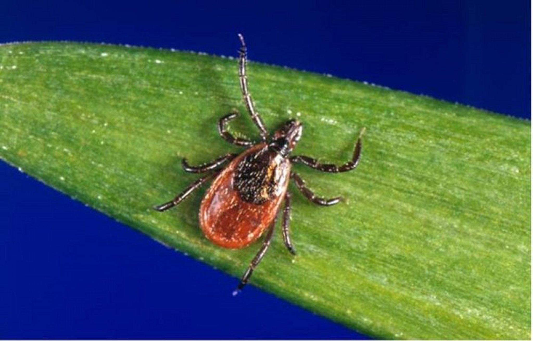
This image shows an adult deer tick, Ixodes scapularis, which transmits Babesia microti, the causative agent of babesiosis, and Borrelia burgdorferi, a causative agent of Lyme disease.
Image courtesy of James Gathany via the Public Health Image Library of the Centers for Disease Control and Prevention.
Symptoms and Signs of Babesiosis
Asymptomatic Babesia infection may persist for months to years and remain subclinical throughout its course in otherwise healthy people, especially those < 40 years.
When symptomatic, babesiosis usually starts after a 1- to 2-week incubation period with nonspecific symptoms including malaise, fatigue, chills, fever, headache, myalgia, and arthralgia. In healthy people, symptoms usually resolve after a week. In others, hepatosplenomegaly with jaundice, mild to moderately severe hemolytic anemia, mild neutropenia, and thrombocytopenia may occur. Noncardiac pulmonary edema can develop in people with severe disease.
Babesiosis is sometimes fatal, particularly in older adults, patients with asplenia, and patients with advanced HIV infection or another form of immunosuppression, such as patients who have received transplantation. In such patients, babesiosis may resemble Plasmodium falciparum malaria, with high fever, hemolytic anemia, hemoglobinuria, jaundice, and renal failure. Splenectomy may cause previously acquired asymptomatic parasitemia to become symptomatic.
Babesiosis can be transmitted transplacentally from infected pregnant patient to fetus. This vertical transmission can lead to congenital babesiosis, which may manifest with symptoms such as fever, anemia, and thrombocytopenia in the neonate.
Diagnosis of Babesiosis
Light microscopy of blood smears
Serologic and polymerase chain reaction (PCR)–based tests
Most patients with babesiosis do not remember a tick bite, but they may reside in or report a history of travel to an endemic region.
Babesiosis is usually diagnosed by finding Babesia in red blood cells in blood smears, but differentiation from Plasmodium species can be difficult. Tetrad forms (the so-called Maltese cross formation), although not common, are unique to Babesia and helpful diagnostically (1).


This image is a photomicrograph of a blood smear showing tetrad configurations of Babesia species trophozoites, resembling Plasmodium falciparum. Babesia species trophozoites exhibit variability in shape and size and do not produce pigment.
CDC/ Steven Glenn, Laboratory & Consultation Division
Serologic and PCR-based tests are available. Antibody detection by indirect fluorescent antibody (IFA) testing using B. microti antigens can be helpful in patients with low-level parasitemia but may be falsely negative in those infected with other Babesia species. PCR-based assays can help differentiate Babesia from P. falciparum if blood smear findings are ambiguous, confirm infection in patients with low parasitemia, and identify the Babesia species. For patients with a positive Babesia antibody test, confirmation with a blood smear or PCR-based test is recommended because Babesia antibodies can persist in blood for a year or more after apparent clearance of infection, with or without treatment.
In acutely ill patients, nonspecific laboratory findings may include proteinuria, hemoglobinuria, and elevated levels of liver enzymes, blood urea nitrogen, and creatinine.
Diagnosis reference
Centers for Disease Control and Prevention (CDC). Babesiosis. Laboratory Diagnosis. Accessed February 11, 2025.
Treatment of Babesiosis
Atovaquone plus azithromycin
Quinine plus clindamycin
Asymptomatic patients usually require no treatment (except when parasitemia is present for 1 month or longer).
Treatment is indicated for patients with persistent high fever, rapidly increasing parasitemia, and falling hematocrit (1).
The combination of atovaquone and azithromycin given for 7 to 10 days has fewer adverse effects and is as effective as traditional therapy with quinine plus clindamycin in all patients with babesiosis (2). The duration of therapy may be extended beyond 10 days for immunocompromised patients.
Oral atovaquone along with IV azithromycin given every 24 hours is the preferred regimen until symptoms abate; after abatement, oral therapy with both atovaquone and azithromycin is preferred.
Combinations of atovaquone and azithromycin are preferred, andquinine plus clindamycin is considered an alternative regimen for severely ill patients. Recipients of quinine must be monitored closely for adverse effects (also called cinchonism). In immunocompetent patients, most symptoms resolve and blood smears become negative during the standard 7- to 10-day treatment course. PCR test results may remain positive despite completion of treatment; however, relapse is rare (1, 3).
Exchange transfusion has been used in severely ill patients with high-grade (> 10% of erythrocytes) parasitemia.
Treatment references
1. Krause PJ, Auwaerter PG, Bannuru RR, et al. Clinical Practice Guidelines by the Infectious Diseases Society of America (IDSA): 2020 Guideline on Diagnosis and Management of Babesiosis [published correction appears in Clin Infect Dis. 2021 Jul 1;73(1):172-173. doi: 10.1093/cid/ciab275.]. Clin Infect Dis. 2021;72(2):e49-e64. doi:10.1093/cid/ciaa1216
2. Krause PJ, Lepore T, Sikand VK, et al. Atovaquone and azithromycin for the treatment of babesiosis. N Engl J Med. 2000;343(20):1454-1458. doi:10.1056/NEJM200011163432004
3. Centers for Disease Control and Prevention (CDC): Clinical Care of Babesiosis. Accessed February 11, 2025.
Prevention of Babesiosis
To prevent babesiosis, standard tick precautions should be taken by all people in endemic areas. Patients with asplenia and patients with HIV infection should be particularly cautious.
People who have had babesiosis are deferred from donating blood and potentially organs to prevent transmission until treatment is completed and parasitemia is no longer detected. Screening of blood and organ donors is now performed in areas within the United States with relatively high incidences of infection.
Tick bite prevention
Preventing tick access to skin includes the following:
Staying on paths and trails
Tucking trousers into boots or socks
Wearing long-sleeved shirts
Applying repellents with diethyltoluamide (DEET) to skin surfaces
DEET should be used cautiously in very young children because toxic reactions have been reported. Permethrin on clothing effectively kills ticks. Frequent searches for ticks, particularly in hairy areas of the body and on children, are essential in endemic areas.
Engorged ticks should be removed with care and not crushed between the fingers because crushing the tick may result in disease transmission. The tick’s body should not be grasped or squeezed. Gradual traction on the head of the tick with a small forceps dislodges it. The point of attachment should be swabbed with alcohol. Petroleum jelly, alcohol, lit matches, and other irritants are not effective ways to remove ticks and should not be used.
No practical means are available to rid entire areas of ticks, but tick populations may be reduced in endemic areas indirectly by controlling small-animal populations, especially rodents.
Key Points
Endemic areas of babesiosis in the United States include the coast and islands of southern New England and New Jersey as well as parts of the upper Midwest and Northwest.
Babesiosis ranges from a mild, asymptomatic infection to a severe, life-threatening illness (mainly in patients who are older or who have asplenia or in patients with HIV or other immunocompromising conditions).
Symptoms are nonspecific and may resemble those of P. falciparum malaria, with prolonged fever, headache, myalgias, and sometimes jaundice.
Diagnose using light microscopy of blood smears or with serologic and PCR-based tests.
Treat symptomatic patients with atovaquone plus azithromycin or, as an alternative, quinine plus clindamycin.
More Information
The following English-language resource may be useful. Please note that The Manual is not responsible for the content of this resource.
MSD Veterinary Manual: Babesiosis in Animals
Drug Information for the Topic


