


A perilunate dislocation is disruption of the normal relationship between the lunate and capitate. A lunate dislocation is separation of the lunate from both the capitate and the radius.
Perilunate and lunate dislocations result when great force is applied to a hyperextended wrist. They usually result from a fall on an outstretched hand or occur in a motor vehicle crash. Perilunate dislocations are more common than lunate dislocations.
These dislocations cause pain, swelling, and deformity in the wrist and proximal hand.
If a perilunate or lunate dislocation is not diagnosed and treated promptly, complications can develop. They include:
Median nerve injury
Avascular necrosis of the scaphoid or lunate and deterioration of the joint (scapholunate advanced collapse).
(See Overview of Dislocations.)
Diagnosis of Perilunate and Lunate Dislocations
Radiographs
Plain radiographs (anteroposterior, lateral, and oblique views) are performed. To avoid missing the diagnosis, clinicians should assess the relationship between the radius, lunate, and capitate bones on a true lateral view.

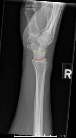
This lateral view of a normal wrist demonstrates the normal interface between the radius and lunate (red line) and the lunate and capitate (yellow line).
This lateral view of a normal wrist demonstrates the normal interface between the radius and lunate (red line) and the
Image courtesy of Danielle Campagne, MD.

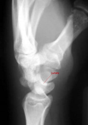
On a lateral view of a perilunate dislocation, the capitate does not articulate with the lunate.
On a lateral view of a perilunate dislocation, the capitate does not articulate with the lunate.
Image courtesy of Danielle Campagne, MD.

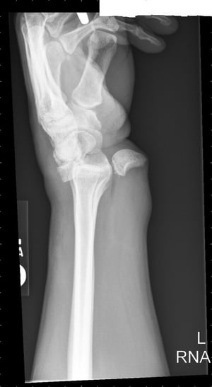
Lunate dislocation results in a spilled teacup configuration where the lunate is rotated and displaced volarly.
Lunate dislocation results in a spilled teacup configuration where the lunate is rotated and displaced volarly.
Image courtesy of Danielle Campagne, MD.
This lateral view of a normal wrist demonstrates the normal interface between the radius and lunate (red line) and the lunate and capitate (yellow line).
This lateral view of a normal wrist demonstrates the normal interface between the radius and lunate (red line) and the
Image courtesy of Danielle Campagne, MD.
On a lateral view of a perilunate dislocation, the capitate does not articulate with the lunate.
On a lateral view of a perilunate dislocation, the capitate does not articulate with the lunate.
Image courtesy of Danielle Campagne, MD.
Lunate dislocation results in a spilled teacup configuration where the lunate is rotated and displaced volarly.
Lunate dislocation results in a spilled teacup configuration where the lunate is rotated and displaced volarly.
Image courtesy of Danielle Campagne, MD.
In a perilunate dislocation, the capitate is not vertically aligned with the lunate and radius on a lateral view of the wrist. The lunate and radius remain correctly aligned.
In a lunate dislocation, the lunate is rotated and displaced volarly out of alignment and appears similar to a spilled teacup.
Treatment of Perilunate and Lunate Dislocations
Closed reduction and splinting
Usually surgical repair
Treatment of both perilunate and lunate dislocations is closed reduction and splinting in the emergency department. Closed reduction requires procedural sedation.
To perform closed reduction, longitudinal traction is applied to the fingers and wrist to disengage the carpal bones from the wrist. Applying direct pressure to the capitate while at the same time flexing and extending the wrist can reduce a perilunate dislocation. Applying direct pressure to the lunate while extending the wrist can guide the lunate dorsally back into proper rotation and alignment. Reduction should be confirmed with radiographs. Both the wrist and elbow should be immobilized in the neutral position (eg, with a sugar tong splint).
Patients should be immediately referred to an orthopedic surgeon; most dislocations must be surgically repaired because function is better after surgical repair (1).
Treatment reference
1. Goodman AD, Harris AP, Gil JA, et al. Evaluation, Management, and Outcomes of Lunate and Perilunate Dislocations. Orthopedics. 2019;42(1):e1-e6. doi:10.3928/01477447-20181102-05
Key Points
Perilunate and lunate dislocations usually result from a fall on an outstretched hand or a motor vehicle crash.
Treat these dislocations promptly to prevent complications (eg, nerve damage, deterioration of the joint).
Diagnose by plain radiographs (anteroposterior, lateral, and oblique views), with particular attention to the relationship between the radius, lunate, and capitate bones on a true lateral view.
Reduce manually and splint the wrist and elbow in the neutral position.
Refer patients immediately to an orthopedic surgeon because most of these dislocations must be surgically repaired to optimize function.


