


Tension pneumothorax is a life-threatening condition that occurs either in patients on mechanical ventilation with barotrauma or as a result of thoracic trauma. Damaged lung tissue or a chest wound form a one-way valve mechanism, resulting in progressive air accumulation, mediastinal shift, and decreased venous return to the heart. Initial symptoms and signs are dyspnea and pleuritic chest pain, then as intrathoracic pressure increases, hypotension, tracheal deviation, and jugular venous distention develop. The affected hemithorax is hyperresonant to percussion with absent breath sounds. Diagnosis is by vital signs and physical examination. Treatment is with immediate needle decompression followed by tube thoracostomy.
Tension pneumothorax develops when a lung or chest wall injury acts as a one-way valve mechanism that allows air into the pleural space but not out of it. As a result, air accumulates and compresses the lung, eventually shifting the mediastinum, compressing the contralateral lung, increasing intrathoracic pressure, and decreasing venous return to the heart, resulting in shock. These effects can develop rapidly, particularly in patients undergoing positive pressure ventilation.


This illustration shows a tension pneumothorax, which develops when a lung injury (shown in the illustration as a small dark area on the left lung) or chest wall injury acts as a one-way valve mechanism that allows air into the pleural space but not out of it. (Small black arrows indicate air leaking out of the lung; large black arrows indicate pressure from air in the pleural space.) As a result, air accumulates and intrathoracic pressure increases, compressing the affected lung. Eventually, all the midline thoracic structures shift, including the mediastinum; the contralateral lung is compressed; and venous return to the heart decreases, resulting in shock. These complications can develop rapidly, particularly in patients receiving positive pressure ventilation.
Pepermpron/stock.adobe.com
Causes include mechanical ventilation (most commonly) and simple (uncomplicated) pneumothorax with lung injury that fails to seal following penetrating or blunt chest trauma or failed central venous cannulation.
Symptoms and Signs of Tension Pneumothorax
Symptoms and signs initially are those of simple pneumothorax (dyspnea, pleuritic chest pain, decreased breath sounds). As intrathoracic pressure increases, patients develop hypotension, tracheal deviation, and jugular venous distention. The affected hemithorax is hyperresonant to percussion and often feels somewhat distended, tense, and poorly compressible to palpation, with absent breath sounds on the affected side.
Diagnosis of Tension Pneumothorax
History and physical examination
Tension pneumothorax is diagnosed in patients on mechanical ventilation or thoracic trauma based on vital signs and physical examination.It is a life-threatening emergency and treatment should not be delayed pending radiographic confirmation. Although cardiac tamponade can also cause hypotension, neck vein distention, and sometimes respiratory distress, tension pneumothorax can be differentiated clinically by its unilateral absence of breath sounds and hyperresonance to percussion.

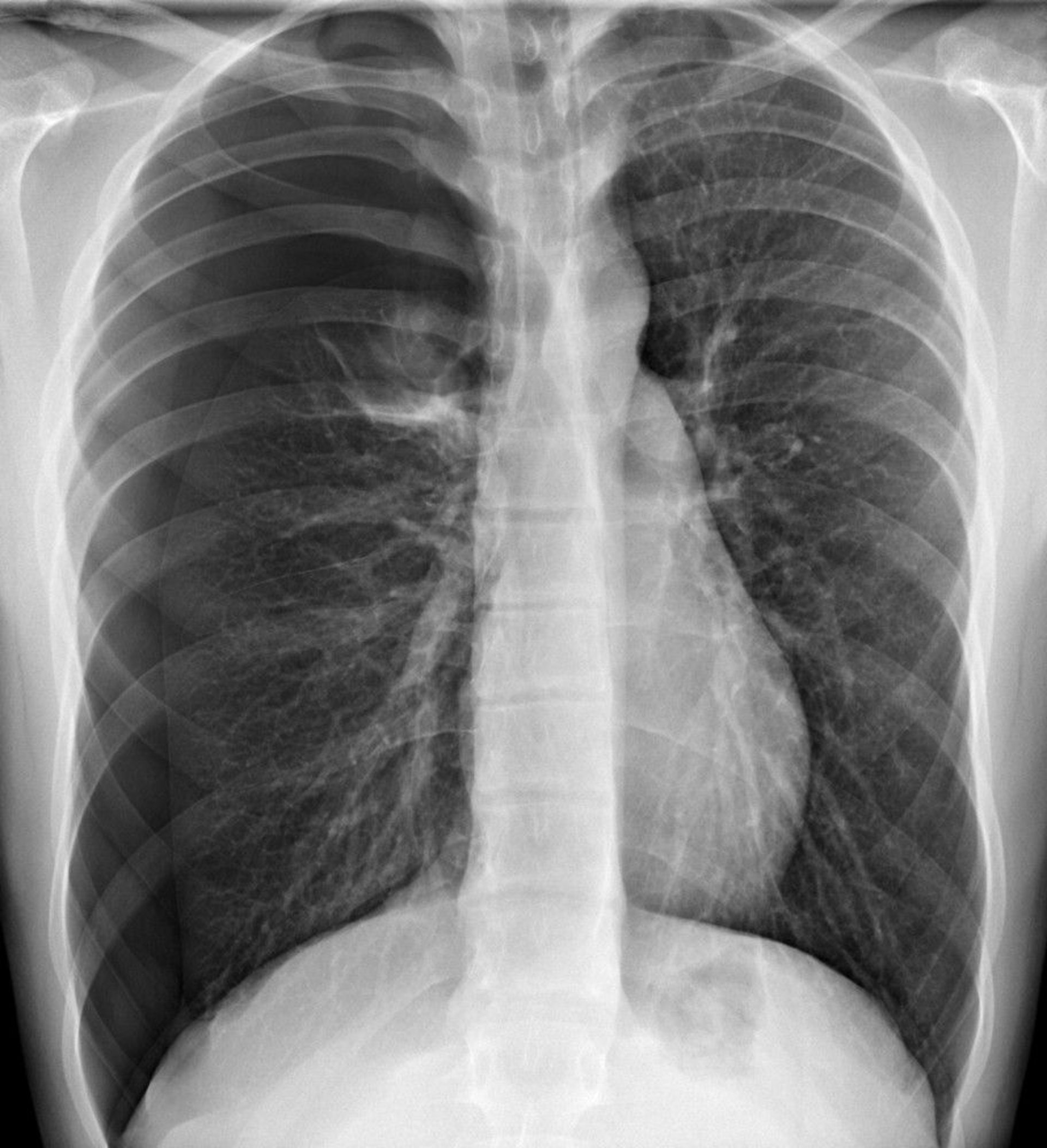
In this radiograph, a pneumothorax is visible in the right hemithorax, where, in the periphery, there are no lung markings. A shift of the heart and mediastinum toward the left side can cause tension pneumothorax physiology. Tension pneumothorax, however, should be diagnosed clinically and not await radiographic confirmation.
DU CANE MEDICAL IMAGING LTD/SCIENCE PHOTO LIBRARY
Pearls & Pitfalls
|
Treatment of Tension Pneumothorax
Needle decompression followed by tube thoracostomy
Tension pneumothorax is treated with immediate needle decompression by inserting a large-bore (eg, 14- or 16-gauge) needle or with finger thoracostomy, into the fourth or fifth intercostal space along the midaxillary line (1, 2, 3, 4). Air will rapidly escape the pleural space and decompression can sometimes be heard. Because needle decompression causes a simple pneumothorax, tube thoracostomy should be performed immediately afterwards.


Treatment references
1. Inaba K, Karamanos E, Skiada D, et al. Cadaveric comparison of the optimal site for needle decompression of tension pneumothorax by prehospital care providers. J Trauma Acute Care Surg. 2015;79(6):1044-1048. doi:10.1097/TA.0000000000000849
2. Nelson M, Chavda Y, Stankard B, et al. Using Ultrasound to Determine Optimal Location for Needle Decompression of Tension Pneumothorax: A Pilot Study. J Emerg Med. 2022;63(4):528-532. doi:10.1016/j.jemermed.2022.08.004
3. Inaba K, Karamanos E, Skiada D, et al. Cadaveric comparison of the optimal site for needle decompression of tension pneumothorax by prehospital care providers. J Trauma Acute Care Surg. 2015;79(6):1044-1048. doi:10.1097/TA.0000000000000849
4. Nelson M, Chavda Y, Stankard B, et al. Using Ultrasound to Determine Optimal Location for Needle Decompression of Tension Pneumothorax: A Pilot Study. J Emerg Med. 2022;63(4):528-532. doi:10.1016/j.jemermed.2022.08.004


