


Pleural effusions are accumulations of fluid within the pleural space. They have multiple causes and are usually classified as transudates or exudates. Detection is by physical examination, chest radiograph, and bedside thoracic ultrasound. Thoracentesis and pleural fluid analysis are often required to determine cause. Asymptomatic bilateral transudates require no treatment. Symptomatic transudates and most exudates require thoracentesis, chest tube drainage, and definitive pleural management (indwelling pleural catheter, talc pleurodesis, thoracic surgery, or a combination).
Normally, 10 to 20 mL of pleural fluid, similar in composition to plasma but lower in protein (< 1.5 g/dL [< 15 g/L]), is spread thinly over the visceral and parietal pleurae, facilitating movement between the lungs and chest wall. The fluid enters the pleural space from systemic capillaries in the parietal pleurae and exits via parietal pleural stomas and lymphatics. The fluid ultimately drains into the right atrium, so clearance is dependent on capillary and, in part, right-sided pressures. Pleural fluid accumulates as an effusion when too much fluid enters or too little fluid exits the pleural space.
Etiology of Pleural Effusion
Pleural effusions are usually categorized as:
Transudates
Exudates
Categorization of the effusions is based on laboratory characteristics of the fluid (see table ). Whether unilateral or bilateral, a transudate can usually be treated without extensive evaluation, whereas the cause of an exudate requires investigation. There are numerous (> 50) causes (see table ).
Transudative effusions are caused by some combination of increased hydrostatic pressure and decreased plasma oncotic pressure. Heart failure is the most common cause, followed by cirrhosis with ascites, and by hypoalbuminemia in conjunction with increased hydrostatic pressure as occurs in the nephrotic syndrome (1, 2).
Exudative effusions are caused by local processes that lead to increased capillary permeability, resulting in exudation of fluid, protein, cells, and other serum constituents. Causes are numerous; the most common are infections, including bacterial pneumonia(parapneumonic effusion), viral infection, and tuberculosis (TB), cancer, and pulmonary embolism (1).
Empyema is frank pus in the pleural space. It can occur as a complication of pneumonia, thoracotomy, abscesses (lung, hepatic, or subdiaphragmatic), or penetrating trauma with secondary infection. Empyema necessitates soft-tissue extension of empyema, leading to chest wall infection and external drainage.
Criteria for Identifying Exudative Pleural Effusions
Test | Exudate | Sensitivity (%) | Specificity (%) |
|---|---|---|---|
Light’s criteria (≥ 1 of the following 3): | 98 | 77 | |
| ≥ 2/3 ULN for serum LDH | 66 | 100 |
| ≥ 0.5 | 91 | 89 |
| ≥ 0.6 | 93 | 82 |
Fluid total protein | ≥ 3 g/dL (30 g/L) | 90 | 90 |
Fluid cholesterol | ≥ 60 mg/dL (1.55 mmol/L) ≥ 43 mg/dL (1.11 mmol/L) | 54 75 | 92 80 |
Pleural fluid:serum cholesterol ratio | ≥ 0.3 | 89 | 71 |
Serum protein – pleural fluid protein† | ≤ 3.1 g/dL (31 g/L) | 87 | 92 |
* Correction for increase in LDH due to red blood cell lysis = measured LDH − 0.0012 × red blood cell count/mcL. | |||
† Preferred test for patients who are prescribed diuretics after development of effusion if Light’s exudative criteria are met, but none of the biochemical measurements are > 15% above the cutoff levels for Light’s criteria. | |||
LDH = lactate dehydrogenase; ULN = upper limit of normal. | |||
Data modified from Light RW: Clinical practice: Pleural effusion. N Engl J Med 346:1971–1977, 2002. doi:10.1056/NEJMcp010731 | |||
Chylous effusion (chylothorax) is a milky white effusion high in triglycerides caused by traumatic, intraoperative, or neoplastic (most often lymphomatous) damage to the thoracic duct. Chylous effusion also occurs with the superior vena cava syndrome.
Chyliform (cholesterol or pseudochylous) effusions resemble chylous effusions but are low in triglycerides and high in cholesterol. Chyliform effusions are thought to be due to release of cholesterol from lysed red blood cells and neutrophils in long-standing effusions when absorption is blocked by the thickened pleura. The most common causes are rheumatoid pleuritis and chronic TB.
Hemothorax is bloody fluid (pleural fluid hematocrit > 50% peripheral hematocrit) in the pleural space due to trauma or, rarely, as a result of coagulopathy or after rupture of a major blood vessel, such as the aorta or pulmonary artery.
Trapped lung is a lung encased by a fibrous peel caused by empyema or tumor. Because the lung cannot expand, the pleural pressure becomes more negative than normal, increasing transudation of fluid from parietal pleural capillaries. The fluid characteristically is borderline between a transudate and an exudate; ie, the biochemical values are within 15% of the cutoff levels for Light’s criteria (see table ).
Iatrogenic effusions can be caused by migration or misplacement of a feeding tube into the trachea or perforation of the superior vena cava by a central venous catheter, leading to infusion of tube feedings or IV solution into the pleural space.
Some Causes of Pleural Effusiona
Causeb | Comments |
|---|---|
Transudate | |
Bilateral effusions in 81%; right-sided in 12%; left-sided in 7% c With left ventricular failure, there is increased interstitial fluid, which crosses the visceral pleura and enters the pleural space | |
Cirrhosis with ascites (hepatic hydrothorax) | Right-sided effusions in 70%; left-sided in 15%; bilateral in 15% d Ascitic fluid migration to the pleural space through diaphragmatic defects Effusion present in approximately 5% of patients with clinically apparent ascites |
Nephrotic syndrome(or other hypoalbuminemia) | Usually bilateral effusions; commonly subpulmonic Decreased intravascular oncotic pressure plus hypervolemia causing transudation into the pleural space Associated with edema or anasarca elsewhere |
Retroperitoneal urine dissection into the pleural space, causing urinothorax | |
Constrictive pericarditis | Increases in right- and left-sided IV hydrostatic pressure In some patients, accompanied by massive anasarca and ascites due to a mechanism similar to that for hepatic hydrothorax |
Mechanism similar to that for hepatic hydrothorax Pleural fluid with characteristics similar to dialysate | |
Systemic capillary leak syndrome | Rare Accompanied by anasarca and pericardial effusion |
Myxedema (hypothyroidism) | Usually transudate if pericardial effusion is also present, due to elevated hydrostatic pressures; either transudate or exudate if pleural effusion is isolated |
Increases negative intrapleural pressure | |
Exudate | |
Pneumonia (parapneumonic effusion) | May be uncomplicated (not frankly infected), or complicated with loculations or septations, or purulent (empyema) Thoracentesis necessary to differentiate Pleural fluid chemistry typically shows very high LDH (eg, > 900 U/L [15 microkat/L]) and low glucose |
Cancer | Most commonly lung cancer, breast cancer, or lymphoma but possible with any tumor metastatic to pleurae Typically causing dull, aching chest pain |
Effusion present in approximately 40%e: Almost always exudative; often hemorrhagicf Pulmonary embolism suspected when dyspnea is disproportionate to size of effusion | |
Viral infection | Effusion usually small with or without parenchymal infiltrate Predominantly systemic symptoms rather than pulmonary symptoms |
Small left-sided effusion is very common, but larger in 10% of patientsg Early (< 30 days) effusions are bloody with eosinophils Late (> 30 days) effusions are clear and lymphocytic; may recur | |
Effusion usually unilateral and ipsilateral to parenchymal infiltrates if present Effusion due to hypersensitivity reaction to TB protein Pleural fluid TB cultures positive in 45% h. Typically, pleural fluid glucose level low (in the low nearly normal range) compared with serum glucose | |
Effusion in 1–3% i Extensive parenchymal sarcoid and often extrathoracic sarcoid Pleural granulomas in many patients without effusion Pleural fluid predominantly lymphocytic | |
Infradiaphragmatic abscess | Causes sympathetic subpulmonic effusion Neutrophils predominant in pleural fluid pH and glucose normal |
Many possible etiologic factors: Pneumonias (parapneumonic), including Pneumocystis jirovecii pneumonia, other opportunistic infections, TB, and pulmonary Kaposi sarcoma | |
Effusion typically in older males with rheumatoid nodules and deforming arthritis Must differentiate from parapneumonic effusion (both characterized by low glucose, low pH, and high LDH) | |
Effusion possibly first manifestation of SLE Common with medication-induced SLE Diagnosis established by serologic tests of blood, not of pleural fluid | |
Medications | Many medications, most notably bromocriptine, dantrolene, nitrofurantoin, interleukin-2 (for treatment of renal cell cancer and melanoma), tyrosine kinase inhibitors (eg, dasatinib), amiodarone, and methysergide |
Ovarian hyperstimulation syndrome | Syndrome occurring as a complication of ovulation induction with hCG and occasionally clomiphene Effusion developing 7–14 days after hCG injection Right-sided or bilateral |
Acute: Effusion present in approximately 35%, unilateral in about halfj Effusion due to transdiaphragmatic transfer of the exudative inflammatory fluid and diaphragmatic inflammation Chronic: Effusion due to sinus tract from pancreatic pseudocyst through diaphragm into pleural space Predominantly chest symptoms rather than abdominal symptoms Patients presenting with cachexia that resembles cancer | |
Superior vena cava syndrome | Effusion usually caused by blockage of intrathoracic venous and lymphatic flow by cancer or thrombosis in a central catheter May be an exudate or a chylothorax |
Patients extremely sick Medical emergency Morbidity and mortality due to infection of the mediastinum and pleural space | |
Benign asbestos pleural effusion | Effusion occurring decades after initial exposure Frequently asymptomatic Tends to come and go Diagnosis of exclusion; must rule out mesothelioma |
Benign ovarian tumor (Meigs syndrome) | Mechanism similar to that for hepatic hydrothorax Surgery sometimes indicated for patients with ovarian mass, ascites, and pleural effusion For diagnosis, disappearance of ascites and effusion postoperatively required |
Yellow nail syndrome | Triad of pleural effusion, lymphedema, and yellow nails, sometimes appearing decades apart Pleural fluid with relatively high protein but low lactate dehydrogenase Tendency for effusion to recur No pleuritic chest pain |
Transudative or Exudative | |
Trapped lung | Encasement with fibrous peel increasing negative intrapleural pressure May be exudative or borderline exudate |
Kidney failure requiring dialysis | Effusion in up to 20%k Transudative or exudative Often symptomatic Diagnosis of exclusion |
a Causes are listed in approximate order of greatest frequency first. | |
bMummadi SR, Stoller JK, Lopez R, Kailasam K, Gillespie CT, Hahn PY. Epidemiology of Adult Pleural Disease in the United States. Chest 2021;160(4):1534-1551. doi:10.1016/j.chest.2021.05.026 and Vakil E, Taghizadeh N, Tremblay A. The Global Burden of Pleural Diseases. Semin Respir Crit Care Med 2023;44(4):417-425. doi:10.1055/s-0043-1769614 | |
cMorales-Rull JL, Bielsa S, Conde-Martel A, et al. Pleural effusions in acute decompensated heart failure: Prevalence and prognostic implications. Eur J Intern Med 2018;52:49-53. doi:10.1016/j.ejim.2018.02.004 | |
dAlonso JC. Pleural effusion in liver disease. Semin Respir Crit Care Med 2010;31(6):698-705. doi:10.1055/s-0030-1269829 | |
eLi P, An J, Wang S, et al. Incidence and Prognostic Role of Pleural Effusion in Patients with Pulmonary Embolism: A Systematic Review and Meta-Analysis. J Clin Med 2023;12(6):2315. doi:10.3390/jcm12062315 | |
fFindik S. Pleural effusion in pulmonary embolism. Curr Opin Pulm Med 2012;18(4):347-354. doi:10.1097/MCP.0b013e32835395d5 | |
g Light RW. Pleural effusions after coronary artery bypass graft surgery. Curr Opin Pulm Med 2002;8(4):308-311. doi:10.1097/00063198-200207000-00011 | |
hLo Cascio CM, Kaul V, Dhooria S, Agrawal A, Chaddha U. Diagnosis of tuberculous pleural effusions: A review. Respir Med. 2021;188:106607. doi:10.1016/j.rmed.2021.106607 | |
iChopra A, Foulke L, Judson MA. Sarcoidosis associated pleural effusion: Clinical aspects. Respir Med 2022;191:106723. doi:10.1016/j.rmed.2021.106723 and Huggins JT, Doelken P, Sahn SA, King L, Judson MA. Pleural effusions in a series of 181 outpatients with sarcoidosis. Chest 2006;129(6):1599-1604. doi:10.1378/chest.129.6.1599 | |
jZeng T, An J, Wu Y, et al. Incidence and prognostic role of pleural effusion in patients with acute pancreatitis: a meta-analysis. Ann Med 2023;55(2):2285909. doi:10.1080/07853890.2023.2285909 | |
kBakirci T, Sasak G, Ozturk S, Akcay S, Sezer S, Haberal M. Pleural effusion in long-term hemodialysis patients. Transplant Proc 2007;39(4):889-891. doi:10.1016/j.transproceed.2007.02.020 | |
hCG = human chorionic gonadotropin; IV = intravenous; LDH = lactate dehydrogenase. | |
Effusions with no obvious cause are often due to occult pulmonary emboli, tuberculosis, or cancer (3). Etiology is unknown for some effusions even after extensive study (also referred to as non-specific pleuritis); many of these effusions are thought to be due to viral infection.
Etiology references
1. Feller-Kopman D, Light R. Pleural Disease. N Engl J Med 2018;378(8):740-751. doi:10.1056/NEJMra1403503
2. Mummadi SR, Stoller JK, Lopez R, Kailasam K, Gillespie CT, Hahn PY. Epidemiology of Adult Pleural Disease in the United States. Chest 2021;160(4):1534-1551. doi:10.1016/j.chest.2021.05.026
3. Roberts ME, Rahman NM, Maskell NA, et al. British Thoracic Society Guideline for pleural disease. Thorax 2023;78(Suppl 3):s1-s42. doi:10.1136/thorax-2022-219784
Symptoms and Signs of Pleural Effusion
Some pleural effusions are asymptomatic and are discovered incidentally during physical examination or on chest radiograph.
Many effusions cause dyspnea, pleuritic chest pain, or both. Pleuritic chest pain, a vague discomfort or sharp pain that worsens during inspiration, indicates inflammation of the parietal pleura. Pain is usually felt over the inflamed site, but referred pain is possible. The posterior and peripheral portions of the diaphragmatic pleura are supplied by the lower 6 intercostal nerves, and irritation there may cause pain in the lower chest wall or abdomen that may simulate intra-abdominal disease. Irritation of the central portion of the diaphragmatic pleura, innervated by the phrenic nerves, causes pain that is referred to the neck and shoulder.
Physical examination may reveal decreased breath sounds on the side of the effusion, dullness to percussion, and absent tactile fremitus. These findings can also be caused by pleural thickening. With large-volume effusions, respiration may be rapid and shallow.
A pleural friction rub, although infrequent, is the classic physical sign. The friction rub varies from a few intermittent sounds that may simulate crackles to a fully developed harsh grating, creaking, or leathery sound synchronous with respiration, heard during inspiration and expiration. Friction sounds adjacent to the heart (pleuropericardial rub) may vary with the heartbeat and may be confused with the friction rub of pericarditis. (Pericardial rub is best heard over the left border of the sternum in the third and fourth intercostal spaces, is characteristically a to-and-fro sound synchronous with the heartbeat, and is not influenced significantly by respiration).
Sensitivity and specificity of the physical examination for detecting effusion are probably low.

A leathery sound that fluctuates with the respiratory cycle.
Audio file courtesy of David W. Cugell, MD.
Friction rubs are frequently described as creaking or scratching but may sound like more common murmurs. This rub is of
Recording provided by Jules Constant, MD.
A leathery sound that fluctuates with the respiratory cycle.
Audio file courtesy of David W. Cugell, MD.
Friction rubs are frequently described as creaking or scratching but may sound like more common murmurs. This rub is often triphasic, with a systolic component and 2 components in quick succession during diastole (representing early diastolic filling and atrial systole). The rub may also have 1 component only, in ventricular systole.
Recording provided by Jules Constant, MD.
Diagnosis of Pleural Effusion
Chest radiograph
Thoracic ultrasound
Pleural fluid analysis
Sometimes CT with venous contrast, CT angiography, or other tests
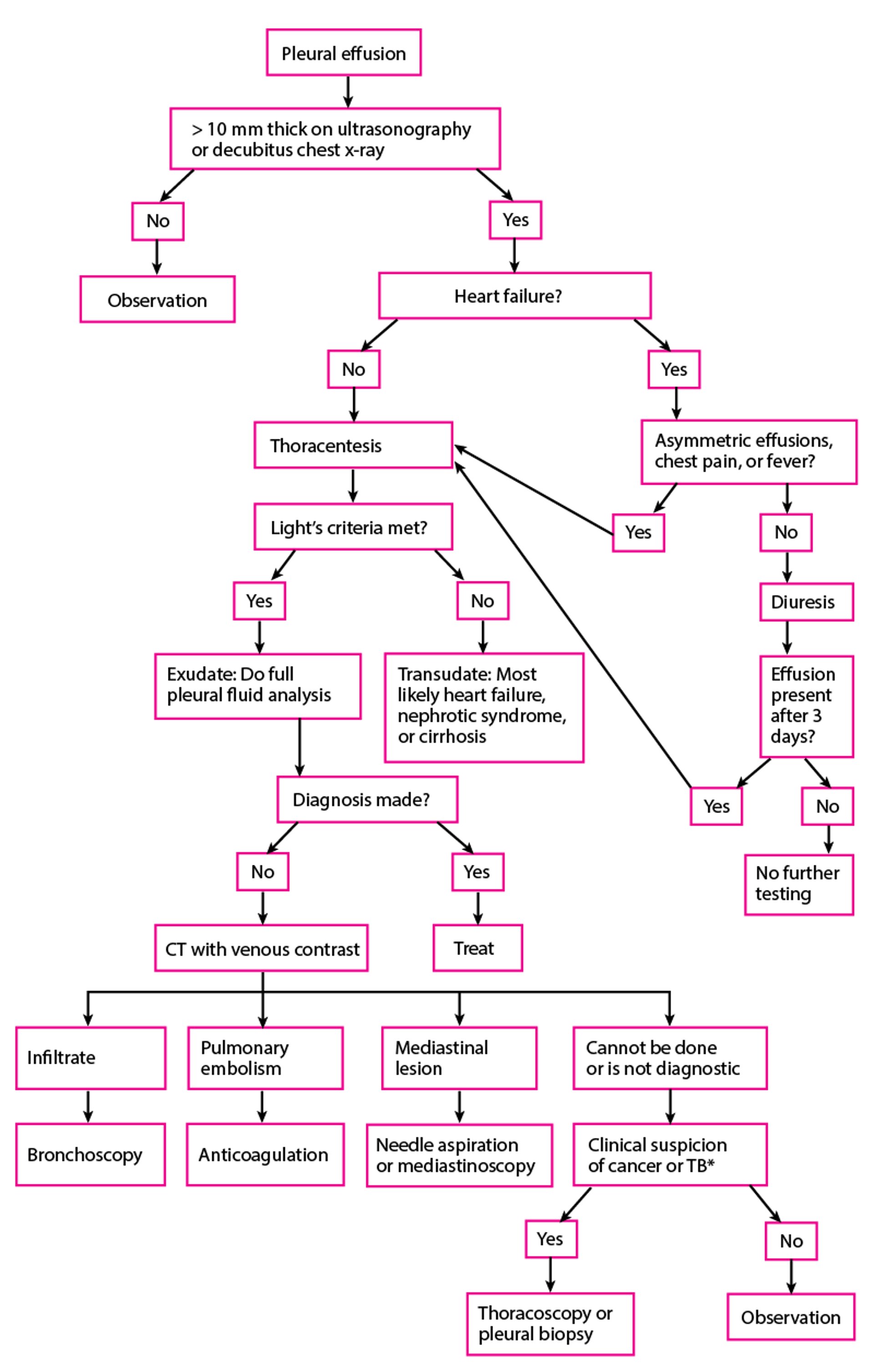
Pleural effusion is suspected in patients with pleuritic pain, unexplained dyspnea, or suggestive signs. Diagnostic tests are indicated to document the presence of pleural fluid and to determine its cause (see figure ).
Presence of effusion

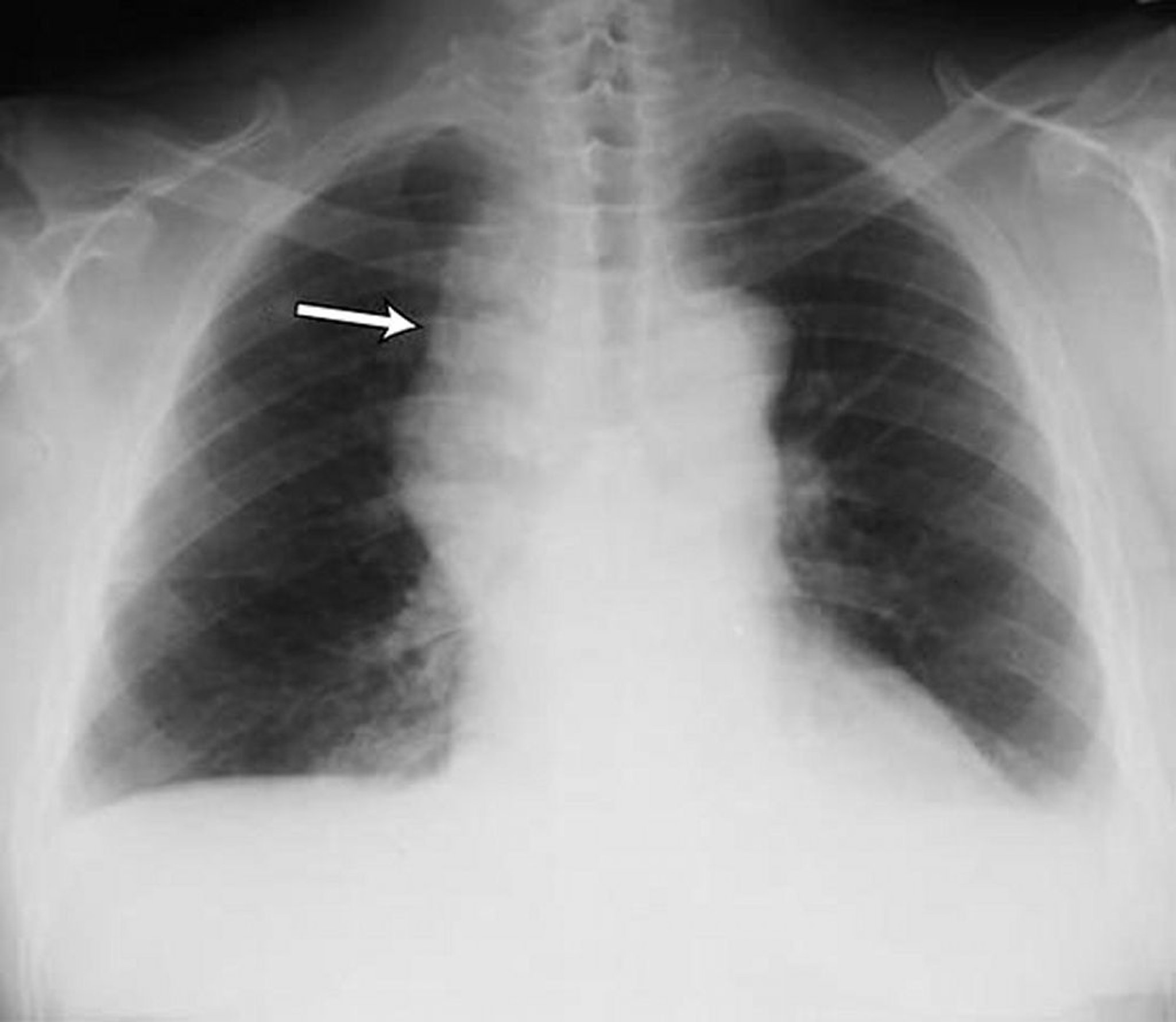
Small bilateral pleural effusions in a patient with non-Hodgkin lymphoma. Both costophrenic angles are blunted. The arrow points to widened mediastinum due to lymphoma.
By permission of the publisher. From Huggins J, Sahn S. In Bone's Atlas of Pulmonary and Critical Care Medicine. Edited by J Crapo. Philadelphia, Current Medicine, 2005.
Chest radiograph is the first test performed to confirm the presence of pleural fluid. The lateral upright chest radiograph should be examined when a pleural effusion is suspected. In an upright radiograph, 75 mL of fluid blunts the posterior costophrenic angle. Blunting of the lateral costophrenic angle usually requires about 175 mL but may take as much as 500 mL. Lateral decubitus radiographs (with the side of the effusion down) may be able to detect small fluid volumes more easily than standard upright radiographs, particularly with a free-flowing effusion. Larger pleural effusions opacify portions of the hemithorax and may cause mediastinal shift; effusions > 4 L may cause complete opacification of the hemithorax and mediastinal shift to the contralateral side.

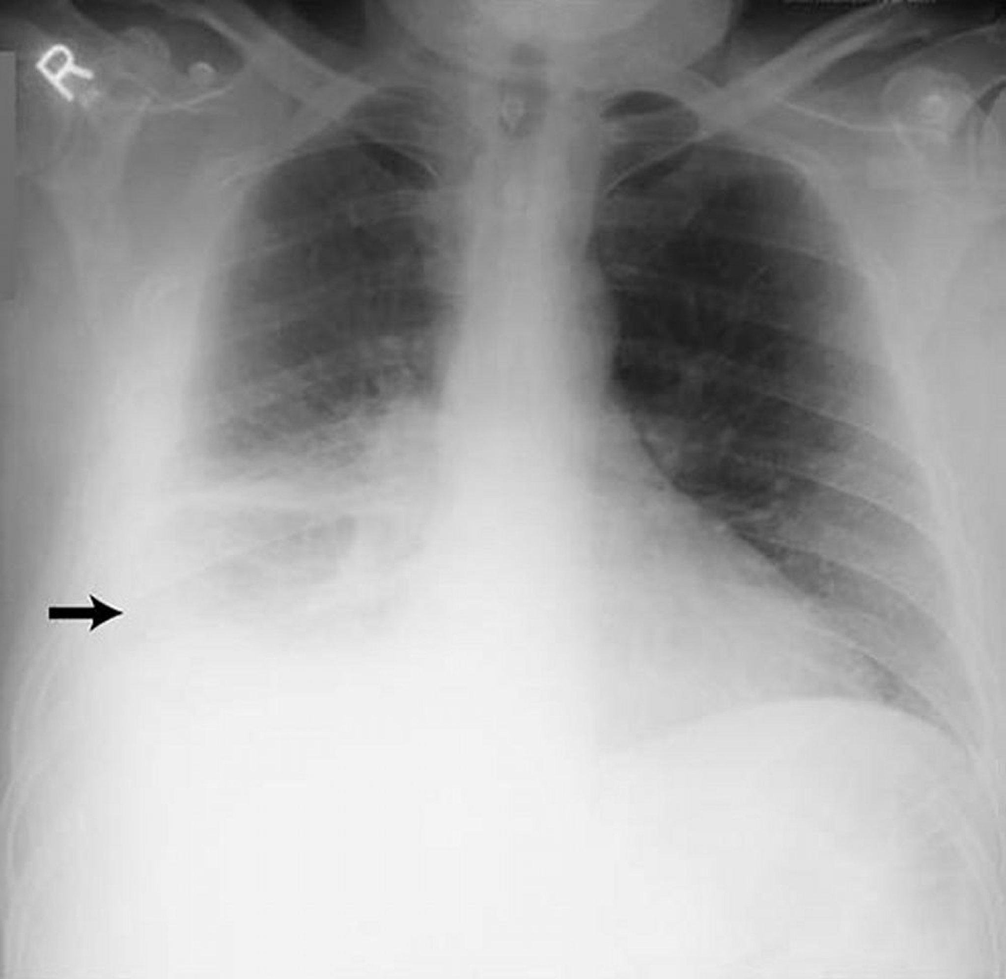
This image shows large right-sided pleural effusion (arrow) in a patient with rheumatoid pleuritis.
By permission of the publisher. From Huggins J, Sahn S. In Bone's Atlas of Pulmonary and Critical Care Medicine. Edited by J Crapo. Philadelphia, Current Medicine, 2005.

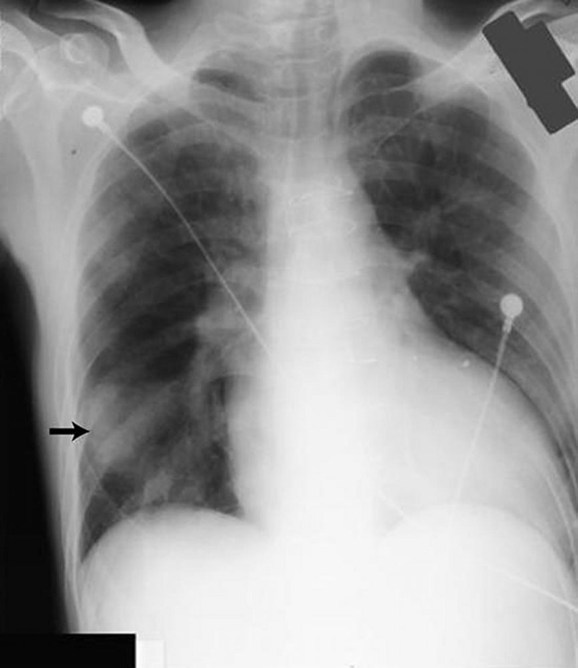
Opacity in the right lower lung field (arrow) resembles a solid mass but was caused by fluid in the major fissure in a patient with heart failure.
By permission of the publisher. From Huggins J, Sahn S. In Bone's Atlas of Pulmonary and Critical Care Medicine. Edited by J Crapo. Philadelphia, Current Medicine, 2005.
Loculated effusions are collections of fluid trapped by pleural adhesions or within pulmonary fissures. Further imaging (lateral decubitus radiographs, chest CT, or ultrasound) should be performed if it is unclear whether a radiographic density represents fluid or parenchymal infiltrates or whether suspected fluid is loculated or free-flowing; these tests are more sensitive than upright radiographs and can detect small fluid volumes. Loculated effusions, particularly those in the horizontal or oblique fissure, can be confused with a solid pulmonary mass (pseudotumor). They may change shape and size with changes in the patient’s position and the amount of pleural fluid.
Thoracic ultrasound is considered standard of care for diagnosis of pleural effusion and is performed and interpreted by physicians at the bedside. It is highly accurate for the detection of small volumes of pleural fluid and provides additional diagnostic information (eg, the presence of septation, pleural thickening).
CT with venous contrast is a valuable next investigation that provides information on pleural enhancement and potential pleural nodularity. (Noncontrast CT may be used for initial evaluation but cannot exclude malignancy or infection.) CT with venous contrast is valuable for evaluating the underlying lung parenchyma for infiltrates or masses when the lung is obscured by the effusion or when the detail on chest radiographs is insufficient for distinguishing loculated fluid from a solid mass.


This image of an axial noncontrast enhanced CT scan of the chest reveals a small- to moderate-sized right pleural effusion in a female patient with breast cancer. (Mediastinal CT scan windows)
Steven Needell/SCIENCE PHOTO LIBRARY

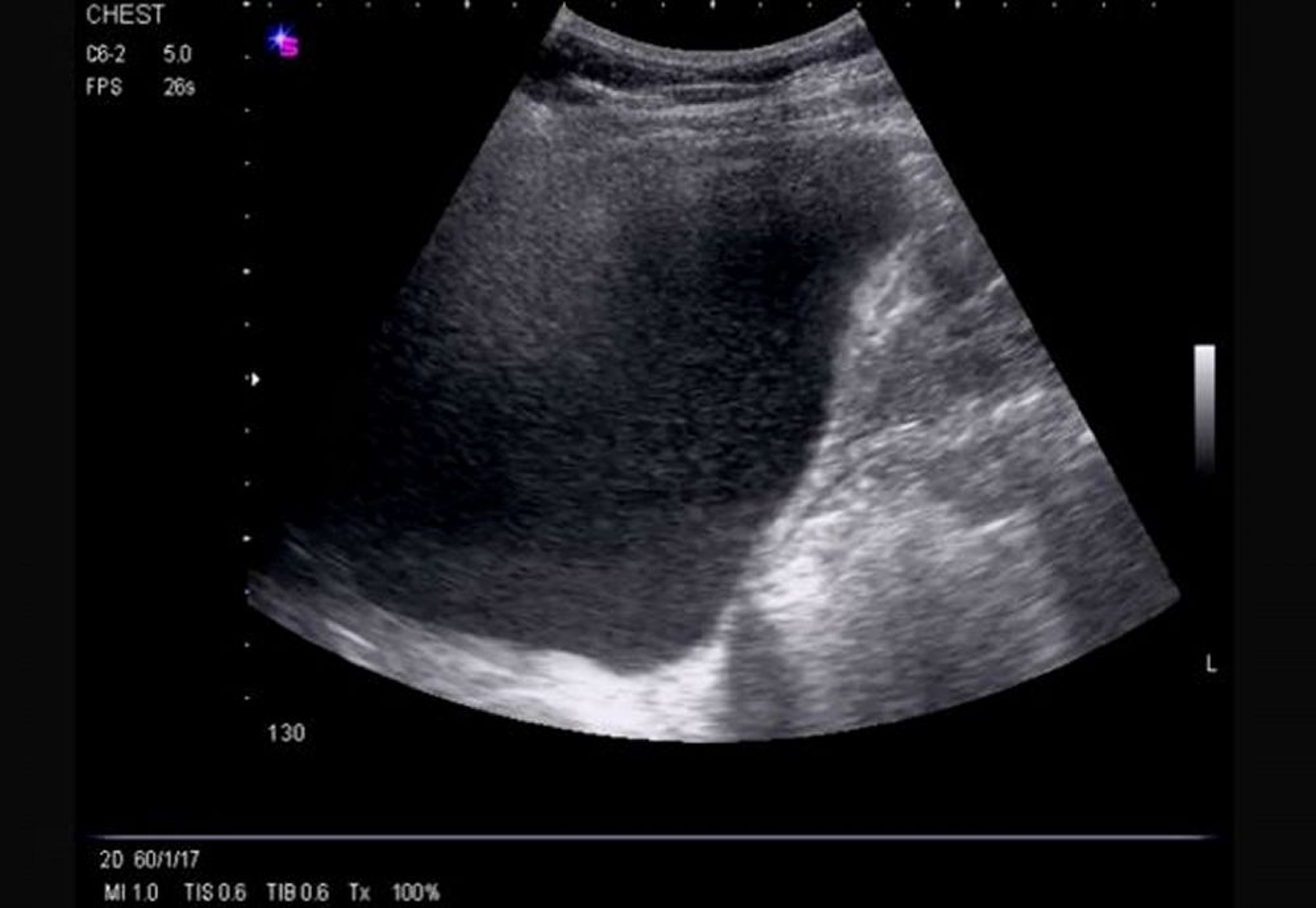
This ultrasound shows a large, left-sided, free-flowing effusion causing compressive left lower lobe atelectasis and revealing the left ventricle.
Image courtesy of Najib M. Rahman, BMBCh MA (oxon) DPhil.
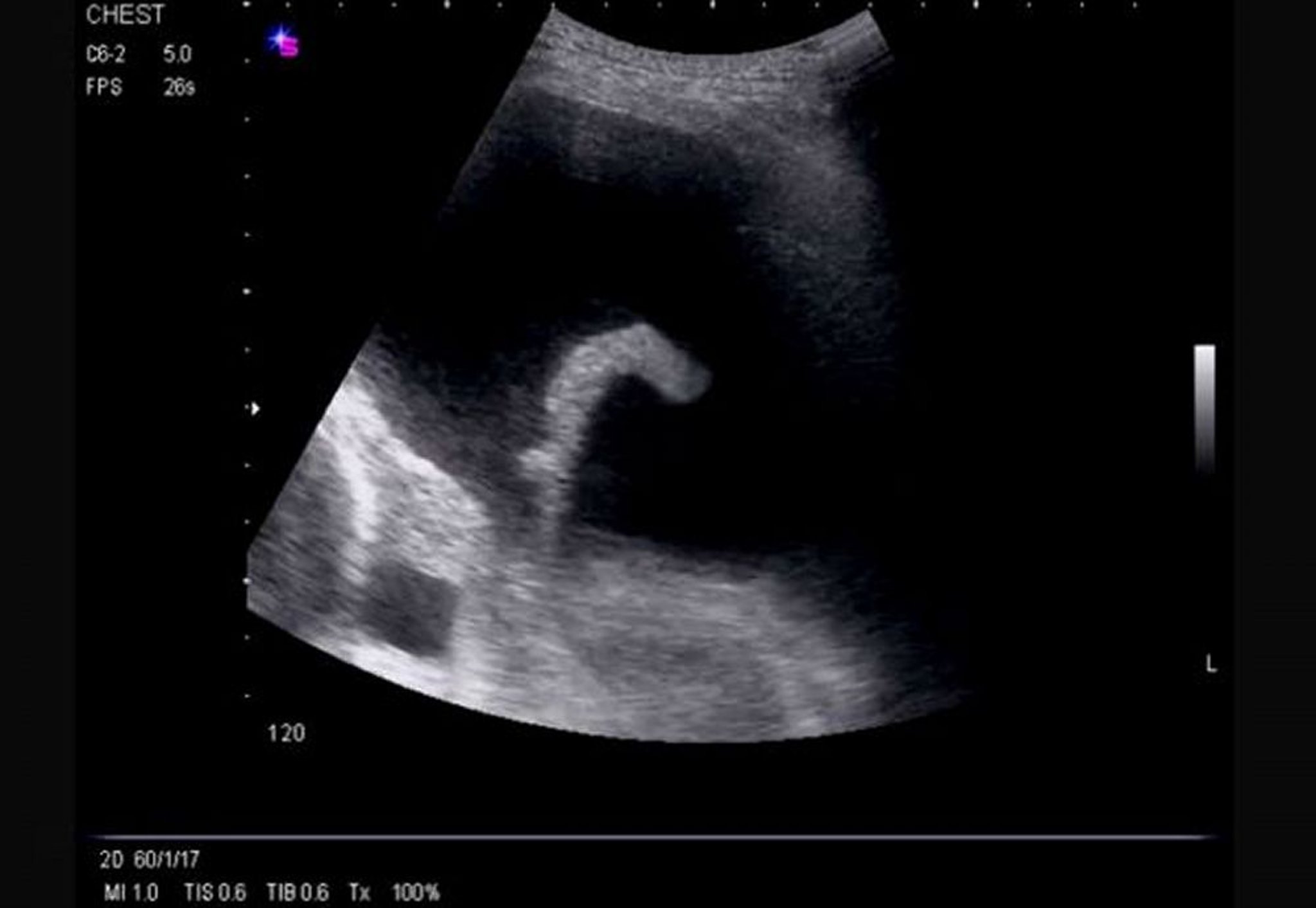
This ultrasound shows a massive, right-sided, free-flowing effusion causing inversion of the hemidiaphragm and demonstrating echogenicity.
Image courtesy of Najib M. Rahman, BMBCh MA (oxon) DPhil.
Cause of effusion
Thoracentesis should be performed in almost all patients who have pleural fluid that is ≥ 10 mm in thickness on CT, ultrasound, or lateral decubitus radiograph and that is new or of uncertain etiology. In general, the only patients who do not require thoracentesis are those who have heart failure with symmetric pleural effusions and no chest pain or fever; in these patients, diuresis can be attempted and thoracentesis avoided unless effusions persist for ≥ 3 days. Thoracentesis and subsequent pleural fluid analysis are also often unnecessary for pleural effusions that are chronic, have a known cause, and cause no symptoms.
Thoracentesis should be performed using ultrasonographic guidance in all cases, to improve procedural success and reduce complications (1).
Pearls & Pitfalls
|
Pleural fluid analysis is performed to diagnose the cause of pleural effusion. Analysis begins with visual inspection, which can:
Distinguish bloody and chylous (or chyliform) from other effusions
Identify purulent effusions strongly suggestive of empyema
Identify viscous fluid, which is characteristic of some mesotheliomas
Fluid should always be sent for total protein, glucose, lactate dehydrogenase (LDH), cell count and cell differential, and Gram stain. Other tests such as pleural fluid pH, aerobic and anaerobic bacterial cultures, cytology, tuberculosis fluid markers [adenosine deaminase or interferon-gamma], amylase, mycobacterial and fungal stains and cultures, triglycerides, and cholesterol are used in appropriate clinical settings.
Fluid analysis helps distinguish transudates from exudates; multiple criteria exist, but not one perfectly discriminates between the 2 types. When Light’s criteria are used (see table ), serum LDH and total protein levels should be measured as close as possible to the time of thoracentesis for comparison with those in pleural fluid. Light’s criteria correctly identify almost all exudates but misidentify approximately 20% of transudates as exudates (2). If transudative effusion is suspected (eg, due to heart failure or cirrhosis) and none of the biochemical measurements are < 15% above the cutoff levels for Light’s criteria, the difference between serum and the pleural fluid protein is measured. If the difference is > 3.1 g/dL (> 31 g/L), the patient probably has a transudative effusion.
Imaging may also help. If the diagnosis remains unclear after pleural fluid analysis, CT with venous contrast enhancement is indicated to assess for pleural enhancement, pleural nodularity, pulmonary infiltrates, or mediastinal lesions. CT pulmonary angiography can evaluate for a suspected pulmonary embolus. Findings of pulmonary emboli indicate the need for long-term anticoagulation. Pleural nodularity and thickening indicate the need for pleural biopsy (thoracoscopic or image guided). The presence of lung infiltrates or lesions, depending on suspected causes, may indicate the need for bronchoscopy or image-guided lung biopsy.
When tuberculous pleuritis is suspected, the level of adenosine deaminase in the pleural fluid is measured. A level > 40 U/L (667 nkat/L) has a 95% sensitivity and specificity for the diagnosis of tuberculous pleuritis (1); however, the adenosine deaminase level can also be elevated in patients with cancer.
Diagnosis of Pleural Effusion
* Based on presence of fever, weight loss, history of cancer, or other suggestive symptoms. TB = tuberculosis. |

Diagnosis references
1. Roberts ME, Rahman NM, Maskell NA, et al. British Thoracic Society Guideline for pleural disease. Thorax 2023;78(Suppl 3):s1-s42. doi:10.1136/thorax-2022-219784
2. Feller-Kopman D, Light R. Pleural Disease. N Engl J Med 2018;378(8):740-751. doi:10.1056/NEJMra1403503
Treatment of Pleural Effusion
Treatment of symptoms and underlying disorder
Drainage of some symptomatic effusions
Other treatments for parapneumonic and malignant effusions
Small (< 10 mm of fluid on lateral decubitus radiograph or ultrasound) and asymptomatic effusions generally do not require treatment because many effusions resorb spontaneously when the underlying disorder is treated, especially effusions due to uncomplicated pneumonias, pulmonary embolism, or surgery. Pleuritic pain can usually be managed with nonsteroidal anti-inflammatory drugs (NSAIDs) or other oral analgesics. At times, a short course of oral opioids is required.
Therapeutic thoracentesis (usually around 1 to 1.5 L in a single treatment) is sufficient for many symptomatic effusions and can be repeated for effusions that reaccumulate. There are no arbitrary limits on the total amount of fluid that can be removed (1). Removal of fluid can be continued until the effusion is drained or the patient develops chest tightness, chest pain, or severe coughing. A rare complication is re-expansion pulmonary edema (REPE) (2). When it occurs, REPE can be life threatening, but it appears to be an unpredictable event.
Effusions that are chronic, recurrent, and causing symptoms can be treated with pleurodesis or by intermittent drainage with an indwelling catheter. Effusions caused by pneumonia or cancer may require additional specific measures.
Parapneumonic effusion and empyema
In patients with adverse prognostic factors (pH < 7.20, glucose < 60 mg/dL [< 3.33 mmol/L], positive Gram stain or culture, loculations), the effusion should be completely drained via thoracentesis or tube thoracostomy (3). If complete drainage is impossible, a thrombolytic (fibrinolytic) medication (eg, alteplase) plus a DNAse (eg, dornase alfa) can be administered intrapleurally. If intrapleural treatment is unsuccessful, thoracic surgical intervention is indicated. Video-assisted thoracoscopic surgery (VATS) is usually tried first, although thoracotomy may be required.
Malignant pleural effusion
If dyspnea caused by malignant pleural effusion is relieved by thoracentesis but fluid and dyspnea redevelop, chronic (intermittent) drainage or pleurodesis is indicated. Asymptomatic effusions and effusions causing dyspnea unrelieved by thoracentesis do not require additional procedures.
Indwelling pleural catheters and pleurodesis are equally effective for treating malignant pleural effusion. Choice depends on patient preference. Both decrease dyspnea and improve quality of life effectively. Chemical pleurodesis requires longer hospital stay but long-term complications such as cellulitis are more frequent with indwelling catheters (4).
Pleurodesis is performed by instilling a sclerosing agent into the pleural space to fuse the visceral and parietal pleura and eliminate the space. The most effective and commonly used sclerosing agents are talc, doxycycline, or bleomycin delivered via chest tube or thoracoscopy. Pleurodesis is contraindicated if the lung does not expand after full drainage with a chest tube (entrapped lung), in which case an indwelling pleural catheter is indicated.
Shunting of pleural fluid to the peritoneum (pleuroperitoneal shunt) is rarely performed but is an option in patients who have trapped lung.
Surgical treatment (pleurectomy or decortication) should be considered in patients with trapped lung who have good performance status and a good prognosis.
Treatment references
1. Feller-Kopman D, Berkowitz D, Boiselle P, et al. Large-volume thoracentesis and the risk of reexpansion pulmonary edema. Ann Thoracic Surg 2007;84:1656–1662. doi:10.1016/j.athoracsur.2007.06.038
2. Cusumano G, La Via L, Terminella A, Sorbello M. Re-Expansion Pulmonary Edema as a Life-Threatening Complication in Massive, Long-Standing Pneumothorax: A Case Series and Literature Review. J Clin Med 2024;13(9):2667. Published 2024 May 2. doi:10.3390/jcm13092667
3. Roberts ME, Rahman NM, Maskell NA, et al. British Thoracic Society Guideline for pleural disease. Thorax 2023;78(Suppl 3):s1-s42. doi:10.1136/thorax-2022-219784
4. Iyer NP, Reddy CB, Wahidi MM, et al. Indwelling Pleural Catheter versus Pleurodesis for Malignant Pleural Effusions. A Systematic Review and Meta-Analysis. Ann Am Thorac Soc 2019;16(1):124-131. doi:10.1513/AnnalsATS.201807-495OC
Key Points
Transudative effusions are caused by some combination of increased hydrostatic pressure and decreased plasma oncotic pressure.
Exudative effusions result from increased capillary permeability, leading to leakage of protein, cells, and other serum constituents.
The most common causes of transudative effusions are heart failure, cirrhosis with ascites, and nephrotic syndrome.
The most common causes of exudative effusions are pneumonia, cancer, pulmonary embolism, and tuberculosis.
Evaluation requires imaging (usually chest radiograph and thoracic ultrasound) to confirm presence of fluid and pleural fluid analysis to help determine cause.
Lateral decubitus radiographs, chest CT, or ultrasound should be performed if it is unclear whether a radiographic density represents fluid or parenchymal infiltrates or whether suspected fluid is loculated or free-flowing.
Larger effusions that are symptomatic require thoracentesis, except for recurrent effusions with a known cause.
Effusions that are chronic or recurrent and causing symptoms can be treated with pleurodesis or by intermittent drainage with an indwelling catheter.
Drug Information for the Topic


