


In ultrasound, a signal generator is combined with a transducer. Piezoelectric crystals in the signal generator convert electricity into high-frequency sound waves, which are sent into tissues. The tissues scatter, reflect, and absorb the sound waves to varying degrees. The sound waves that are reflected back (echoes) are converted into electric signals. A computer analyzes the signals and displays an anatomic image on a screen.
Ultrasound is portable, widely available, relatively inexpensive, and safe imaging modality, as it does not involve ionizing radiation. Among its practical advantages, it can be performed by health care professionals after a relatively brief period of focused training. In many clinical settings, ultrasound examinations can be performed without the direct supervision of a radiologist or specialized sonographer, enabling rapid point-of-care assessment.

Procedure demonstrated by Robert Strony, DO, MBA, RDCS, FACEP, Medical Director, Point of Care Ultrasound, Geisinger; Clinical Associate Professor of Medicine, Geisinger Commonwealth School of Medicine; Associate Professor (Adjunct) Lewis Katz School of Medicine, Temple University.

Procedure demonstrated by Robert Strony, DO, MBA, RDCS, FACEP, Medical Director, Point of Care Ultrasound, Geisinger; Clinical Associate Professor of Medicine, Geisinger Commonwealth School of Medicine; Associate Professor (Adjunct) Lewis Katz School of Medicine, Temple University.

Procedure demonstrated by Robert Strony, DO, MBA, RDCS, FACEP, Medical Director, Point of Care Ultrasound, Geisinger; Clinical Associate Professor of Medicine, Geisinger Commonwealth School of Medicine; Associate Professor (Adjunct) Lewis Katz School of Medicine, Temple University.


Procedure demonstrated by Robert Strony, DO, MBA, RDCS, FACEP, Medical Director, Point of Care Ultrasound, Geisinger; Clinical Associate Professor of Medicine, Geisinger Commonwealth School of Medicine; Associate Professor (Adjunct) Lewis Katz School of Medicine, Temple University.

Procedure demonstrated by Robert Strony, DO, MBA, RDCS, FACEP, Medical Director, Point of Care Ultrasound, Geisinger; Clinical Associate Professor of Medicine, Geisinger Commonwealth School of Medicine; Associate Professor (Adjunct) Lewis Katz School of Medicine, Temple University.

Procedure demonstrated by Robert Strony, DO, MBA, RDCS, FACEP, Medical Director, Point of Care Ultrasound, Geisinger; Clinical Associate Professor of Medicine, Geisinger Commonwealth School of Medicine; Associate Professor (Adjunct) Lewis Katz School of Medicine, Temple University.
Uses of Ultrasound
Ultrasound can identify superficial growths and foreign bodies (eg, in the thyroid gland, breasts, testes, limbs, and some lymph nodes). With deeper structures, other tissues and densities (eg, bone, gas) can interfere with images.
Ultrasound is commonly used to evaluate the following:
Heart (echocardiography): For example, to detect valvular and chamber size abnormalities and to estimate ejection fraction and myocardial strain (see Echocardiography)
Gallbladder and biliary tract: For example, to detect gallstones and biliary tract obstruction (see Imaging Tests of the Liver and Gallbladder: Ultrasonography)
Urinary tract: For example, to distinguish cysts (usually benign) from solid masses (often malignant) in the kidneys or to detect obstruction such as calculi or other structural abnormalities in the kidneys, ureters, or bladder (see Genitourinary Imaging Tests: Ultrasonography)
Female reproductive organs: For example, to detect tumors and inflammation in the ovaries, fallopian tubes, or uterus (see Introduction to Gynecologic Tumors)
Pregnancy: For example, to evaluate the growth and development of the fetus and to detect abnormalities of the placenta (eg, placenta previa—see Evaluation of the Obstetric Patient: Ultrasonography)
Musculoskeletal: To evaluate muscles, tendons, and nerves

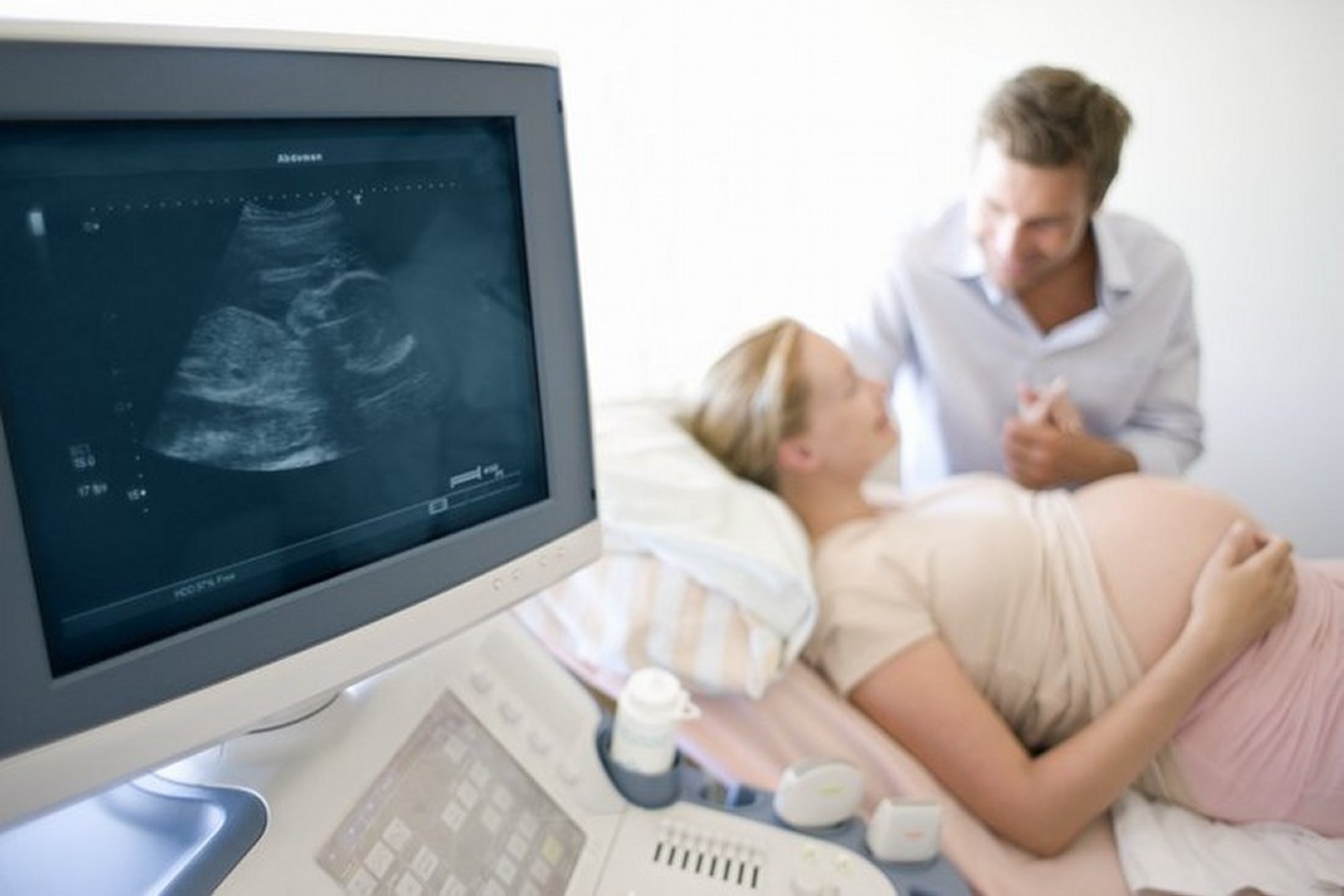
This photo shows an ultrasound image of a fetus.
IAN HOOTON/SCIENCE PHOTO LIBRARY
Bedside ultrasound (also called point-of-care ultrasound [POCUS]) is often used in acute care settings to assist both diagnosis (eg, volume status, cause for hypotension, foreign bodies) and treatment (eg, intravenous catheterization, arthrocentesis).
Ultrasound can also be used to guide biopsy sampling and place intravenous catheters.
Ultrasound is sometimes performed using a small internal transducer on the tip of an endoscope or vascular catheter.
Variations of Ultrasound
Ultrasound information can be displayed in several ways.
A-mode
A-mode is the simplest; signals are recorded as spikes on a graph. The vertical (Y) axis of the display shows the echo amplitude, and the horizontal (X) axis shows depth or distance into the patient.
This type of ultrasound is used for ophthalmologic scanning.
B-mode (gray-scale)
B-mode is most often used in diagnostic imaging; signals are displayed as a 2-dimensional anatomic image. Additionally, B-mode ultrasound can also produce 3-dimensional (volumetric) and 4-dimensional (3-dimensional with time) images. Three-dimensional ultrasound in commonly used for visualizing fetal anatomy in obstetric/gynecologic examinations. Four-dimensional ultrasound enables dynamic imaging of moving structures, such as fetal movements or cardiac motion.
B-mode is commonly used to evaluate the developing fetus and to evaluate organs, including the liver, spleen, kidneys, thyroid gland, testes, breasts, uterus, ovaries, and prostate gland.
B-mode ultrasound is fast enough to show real-time motion, such as the motion of the beating heart or pulsating blood vessels. Real-time imaging provides anatomic and functional information.
M-mode
M-mode is used to image moving structures; signals reflected by the moving structures are converted into waves that are displayed continuously across a vertical axis.
M-mode is used primarily for assessment of fetal heartbeat and, in cardiac imaging, to evaluate valvular disorders.
Doppler
Doppler ultrasound is used to assess blood flow. It uses the Doppler effect (alteration of sound frequency by reflection off a moving object). The moving objects are red blood cells in blood.
Direction and velocity of blood flow can be determined by analyzing changes in the frequency of sound waves:
If a reflected sound wave is lower in frequency than the transmitted sound wave, blood flow is away from the transducer.
If a reflected sound wave is higher in frequency than the transmitted sound wave, blood flow is toward the transducer.
The magnitude of the change in frequency is proportional to blood flow velocity.
Changes in frequency of the reflected sound waves are converted into images showing blood flow direction and velocity.

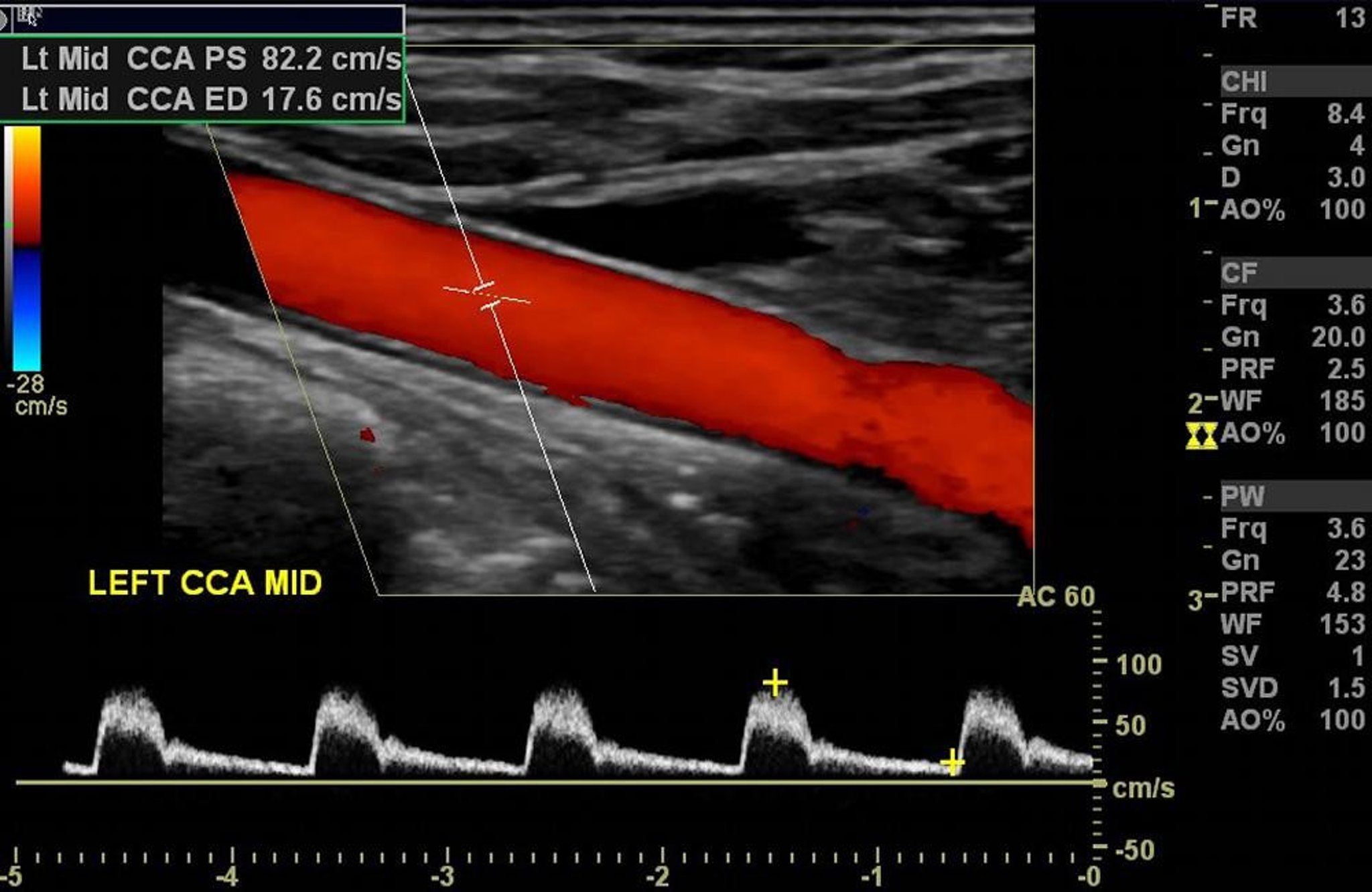
Color Doppler ultrasound of the left carotid artery shows normal arterial flow without significant stenosis.
Image courtesy of Hakan Ilaslan, MD.
Doppler ultrasound is also used:
To evaluate vascularity of tumors and organs
To evaluate heart function (eg, as for echocardiography)
To detect occlusion and stenosis of blood vessels
To detect blood clots in blood vessels (eg, in deep venous thrombosis)
To detect synovitis in joints
Spectral Doppler ultrasound displays blood flow information as a graph with velocity on the vertical axis and time on the horizontal axis. Specific velocities can be measured if the Doppler angle (the angle between the direction of the ultrasound beam and the direction of blood flow) can be determined. Velocity measurements and the appearance of the spectral Doppler tracing can indicate the severity of vascular stenoses.
Duplex Doppler ultrasound combines the graphic display of spectral ultrasound with the images of B-mode.
Color Doppler ultrasound converts the Doppler blood flow information into a color image with blood flow in color; it is displayed on a gray-scale anatomic ultrasound image. Direction of blood flow is indicated by the shade of color (eg, red for blood flow toward the transducer, blue for blood flow away from the transducer). Average blood flow velocity is indicated by the brightness of the color (eg, bright red indicates high-velocity flow toward the transducer; dark blue indicates low-velocity flow away from the transducer).
Disadvantages of Ultrasound
Quality of images depends on the skills of the operator.
Obtaining clear images of the target structures can be technically difficult in patients with excess adipose tissue.
Ultrasound cannot be used to image through bone or gas, so certain images may be difficult to obtain.


