Coronary artery disease (CAD) involves impairment of blood flow through the coronary arteries, most commonly by atheromas. Clinical presentations include silent ischemia, stable angina, acute coronary syndromes (unstable angina, myocardial infarction), and sudden cardiac death. Diagnosis is by symptoms, ECG, stress testing, and sometimes coronary angiography. Prevention consists of modifying reversible risk factors (eg, hypercholesterolemia, hypertension, physical inactivity, obesity, diabetes, smoking). Treatment includes medications and procedures to reduce ischemia and restore or improve coronary blood flow.
Coronary artery disease is the leading cause of death in both sexes, accounting for approximately 15% of deaths worldwide (and about one-third of all deaths in people 35 years old); and substantial numbers of deaths occur in low resource areas (1, 2). Mortality from coronary artery disease is approximately 5 times higher in males than in females, but the mortality difference declines with age (3).
General references
1. GBD 2023 Causes of Death Collaborators. Global burden of 292 causes of death in 204 countries and territories and 660 subnational locations, 1990-2023: a systematic analysis for the Global Burden of Disease Study 2023. Lancet. 2025;406(10513):1811-1872. doi:10.1016/S0140-6736(25)01917-8
2. Ralapanawa U, Sivakanesan R. Epidemiology and the Magnitude of Coronary Artery Disease and Acute Coronary Syndrome: A Narrative Review. J Epidemiol Glob Health. 2021;11(2):169-177. doi:10.2991/jegh.k.201217.001
3. Bots SH, Peters SAE, Woodward M. Sex differences in coronary heart disease and stroke mortality: a global assessment of the effect of ageing between 1980 and 2010. BMJ Glob Health. 2017;2(2):e000298. doi:10.1136/bmjgh-2017-000298
Coronary Artery Anatomy
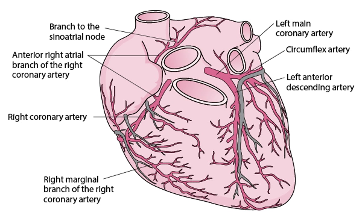
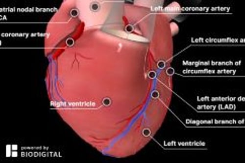
The right and left coronary arteries arise from the right and left coronary sinuses in the root of the aorta just above the aortic valve orifice (see figure ). The coronary arteries divide into large and medium-sized arteries that run along the heart’s surface (epicardial coronary arteries) and subsequently send smaller arterioles into the myocardium.
The left coronary artery begins as the left main artery and quickly divides into the left anterior descending (LAD), circumflex, and sometimes an intermediate artery (ramus intermedius). The LAD artery usually follows the anterior interventricular groove and, in some people, continues over the apex. This artery supplies the anterior septum (including the proximal conduction system) and the anterior free wall of the left ventricle (LV). The circumflex artery, which is usually smaller than the LAD artery, supplies the lateral LV free wall.
The dominant coronary artery refers to the one that gives rise to the posterior descending artery. Most people have right dominance: The right coronary artery passes along the atrioventricular (AV) groove over the right side of the heart; it supplies the sinus node (in 55%), right ventricle, and usually the AV node and inferior myocardial wall. Approximately 10 to 15% of people have left dominance: The circumflex artery is larger and continues along the posterior AV groove to supply the posterior wall and AV node.
Arteries of the Heart

Etiology of Coronary Artery Disease
Usually, coronary artery disease is due to:
Coronary artery atherosclerosis: Subintimal deposition of atheromas in large and medium-sized coronary arteries, resulting in obstructive coronary artery disease.
Less often, coronary artery disease manifests as:
Coronary artery spasm (Vasospastic Angina)
Vascular endothelial dysfunction can promote atherosclerosis and contribute to coronary artery spasm, and is also recognized as a cause of microvascular angina. Both coronary artery spasm and microvascular angina can cause ischemia or myocardial infarction with non-obstructive coronary arteries (MINOCA). The term MINOCA encompasses all causes of myocardial ischemia and infarction in the absence of significant (≥ 50%) coronary artery occlusion (1). Other disease entities classified as MINOCA include coronary artery dissection and embolism.
Other, rarer types of coronary artery disease include coronary artery aneurysm (eg, in Kawasaki disease) and vasculitis (eg, in Takayasu arteritis).
Etiology reference
1. Tamis-Holland JE, Jneid H, Reynolds HR, et al. Contemporary Diagnosis and Management of Patients With Myocardial Infarction in the Absence of Obstructive Coronary Artery Disease: A Scientific Statement From the American Heart Association. Circulation. 2019;139(18):e891-e908. doi:10.1161/CIR.0000000000000670
Pathophysiology of Coronary Artery Disease
Coronary atherosclerosis
Coronary atherosclerosis is often irregularly distributed in different vessels but clusters in the proximal portions of the coronary arteries and at points of turbulence such as vessel bifurcations (1, 2). As the atheromatous plaque grows, the arterial lumen progressively narrows, resulting in ischemia (often causing stable angina). The degree of stenosis required to cause ischemia varies with oxygen demand.
Occasionally, an atheromatous plaque ruptures or splits. Reasons are unclear but probably relate to plaque morphology, plaque calcium content, and plaque softening due to an inflammatory process. Rupture exposes collagen and other thrombogenic material, which activate platelets and the coagulation cascade (see figure ), resulting in an acute thrombus that interrupts coronary blood flow and causes some degree of myocardial ischemia. The consequences of acute ischemia, collectively referred to as acute coronary syndromes (ACS), depend on the location and degree of obstruction and range from unstable angina, to non–ST-segment elevation myocardial infarction (NSTEMI), to ST-segment elevation myocardial infarction (STEMI), which can result in transmural infarction, and other complications including malignant ventricular arrhythmias, conduction defects, heart failure, and sudden death.
See Pathophysiology of Atherosclerosis for more detail.
Coronary artery spasm
Coronary artery spasm is a transient, focal increase in vascular tone, markedly narrowing the lumen and reducing blood flow; symptomatic ischemia (vasospastic angina) may result. Marked narrowing can trigger thrombus formation, causing infarction or life-threatening arrhythmia. Spasm can occur in arteries with or without atheroma.
In arteries without atheroma, basal coronary artery tone is probably increased, and response to vasoconstricting stimuli is probably exaggerated. The exact mechanism is unclear but may involve endothelial cell abnormalities of nitric oxide production or an imbalance between endothelium-derived contracting and relaxing factors (3, 4).
In arteries with atheroma, the atheroma is thought to play a causative role in both endothelial dysfunction and smooth muscle hyperreactivity (5). Recurrent spasm may also damage the intima and lead to atheroma formation.
Use of vasoconstricting substances (eg, cocaine, nicotine) and emotional stress also can trigger coronary spasm.
Coronary artery dissection
Coronary artery dissection is a rare, non-traumatic tear in the coronary intima with creation of a false lumen. Blood flowing through the false lumen expands it, which restricts blood flow through the true lumen, sometimes causing coronary ischemia or infarction. Dissection may occur in atherosclerotic or non-atherosclerotic coronary arteries. Non-atherosclerotic (spontaneous) coronary artery dissection is an important cause of myocardial infarction in younger and middle-aged females, including pregnant or postpartum people, as well as patients with fibromuscular dysplasia or other connective tissue disorders (6, 7).
Pathophysiology references
1. Bax AM, Yoon YE, Gianni U, et al. Plaque Character and Progression According to the Location of Coronary Atherosclerotic Plaque. Am J Cardiol. 2021;158:15-22. doi:10.1016/j.amjcard.2021.07.040
2. Yang X, Zhang J, Song Y, et al. Deciphering age- and sex-specific patterns of coronary artery atherosclerosis from a large Chinese cohort. Nat Commun. 2025;16(1):10616. doi:10.1038/s41467-025-64940-8
3. Ford TJ, Ong P, Sechtem U, et al. Assessment of Vascular Dysfunction in Patients Without Obstructive Coronary Artery Disease: Why, How, and When. JACC Cardiovasc Interv. 2020;13(16):1847-1864. doi:10.1016/j.jcin.2020.05.052
4. Tamis-Holland JE, Jneid H, Reynolds HR, et al. Contemporary Diagnosis and Management of Patients With Myocardial Infarction in the Absence of Obstructive Coronary Artery Disease: A Scientific Statement From the American Heart Association. Circulation. 2019;139(18):e891-e908. doi:10.1161/CIR.0000000000000670
5. Peeters D, Woelders E, Jansen T, et al. Association Between Coronary Artery Spasm and Atherosclerotic Disease. JACC Cardiovasc Imaging. 2025;18(2):226-239. doi:10.1016/j.jcmg.2024.05.024
6. Hayes SN, Tweet MS, Adlam D, et al. Spontaneous Coronary Artery Dissection: JACC State-of-the-Art Review. J Am Coll Cardiol. 2020;76(8):961-984. doi:10.1016/j.jacc.2020.05.084
7. Kim ESH. Spontaneous Coronary-Artery Dissection. N Engl J Med. 2020;383(24):2358-2370. doi:10.1056/NEJMra2001524
Risk Factors for Coronary Artery Disease
Risk factors for coronary artery disease are the same as risk factors for atherosclerosis (see table ) and may be modifiable or non-modifiable.
Important non-modifiable risk factors include (1, 2, 3, 4, 5):
Older age
Male sex
Family history of early coronary artery disease (death from coronary artery disease in a first-degree relative prior to age 55 years in males or age 65 years in females)
Certain ethnicities (such as South Asian ancestry)
Premature menopause (< 40 years)
Modifiable risk factors include (6, 7, 8, 9, 10, 11, 12, 13):
Dyslipidemia (particularly apolipoprotein B)
Cardiovascular-kidney-metabolic factors (diabetes, insulin resistance, hypertension, chronic kidney disease)
Inflammatory risk factors (C-reactive protein, autoimmune disease, many infections)
Lifestyle factors (tobacco smoke exposure, exercise, diet, alcohol consumption, chronic stress)
Prothrombotic states
Smoking may be a stronger predictor of myocardial infarction in females (14). Genetic factors play a role, and several systemic disorders in addition to strictly cardiovascular-kidney-metabolic risk factors (eg, hypothyroidism, hyperhomocysteinemia) contribute to risk as well.
A high level of apolipoprotein B may identify increased risk when total cholesterol or low-density lipoprotein (LDL) cholesterol level is normal (15, 16). High blood levels of C-reactive protein indicate plaque instability and inflammation and may be a stronger predictor of risk of ischemic events than high levels of LDL (17), and they can indicate risk even in the presence of a normal lipid profile (residual inflammatory risk) (18). Hypertriglyceridemia is an independent risk factor for atherosclerotic events, even in patients whose LDL levels are adequately controlled by statin therapy (residual triglyceride risk) (19).
CAD risk is also increased by a diet high in saturated fat, sodium, added sugar, and red and processed meat; whereas consumption of fruits, vegetables, nuts, legumes, fish, fiber, and whole grains reduce risk (6, 20, 21, 22, 23, 24, 25, 26, 27).


The Revised Pooled Cohort risk calculator, recommended by some current guidelines, estimates 10-year risk of atherosclerotic cardiovascular disease (6, 28). The PREVENT calculator, recommended by newer guidelines, incorporates additional information, including measures of kidney and metabolic health and socioeconomic variation (as determined by U. S. zip code), to provide 10- and 30- year risk estimates for combined atherosclerotic cardiovascular disease and heart failure (29, 30, 31, 32, 33).
Risk factor references
1. Honigberg MC, Zekavat SM, Aragam K, et al. Association of Premature Natural and Surgical Menopause With Incident Cardiovascular Disease. JAMA. 2019;322(24):2411-2421. doi:10.1001/jama.2019.19191
2. Man JJ, Beckman JA, Jaffe IZ. Sex as a Biological Variable in Atherosclerosis. Circ Res. 2020;126(9):1297-1319. doi:10.1161/CIRCRESAHA.120.315930
3. Nielsen M, Andersson C, Gerds TA, et al. Familial clustering of myocardial infarction in first-degree relatives: a nationwide study. Eur Heart J. 2013;34(16):1198-1203. doi:10.1093/eurheartj/ehs475
4. Patel AP, Wang M, Kartoun U, Ng K, Khera AV. Quantifying and Understanding the Higher Risk of Atherosclerotic Cardiovascular Disease Among South Asian Individuals: Results From the UK Biobank Prospective Cohort Study. Circulation. 2021;144(6):410-422. doi:10.1161/CIRCULATIONAHA.120.052430
5. Savji N, Rockman CB, Skolnick AH, et al. Association between advanced age and vascular disease in different arterial territories: a population database of over 3.6 million subjects. J Am Coll Cardiol. 2013;61(16):1736-1743. doi:10.1016/j.jacc.2013.01.054
6. Arnett DK, Blumenthal RS, Albert MA, et al. 2019 ACC/AHA Guideline on the Primary Prevention of Cardiovascular Disease: A Report of the American College of Cardiology/American Heart Association Task Force on Clinical Practice Guidelines. J Am Coll Cardiol. 2019;74(10):e177-e232. doi:10.1016/j.jacc.2019.03.010
7. Global Cardiovascular Risk Consortium, Magnussen C, Ojeda FM, et al. Global Effect of Modifiable Risk Factors on Cardiovascular Disease and Mortality. N Engl J Med. 2023;389(14):1273-1285. doi:10.1056/NEJMoa2206916
8. Levine GN, Cohen BE, Commodore-Mensah Y, et al. Psychological Health, Well-Being, and the Mind-Heart-Body Connection: A Scientific Statement From the American Heart Association. Circulation. 2021;143(10):e763-e783. doi:10.1161/CIR.0000000000000947
9. Libby P, Buring JE, Badimon L, et al. Atherosclerosis. Nat Rev Dis Primers. 2019;5(1):56. doi:10.1038/s41572-019-0106-z
10. Mechanick JI, Farkouh ME, Newman JD, Garvey WT. Cardiometabolic-Based Chronic Disease, Adiposity and Dysglycemia Drivers: JACC State-of-the-Art Review. J Am Coll Cardiol. 2020;75(5):525-538. doi:10.1016/j.jacc.2019.11.044
11. Ndumele CE, Rangaswami J, Chow SL, et al. Cardiovascular-Kidney-Metabolic Health: A Presidential Advisory From the American Heart Association. Circulation. 2023;148(20):1606-1635. doi:10.1161/CIR.0000000000001184
12. Szwed P, Gąsecka A, Zawadka M, et al. Infections as Novel Risk Factors of Atherosclerotic Cardiovascular Diseases: Pathophysiological Links and Therapeutic Implications. J Clin Med. 2021;10(12):2539. doi:10.3390/jcm10122539
13. Yusuf S, Hawken S, Ounpuu S, et al. Effect of potentially modifiable risk factors associated with myocardial infarction in 52 countries (the INTERHEART study): case-control study. Lancet. 2004;364(9438):937-952. doi:10.1016/S0140-6736(04)17018-9
14. Prescott E, Hippe M, Schnohr P, Hein HO, Vestbo J. Smoking and risk of myocardial infarction in women and men: longitudinal population study. BMJ. 1998;316(7137):1043-1047. doi:10.1136/bmj.316.7137.1043
15. Sniderman AD, Thanassoulis G, Glavinovic T, et al. Apolipoprotein B Particles and Cardiovascular Disease: A Narrative Review. JAMA Cardiol. 2019 Dec 1;4(12):1287-1295. doi: 10.1001/jamacardio.2019.3780
16. Wilkins JT, Li RC, Sniderman A, Chan C, Lloyd-Jones DM. Discordance Between Apolipoprotein B and LDL-Cholesterol in Young Adults Predicts Coronary Artery Calcification: The CARDIA Study. J Am Coll Cardiol. 2016;67(2):193-201. doi:10.1016/j.jacc.2015.10.055
17. Ridker PM, Lei L, Louie MJ, et al. Inflammation and Cholesterol as Predictors of Cardiovascular Events Among 13 970 Contemporary High-Risk Patients With Statin Intolerance. Circulation. 2024;149(1):28-35. doi:10.1161/CIRCULATIONAHA.123.066213
18. Denegri A, Boriani G. High Sensitivity C-reactive Protein (hsCRP) and its Implications in Cardiovascular Outcomes. Curr Pharm Des. 2021;27(2):263-275. doi:10.2174/1381612826666200717090334
19. Nichols GA, Philip S, Reynolds K, Granowitz CB, Fazio S. Increased Cardiovascular Risk in Hypertriglyceridemic Patients With Statin-Controlled LDL Cholesterol. J Clin Endocrinol Metab. 2018;103(8):3019-3027. doi:10.1210/jc.2018-00470
20. Biddinger KJ, Emdin CA, Haas ME, et al. Association of Habitual Alcohol Intake With Risk of Cardiovascular Disease. JAMA Netw Open. 2022;5(3):e223849. doi:10.1001/jamanetworkopen.2022.3849
21. Lichtenstein AH, Appel LJ, Vadiveloo M, et al. 2021 Dietary Guidance to Improve Cardiovascular Health: A Scientific Statement From the American Heart Association. Circulation. 2021;144(23):e472-e487. doi:10.1161/CIR.0000000000001031
22. Micha R, Peñalvo JL, Cudhea F, Imamura F, Rehm CD, Mozaffarian D. Association Between Dietary Factors and Mortality From Heart Disease, Stroke, and Type 2 Diabetes in the United States. JAMA. 2017;317(9):912-924. doi:10.1001/jama.2017.0947
23. Millwood IY, Walters RG, Mei XW, et al. Conventional and genetic evidence on alcohol and vascular disease aetiology: a prospective study of 500 000 men and women in China. Lancet. 2019;393(10183):1831-1842. doi:10.1016/S0140-6736(18)31772-0
24. Satija A, Bhupathiraju SN, Spiegelman D, et al. Healthful and Unhealthful Plant-Based Diets and the Risk of Coronary Heart Disease in U.S. Adults. J Am Coll Cardiol. 2017;70(4):411-422. doi:10.1016/j.jacc.2017.05.047
25. Visseren FLJ, Mach F, Smulders YM, et al. 2021 ESC Guidelines on cardiovascular disease prevention in clinical practice. Eur Heart J. 2021;42(34):3227-3337. doi:10.1093/eurheartj/ehab484
26. Wang DD, Li Y, Chiuve SE, et al. Association of Specific Dietary Fats With Total and Cause-Specific Mortality. JAMA Intern Med. 2016;176(8):1134-1145. doi:10.1001/jamainternmed.2016.2417
27. Wood AM, Kaptoge S, Butterworth AS, et al. Risk thresholds for alcohol consumption: combined analysis of individual-participant data for 599 912 current drinkers in 83 prospective studies. Lancet. 2018;391(10129):1513-1523. doi:10.1016/S0140-6736(18)30134-X
28. Muntner P, Colantonio LD, Cushman M, et al. Validation of the atherosclerotic cardiovascular disease Pooled Cohort risk equations. JAMA. 2014;311(14):1406-1415. doi:10.1001/jama.2014.2630
29. Khan SS, Matsushita K, Sang Y, et al. Development and Validation of the American Heart Association's PREVENT Equations. Circulation. 2024;149(6):430-449. doi:10.1161/CIRCULATIONAHA.123.067626
30. Khan SS, Coresh J, Pencina MJ, et al. Novel Prediction Equations for Absolute Risk Assessment of Total Cardiovascular Disease Incorporating Cardiovascular-Kidney-Metabolic Health: A Scientific Statement From the American Heart Association. Circulation. 2023;148(24):1982-2004. doi:10.1161/CIR.0000000000001191
31. Writing Committee Members, Blumenthal RS, Morris PB, et al. 2026 ACC/AHA/AACVPR/ABC/ACPM/ADA/AGS/APhA/ASPC/NLA/PCNA Guideline on the Management of Dyslipidemia: A Report of the American College of Cardiology/American Heart Association Joint Committee on Clinical Practice Guidelines. Circulation. Published online March 13, 2026. doi:10.1161/CIR.0000000000001423
32. Writing Committee Members, Jones DW, Ferdinand KC, et al. 2025 AHA/ACC/AANP/AAPA/ABC/ACCP/ACPM/AGS/AMA/ASPC/NMA/PCNA/SGIM Guideline for the Prevention, Detection, Evaluation and Management of High Blood Pressure in Adults: A Report of the American College of Cardiology/American Heart Association Joint Committee on Clinical Practice Guidelines. Circulation. 2025;152(11):e114-e218. doi:10.1161/CIR.0000000000001356
33. U. S. Department of Veterans Affairs. VA/DOD Clinical Practice Guidelines. Lipids Management for Cardiovascular Disease Risk Reduction (2025). Version 5.0. Accessed April 9, 2026.
Treatment of Coronary Artery Disease
Medical therapy, including antiplatelet agents, lipid-lowering medications (eg, statins), and beta-blockers
Percutaneous coronary intervention (PCI)
For acute thrombosis, sometimes fibrinolytic medications when immediate PCI is not available
Coronary artery bypass grafting (CABG)
Treatment generally aims to reduce cardiac workload by decreasing oxygen demand and improving coronary artery blood flow, and, over the long term, to halt and reverse the atherosclerotic process. Coronary artery blood flow can be improved by percutaneous coronary intervention (PCI) or coronary artery bypass grafting (CABG). An acute coronary thrombosis may sometimes be dissolved by fibrinolytic medications.
Medical therapy
Medical management of patients with CAD depends on symptoms, cardiac function, and the presence of other disorders (see also Medications for Acute Coronary Syndromes). Recommended therapy includes:
Antiplatelet agents to prevent thrombus formation
Statins to lower LDL cholesterol levels
Beta-blockers to reduce symptoms of angina
Antiplatelet agents and statins improve short-term and long-term outcomes, probably by improving atheromatous plaque stability and endothelial function.
Beta-blockers reduce symptoms of angina by reducing heart rate and contractility and decreasing myocardial oxygen demand. Beta-blockers also reduce mortality post-infarction, especially in the presence of post-myocardial infarction (MI) LV dysfunction.
Calcium channel blockers are also helpful. They often are combined with beta-blockers in managing angina and hypertension but have not been proven to reduce mortality.
Nitrates modestly dilate coronary arteries and decrease venous return, decreasing cardiac work and relieving angina quickly. Longer acting nitrate formulations help decrease angina events but do not decrease mortality.
Angiotensin-converting enzyme (ACE) inhibitors and angiotensin II receptor blockers (ARBs) are most effective at reducing mortality post MI in CAD patients with LV dysfunction (1, 2).
Little evidence exists to guide therapy for patients with endothelial dysfunction. Treatment is generally similar to that for typical large-vessel atherosclerosis, and some evidence suggests that use of beta-blockers may enhance endothelial function (3).
Percutaneous coronary intervention (PCI)
PCI is indicated for patients with acute coronary syndrome (ACS) or with stable ischemic heart disease who have angina despite optimal medical therapy.
Drug-eluting stents, which release an antiproliferative medication (eg, everolimus, zotarolimus) over a period of several weeks, have reduced the rate of restenosis lower than the rate with bare metal stents, to < 10% (4). Most PCI is performed with stents, and most stents used in the United States are drug-eluting.
Patients without significant infarct or complications may return to work and usual activities usually within a few days after stent placement. However, cardiac rehabilitation is recommended for all patients.
In-stent thrombosis occurs because of the inherent thrombogenicity of metallic stents. Most cases occur within the first 24 to 48 hours. However, late stent thrombosis, occurring after 30 days and as late as ≥ 1 year (rarely), can occur with both bare-metal and drug-eluting stents, especially after cessation of antiplatelet therapy. Progressive endothelialization of the bare-metal stent occurs within the first few months and reduces the risk of thrombosis. However, the antiproliferative medications released by drug-eluting stents inhibit this process and prolong the risk of thrombosis. Thus, patients who undergo stent placement are treated with various antiplatelet agents. The current standard regimen for patients with a bare-metal or drug-eluting stent consists of all of the following (5):
Intraprocedural anticoagulation with heparin or a similar agent (eg, bivalirudin, particularly for those at high risk of bleeding)
Aspirin given indefinitely
Clopidogrel, prasugrel, or ticagrelor for at least 3 months and up to 12 months
The best results are obtained when the newer antiplatelet agents (eg, ticagrelor or clopidogrel) are begun before the procedure.
Glycoprotein IIb/IIIa inhibitors are not routinely used in patients who are stable (ie, no comorbidities, no acute coronary syndrome) and are having elective stent placement. They may be beneficial in some patients with an acute coronary syndrome but should not be considered routine. It is unclear whether it is beneficial to give glycoprotein IIb/IIIa inhibitors before arrival in the cardiac catheterization laboratory, but most national organizations do not recommend their use in this situation (5).
A statin is started after stent insertion if one is not already being used because PCI by itself does not cure or prevent the progression of CAD. Statin therapy has been shown to improve long-term event-free survival (6). Patients who receive a statin before the procedure have a lower risk of periprocedural MI.
Overall, risks of undergoing PCI are comparable to those of CABG. Overall mortality rate is < 1% but varies based on individual risk factors, and it tends to be similar to that of CABG; Q wave MI rate is < 1%. In < 1% of patients, intimal dissection causes obstruction requiring emergency CABG. Risk of stroke with PCI is lower than with CABG. A meta-analysis of 19 randomized trials reported a higher risk of stroke in patients undergoing CABG (1.2%) than PCI (0.34%) at 30 days (7). Risk of bleeding is 1 to 2%.
Coronary artery bypass grafting (CABG)
CABG uses arteries (eg, internal mammary, radial) whenever possible to bypass diseased segments of the coronary arteries. If necessary, sections of autologous veins (eg, saphenous) can be used instead. At 1 year, approximately 85% of venous bypass grafts are patent, and after 5 years, one-third or more are completely blocked. However, after 10 years, as many as 97% of internal mammary artery grafts are patent (8). Arteries also hypertrophy to accommodate increased flow. Patients with diabetes and multivessel disease amenable to grafting who undergo CABG have lower rates of MI and all-cause mortality than those undergoing PCI (9).
Coronary artery bypass grafting is typically performed during cardiopulmonary bypass with the heart stopped; a bypass machine pumps and oxygenates blood. Risks of the procedure include stroke and MI. For patients with a normal-sized heart, no history of MI, good ventricular function, and no additional risk factors, risk is 2 to 3% for perioperative MI, 1 to 2% for stroke, and 1 to 3% for mortality; risk increases with age, poor left ventricular function, and presence of underlying disease (10, 11, 12).
After cardiopulmonary bypass, approximately 25 to 30% of patients develop cognitive dysfunction or behavioral changes, possibly caused by microemboli originating in the bypass machine (13). Cognitive or behavioral changes are more prevalent in older patients, prompting suspicion that these changes are most likely due to diminished "neuronal reserve," making older patients more susceptible to minor injuries incurred during cardiopulmonary bypass. Dysfunction ranges from mild to severe and may persist for weeks to years. To minimize this risk, some centers use a beating heart technique (off-pump CABG, which uses no cardiopulmonary bypass), in which a device mechanically stabilizes the part of the heart upon which the surgeon is working. However, long-term studies have failed to demonstrate lasting benefits of this approach in comparison to conventional on-pump CABG.
Coronary artery disease may progress despite bypass surgery. Postoperatively, the rate of proximal obstruction of bypassed vessels increases. Vein grafts become obstructed early if thrombi form and later (several years) if atherosclerosis causes slow degeneration of the intima and media. Aspirin prolongs vein graft patency. Continued smoking has a profound adverse effect on patency. After CABG, a statin should be started or continued at maximally tolerated doses.
Treatment references
1. Indications for ACE inhibitors in the early treatment of acute myocardial infarction: systematic overview of individual data from 100,000 patients in randomized trials. ACE Inhibitor Myocardial Infarction Collaborative Group. Circulation. 1998;97(22):2202-2212. doi:10.1161/01.cir.97.22.2202
2. Düsing R. Mega clinical trials which have shaped the RAS intervention clinical practice. Ther Adv Cardiovasc Dis. 2016;10(3):133-150. doi:10.1177/1753944716644131
3. Peller M, Ozierański K, Balsam P, Grabowski M, Filipiak KJ, Opolski G. Influence of beta-blockers on endothelial function: A meta-analysis of randomized controlled trials. Cardiol J. 2015;22(6):708-716. doi:10.5603/CJ.a2015.0042
4. Bønaa KH, Mannsverk J, Wiseth R, et al. Drug-Eluting or Bare-Metal Stents for Coronary Artery Disease. N Engl J Med. 2016;375(13):1242-1252. doi:10.1056/NEJMoa1607991
5. Writing Committee Members, Lawton JS, Tamis-Holland JE, et al. 2021 ACC/AHA/SCAI Guideline for Coronary Artery Revascularization: A Report of the American College of Cardiology/American Heart Association Joint Committee on Clinical Practice Guidelines [published correction appears in J Am Coll Cardiol. 2022 Apr 19;79(15):1547]. J Am Coll Cardiol. 2022;79(2):e21-e129. doi:10.1016/j.jacc.2021.09.006
6. Grundy SM, Stone NJ, Bailey AL, et al. 2018 AHA/ACC/AACVPR/AAPA/ABC/ACPM/ADA/AGS/APhA/ASPC/NLA/PCNA Guideline on the Management of Blood Cholesterol: A Report of the American College of Cardiology/American Heart Association Task Force on Clinical Practice Guidelines [published correction appears in Circulation. 2019;139(25):e1182-e1186] [published correction appears in Circulation. 2023;148(7):e5]. Circulation. 2019;139(25):e1082-e1143. doi:10.1161/CIR.0000000000000625
7. Palmerini T, Biondi-Zoccai G, Reggiani LB, et al. Risk of stroke with coronary artery bypass graft surgery compared with percutaneous coronary intervention. J Am Coll Cardiol. 2012;60(9):798-805. doi:10.1016/j.jacc.2011.10.912
8. Hillis LD, Smith PK, Anderson JL, et al. 2011 ACCF/AHA Guideline for Coronary Artery Bypass Graft Surgery: a report of the American College of Cardiology Foundation/American Heart Association Task Force on Practice Guidelines [published correction appears in Circulation. 2011;124(25):e957]. Circulation. 2011;124(23):e652-e735. doi:10.1161/CIR.0b013e31823c074e
9. Farkouh ME, Domanski M, Sleeper LA, et al. Strategies for multivessel revascularization in patients with diabetes. N Engl J Med. 2012;367(25):2375-2384. doi:10.1056/NEJMoa1211585
10. Alexander JH, Smith PK. Coronary-Artery Bypass Grafting. N Engl J Med. 2016;374(20):1954-1964. doi:10.1056/NEJMra1406944
11. Gaudino M, Andreotti F, Kimura T. Current concepts in coronary artery revascularisation. Lancet. 2023;401(10388):1611-1628. doi:10.1016/S0140-6736(23)00459-2
12. Peterson ED, Coombs LP, DeLong ER, Haan CK, Ferguson TB. Procedural volume as a marker of quality for CABG surgery. JAMA. 2004;291(2):195-201. doi:10.1001/jama.291.2.195
13. Kulik A, Ruel M, Jneid H, et al. Secondary prevention after coronary artery bypass graft surgery: a scientific statement from the American Heart Association. Circulation. 2015;131(10):927-964. doi:10.1161/CIR.0000000000000182
Prevention of Coronary Artery Disease
The American Heart Association (AHA) currently recommends using the pooled cohort risk assessment equations to estimate lifetime and 10-year risk of atherosclerotic cardiovascular disease (ASCVD). The risk calculator is based on sex, age, race, total and high-density lipoprotein (HDL) cholesterol levels, systolic blood pressure (and whether blood pressure is being treated), diabetes, and smoking status (1). The Predicting Risk of Cardiovascular Disease Events (PREVENT) calculator incorporates chronic kidney disease as a significant risk factor (2). The PREVENT calculator is also recommended by newer guidelines for determining blood pressure thresholds for antihypertensive therapy (3) and as a risk assessment tool in the context of lipid management (1, 4).
Prevention of coronary artery disease involves modifying atherosclerosis risk factors. Preventive measures include both lifestyle modifications and pharmacotherapy. Lifestyle modifications include:
Smoking cessation
Diet and weight management
Physical activity
Mental healthcare
Pharmacotherapy includes:
Lipid-lowering therapies
Antidiabetic or weight loss medications
Antihypertensives
Antiinflammatory therapy
Antiplatelet medications
Anticoagulation
For more detailed information, see Prevention and Treatment of Atherosclerosis.
Modification of serum lipid levels (with statins and other medications) may slow or even partially reverse the progression of CAD. Target cholesterol levels (both LDL and non-high density lipoprotein [HDL]) and therapeutic recommendations for primary prevention of ASCVD are based on estimated 10-year ASCVD risk, age, current lipid profile, and the presence of comorbidities such as diabetes and chronic kidney disease (1). Patients with established ASCVD, including those who have had an acute coronary syndrome, are assigned target cholesterol levels and therapeutic recommendations based upon the number of major ASCVD events and the presence of other risk factors. For both primary and secondary prevention, other lipid-lowering medications such as ezetimibe, PCSK9 inhibitors, or bempedoic acid are indicated for some patients, depending on risk profile and response to statin therapy (see also Lipid-lowering therapy).
Antihypertensive recommendations are relatively uniform. In the United States, for patients who are at low risk (< 7.5% 10-year ASCVD risk based on the PREVENT calculator) with blood pressure > 130/80, lifestyle intervention is the initial strategy followed by antihypertensive therapy if necessary (3). In patients with known coronary artery disease or whose risk of ASCVD is ≥ 7.5% as defined by PREVENT, antihypertensive medication is recommended as the initial therapy for blood pressure > 130/80 mm Hg.
While aspirin therapy is a cornerstone of secondary prevention in patients with established cardiovascular disease, it is not recommended for routine use as primary prevention (5). It can be considered for patients aged 40 to 59 years whose 10-year risk of cardiovascular disease exceeds 10%, if bleeding risk is low, but absolute benefit is likely to be small (6).
Prevention references
1. Writing Committee Members, Blumenthal RS, Morris PB, et al. 2026 ACC/AHA/AACVPR/ABC/ACPM/ADA/AGS/APhA/ASPC/NLA/PCNA Guideline on the Management of Dyslipidemia: A Report of the American College of Cardiology/American Heart Association Joint Committee on Clinical Practice Guidelines. Circulation. Published online March 13, 2026. doi:10.1161/CIR.0000000000001423
2. Khan SS, Matsushita K, Sang Y, et al. Development and Validation of the American Heart Association's PREVENT Equations. Circulation. 2024;149(6):430-449. doi:10.1161/CIRCULATIONAHA.123.067626
3. Writing Committee Members*, Jones DW, Ferdinand KC, et al. 2025 AHA/ACC/AANP/AAPA/ABC/ACCP/ACPM/AGS/AMA/ASPC/NMA/PCNA/SGIM Guideline for the Prevention, Detection, Evaluation and Management of High Blood Pressure in Adults: A Report of the American College of Cardiology/American Heart Association Joint Committee on Clinical Practice Guidelines. Hypertension. 2025;82(10):e212-e316. doi:10.1161/HYP.0000000000000249
4. U. S. Department of Veterans Affairs. VA/DOD Clinical Practice Guidelines. Lipids Management for Cardiovascular Disease Risk Reduction (2025). Version 5.0. Accessed March 16, 2026.
5. Arnett DK, Blumenthal RS, Albert MA, et al. 2019 ACC/AHA Guideline on the Primary Prevention of Cardiovascular Disease: A Report of the American College of Cardiology/American Heart Association Task Force on Clinical Practice Guidelines. J Am Coll Cardiol. 2019;74(10):e177-e232. doi:10.1016/j.jacc.2019.03.010
6. U. S. Preventive Services Task Force, Davidson KW, Barry MJ, et al. Aspirin Use to Prevent Cardiovascular Disease: US Preventive Services Task Force Recommendation Statement. JAMA. 2022;327(16):1577-1584. doi:10.1001/jama.2022.4983
Drug Information for the Topic