

Age-related macular degeneration (AMD) is the most common cause of irreversible central vision loss in older patients. Dilated funduscopic findings are diagnostic; color photographs, fluorescein angiography, and optical coherence tomography assist in confirming the diagnosis and in directing treatment. Treatment is with dietary supplements, intravitreal injection of antivascular endothelial growth factor medications, laser photocoagulation, photodynamic therapy, and low-vision devices.Age-related macular degeneration (AMD) is the most common cause of irreversible central vision loss in older patients. Dilated funduscopic findings are diagnostic; color photographs, fluorescein angiography, and optical coherence tomography assist in confirming the diagnosis and in directing treatment. Treatment is with dietary supplements, intravitreal injection of antivascular endothelial growth factor medications, laser photocoagulation, photodynamic therapy, and low-vision devices.
AMD is the leading cause of permanent, irreversible vision loss in older adults. It is more common among White people (1).
General reference
1. Klein R, Chou CF, Klein BE, et al: Prevalence of age-related macular degeneration in the US population. Arch Ophthalmol 129(1):75-80, 2011. doi: 10.1001/archophthalmol.2010.318
Etiology of Age-Related Macular Degeneration
Risk factors include the following:
Age
Genetic variants (eg, abnormal complement factor H)
Family history
Smoking
Sun exposure
A diet low in omega-3 fatty acids and dark green leafy vegetables
Pathophysiology of Age-Related Macular Degeneration
Two different forms occur:
Dry (nonexudative or atrophic): All age-related macular degeneration (AMD) starts as the dry form. About 85% of people with AMD have only dry AMD (1).
Wet (exudative or neovascular): Wet AMD occurs in about 15% of people.

Image provided by Sunir Garg, MD.

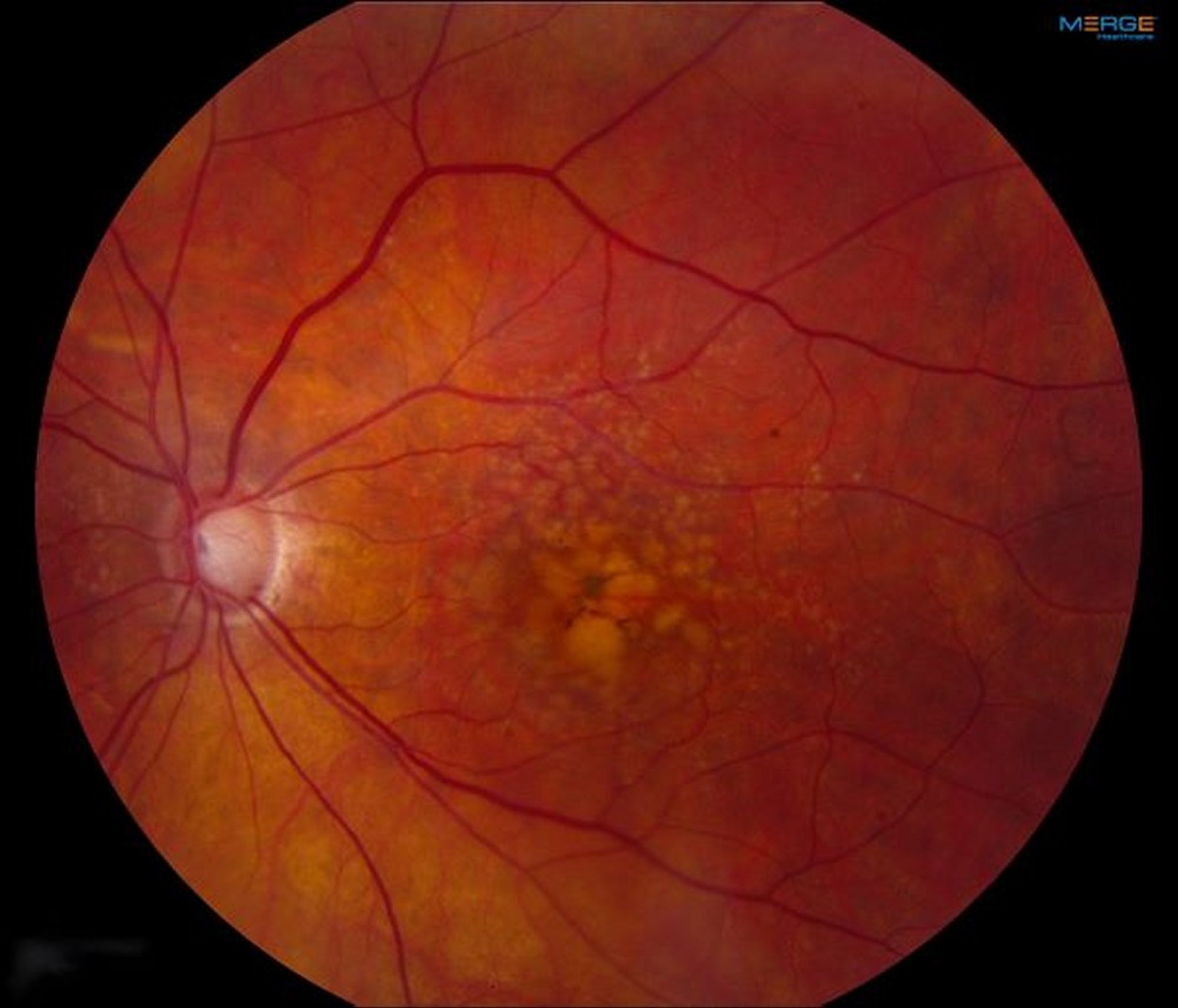
Image provided by Sunir Garg, MD.
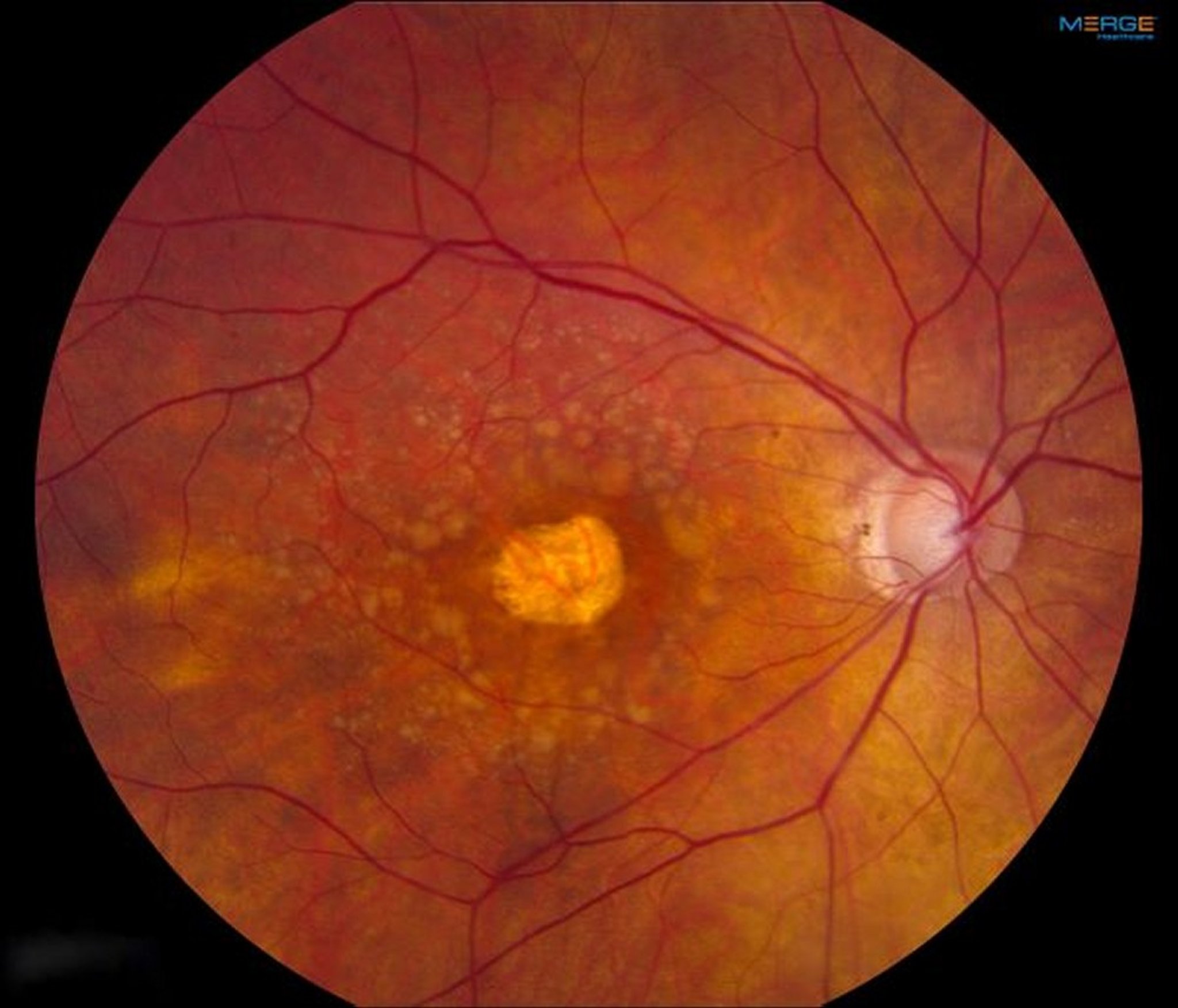
Image provided by Sunir Garg, MD.
Although only 15% of patients with AMD have the wet form, 80 to 90% of the severe vision loss caused by AMD results from wet AMD (1).

PAUL WHITTEN/SCIENCE PHOTO LIBRARY
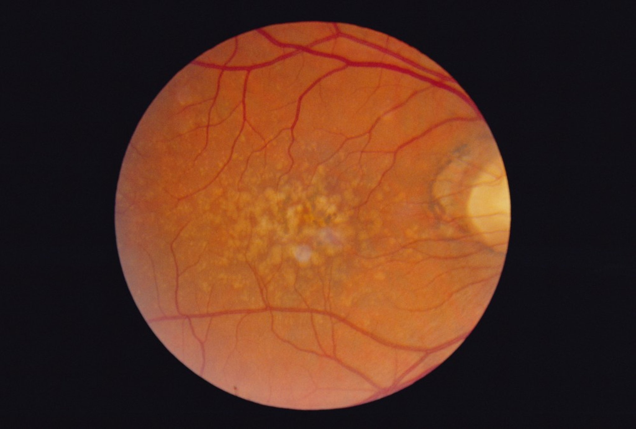
Dry AMD causes changes of the retinal pigment epithelium, typically visible as dark pinpoint areas. The retinal pigment epithelium plays a critical role in keeping the cones and rods healthy and functioning well. Accumulation of waste products from the rods and cones can result in drusen, which appear as yellow spots. Areas of chorioretinal atrophy (referred to as geographic atrophy) occur in more advanced cases of dry AMD. There is no elevated macular scar (disciform scar), edema, hemorrhage, or exudation.
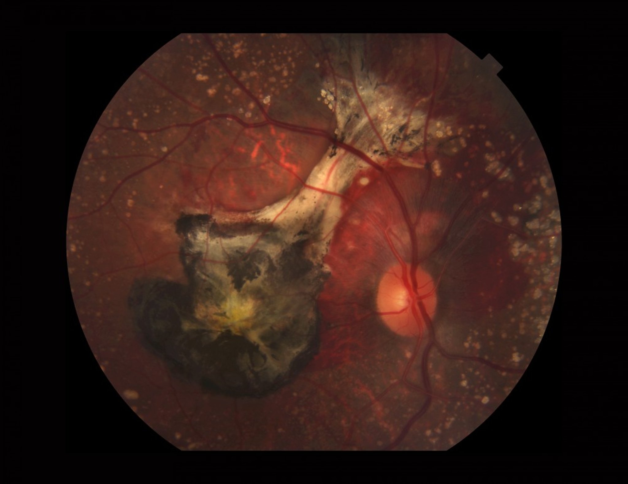
Wet AMD occurs when new abnormal blood vessels develop under the retina in a process called choroidal neovascularization (abnormal new vessel formation). Localized macular edema or hemorrhage may elevate an area of the macula or cause a localized retinal pigment epithelial detachment. Eventually, untreated neovascularization causes a disciform scar under the macula.
Pathophysiology reference
1. Ferris FL 3rd, Fine SL, Hyman L: Age-related macular degeneration and blindness due to neovascular maculopathy. Arch Ophthalmol 102(11):1640-1642, 1984. doi: 10.1001/archopht.1984.01040031330019
Symptoms and Signs of Age-Related Macular Degeneration
Dry AMD
The loss of central vision occurs over years and is painless, and most patients retain enough vision to read and drive. Central blind spots (scotomas) usually occur late in the disease and can sometimes become severe. Symptoms are usually bilateral.
Funduscopic changes include the following:
Changes in the retinal pigment epithelium
Drusen
Areas of chorioretinal atrophy
Wet AMD
Rapid vision loss, usually over days to weeks, is more typical of wet AMD. The first symptom is usually visual distortion, such as a central blind spot (scotoma) or curving of straight lines (metamorphopsia). Peripheral vision and color vision are generally unaffected; however, the patient may become legally blind (< 20/200 vision) in the affected eye, particularly if AMD is not treated. Wet AMD usually affects one eye at a time; thus, symptoms of wet AMD are often unilateral.
Funduscopic changes include the following:
Subretinal fluid, appearing as localized retinal elevation
Retinal edema
Gray-green discoloration under the macula
Exudates in or around the macula
Detachment of retinal pigment epithelium (visible as an area of retinal elevation)
Subretinal hemorrhage in or around the macula

PAUL PARKER/SCIENCE PHOTO LIBRARY
Diagnosis of Age-Related Macular Degeneration
Funduscopic examination
Color fundus photography
Fluorescein angiographyFluorescein angiography
Optical coherence tomography
Both forms of age-related macular degeneration (AMD) are diagnosed by funduscopic examination. Visual changes can often be detected with an Amsler grid. Color photography and fluorescein angiography are done when findings suggest wet AMD. Angiography shows and characterizes subretinal choroidal neovascular membranes and can delineate areas of geographic atrophy. . Color photography and fluorescein angiography are done when findings suggest wet AMD. Angiography shows and characterizes subretinal choroidal neovascular membranes and can delineate areas of geographic atrophy.Optical coherence tomography (OCT) aids in identifying intraretinal and subretinal fluid and can help assess response to treatment.
Treatment of Age-Related Macular Degeneration
Dietary supplements for high-risk dry or unilateral wet age-related macular degeneration (AMD)
Intravitreal antivascular endothelial growth factor (anti-VEGF) medications or laser treatments for wet AMD
Supportive measures
In general, the management of dry AMD focuses on nutritional supplementation to slow disease progression, whereas wet AMD is also treated with intravitreal anti-VEGF or laser therapy.
Dry AMD
There is no way to reverse damage caused by dry AMD. Patients with extensive drusen, pigment changes, and/or geographic atrophy can reduce the risk of developing advanced AMD by 25% by taking daily supplements of the following:
Zinc oxide 80 mgZinc oxide 80 mg
Copper 2 mgCopper 2 mg
Vitamin C 500 mg
Vitamin E 400 IUVitamin E 400 IU
Lutein 10 mg/zeaxanthin 2 mg (or beta-carotene 15 mg or vitamin A 28,000 units for patients who have not smoked)Lutein 10 mg/zeaxanthin 2 mg (or beta-carotene 15 mg or vitamin A 28,000 units for patients who have not smoked)
In patients who currently or formerly smoked, beta-carotene can increase the risk of lung cancer. Substitution of beta-carotene with lutein plus zeaxanthin has been shown to have comparable efficacy (1). Therefore, such a substitution should be considered in patients who currently or formerly smoked. Beta-carotene yellows the skin in some patients. The zinc component of these supplements increases risk of hospitalization for genitourinary tract disorders. Reducing cardiovascular risk factors as well as regularly eating foods high in omega-3 fatty acids and dark green leafy vegetables may help slow disease progression (2); however, randomized trials have not shown that taking supplements of omega-3 fatty acids reduces disease progression (3).
In 2023, intravitreal pegcetacoplan and intravitreal avacincaptad pegol became available for the treatment of advanced dry AMD due to geographic atrophy. These medications inhibit the complement pathway and can slow the progression of geographic atrophy (In 2023, intravitreal pegcetacoplan and intravitreal avacincaptad pegol became available for the treatment of advanced dry AMD due to geographic atrophy. These medications inhibit the complement pathway and can slow the progression of geographic atrophy (4, 5). The primary outcome used in these studies was preservation of retinal tissue, not visual acuity outcomes (the outcome used in studies of wet AMD). These intravitreal medications can be administered every 1 to 2 months.
Wet AMD
Patients with unilateral wet AMD should take the daily nutritional supplements that are recommended for dry AMD to reduce the risk of AMD-induced vision loss in the other eye. The choice of other treatments depends on the size, location, and type of neovascularization. Intravitreal injection of antivascular endothelial growth factor (anti-VEGF) medications (usually ranibizumab, bevacizumab, or aflibercept) can substantially reduce the risk of vision loss and can preserve useful vision in 20% of patients over their lifetime and reading vision in up to one-third of patients (Patients with unilateral wet AMD should take the daily nutritional supplements that are recommended for dry AMD to reduce the risk of AMD-induced vision loss in the other eye. The choice of other treatments depends on the size, location, and type of neovascularization. Intravitreal injection of antivascular endothelial growth factor (anti-VEGF) medications (usually ranibizumab, bevacizumab, or aflibercept) can substantially reduce the risk of vision loss and can preserve useful vision in 20% of patients over their lifetime and reading vision in up to one-third of patients (6).
Newer intravitreal medications are now available that provide extended duration of treatment; these include faricimab as well as high-dose aflibercept (Newer intravitreal medications are now available that provide extended duration of treatment; these include faricimab as well as high-dose aflibercept (7). Faricimab functions as a dual-mechanism medication binding VEGF-A and angiopoietin-2; efficacy is similar to that of aflibercept (). Faricimab functions as a dual-mechanism medication binding VEGF-A and angiopoietin-2; efficacy is similar to that of aflibercept (8). In 2021, an implantable port delivery system for ranibizumab became available for the treatment of wet AMD (). In 2021, an implantable port delivery system for ranibizumab became available for the treatment of wet AMD (9). This is a surgically placed permanent refillable ocular implant that continuously delivers ranibizumab into the vitreous and can be refilled repeatedly. Due to dislocation of the septum inside the device, the company issued a voluntary recall of the implant in October 2022, and it is currently being redesigned. ). This is a surgically placed permanent refillable ocular implant that continuously delivers ranibizumab into the vitreous and can be refilled repeatedly. Due to dislocation of the septum inside the device, the company issued a voluntary recall of the implant in October 2022, and it is currently being redesigned.
In 2020, intravitreal brolucizumab became available for the treatment of wet AMD (In 2020, intravitreal brolucizumab became available for the treatment of wet AMD (10); however, early reports (11) suggest a higher incidence of adverse events with this medication than with other intravitreal anti-VEGF injections, including intraocular inflammation, retinal artery occlusion, and vasculitis.
In a small subset of patients, thermal laser photocoagulation of neovascularization outside the fovea may prevent severe vision loss. Photodynamic therapy, a type of laser treatment, also helps under specific circumstances. Corticosteroids (eg, triamcinolone) are sometimes injected intraocularly along with an anti-VEGF medication. Other treatments, including transpupillary thermotherapy, subretinal surgery, and macular translocation surgery, are seldom used.In a small subset of patients, thermal laser photocoagulation of neovascularization outside the fovea may prevent severe vision loss. Photodynamic therapy, a type of laser treatment, also helps under specific circumstances. Corticosteroids (eg, triamcinolone) are sometimes injected intraocularly along with an anti-VEGF medication. Other treatments, including transpupillary thermotherapy, subretinal surgery, and macular translocation surgery, are seldom used.
Treatment references
1. Age-Related Eye Disease Study 2 Research Group: Lutein + zeaxanthin and omega-3 fatty acids for age-related macular degeneration: The age-related eye disease study 2 (AREDS2) randomized clinical trial. JAMA 309(19):2005-2015, 2013. doi: 10.1001/jama.2013.4997. Clarification and additional information. JAMA 310(2):208, 2013. doi:10.1001/jama.2013.6403
2. Hogg RE, Woodside JV, McGrath A, et al: Mediterranean diet score and its association with age-related macular degeneration: The European Eye Study. Ophthalmology 124(1):82-89, 2017. doi: 10.1016/j.ophtha.2016.09.019
3. Christen WG, Cook NR, Manson JE, et al: Effect of vitamin D and ω-3 fatty acid supplementation on risk of age-related macular degeneration: An ancillary study of the VITAL randomized clinical trial. : Effect of vitamin D and ω-3 fatty acid supplementation on risk of age-related macular degeneration: An ancillary study of the VITAL randomized clinical trial.JAMA Ophthalmol 138(12):1280-1289, 2020. doi: 10.1001/jamaophthalmol.2020.4409
4. Heier JS, Lad EM, Holz FG, et al: Pegcetacoplan for the treatment of geographic atrophy secondary to age-related macular degeneration (OAKS and DERBY): two multicentre, randomised, double-masked, sham-controlled, phase 3 trials.: Pegcetacoplan for the treatment of geographic atrophy secondary to age-related macular degeneration (OAKS and DERBY): two multicentre, randomised, double-masked, sham-controlled, phase 3 trials. Lancet 402(10411):1434-1448, 2023. doi: 10.1016/S0140-6736(23)01520-9
5. Khanani AM, Patel SS, Staurenghi G, et al: Efficacy and safety of avacincaptad pegol in patients with geographic atrophy (GATHER2): 12-month results from a randomised, double-masked, phase 3 trial. : Efficacy and safety of avacincaptad pegol in patients with geographic atrophy (GATHER2): 12-month results from a randomised, double-masked, phase 3 trial.Lancet 402(10411):1449-1458, 2023. doi: 10.1016/S0140-6736(23)01583-0
6. Finger RP, Puth MT, Schmid M, Barthelmes D, et al: Lifetime outcomes of anti-vascular endothelial growth factor treatment for neovascular age-related macular degeneration. JAMA Ophthalmol 138(12):1234-1240, 2020. doi: 10.1001/jamaophthalmol.2020.3989
7. Wykoff CC, Brown DM, Reed K, et al. Effect of high-dose intravitreal aflibercept, 8 mg, in patients with neovascular age-related macular degeneration: The phase 2 CANDELA randomized clinical trial. . Effect of high-dose intravitreal aflibercept, 8 mg, in patients with neovascular age-related macular degeneration: The phase 2 CANDELA randomized clinical trial.JAMA Ophthalmol 141(9):834–842, 2023. doi: 10.1001/jamaophthalmol.2023.2421
8. Heier JS, Khanani AM, Quezada Ruiz C, et al: Efficacy, durability, and safety of intravitreal faricimab up to every 16 weeks for neovascular age-related macular degeneration (TENAYA and LUCERNE): two randomised, double-masked, phase 3, non-inferiority trials. : Efficacy, durability, and safety of intravitreal faricimab up to every 16 weeks for neovascular age-related macular degeneration (TENAYA and LUCERNE): two randomised, double-masked, phase 3, non-inferiority trials.Lancet 399(10326):729-740, 2022. doi: 10.1016/S0140-6736(22)00010-1
9. Holekamp NM, Campochiaro PA, Chang MA, all Archway Investigators: Archway randomized phase 3 trial of the port delivery system with ranibizumab for neovascular age-related macular degeneration. : Archway randomized phase 3 trial of the port delivery system with ranibizumab for neovascular age-related macular degeneration.Ophthalmology S0161-6420(21)00734-X, 2021
10. Dugel PU, Koh A, Ogura Y, HAWK and HARRIER Study Investigators: HAWK and HARRIER: Phase 3, multicenter, randomized, double-masked trials of brolucizumab for neovascular age-related macular degeneration. : HAWK and HARRIER: Phase 3, multicenter, randomized, double-masked trials of brolucizumab for neovascular age-related macular degeneration.Ophthalmology 127(1):72-84, 2020. doi: 10.1016/j.ophtha.2019.04.017
11. Haug SJ, Hien DL, Uludag G, et al: Retinal arterial occlusive vasculitis following intravitreal brolucizumab administration. : Retinal arterial occlusive vasculitis following intravitreal brolucizumab administration.Am J Ophthalmol Case Rep 18:100680, 2020. doi: 10.1016/j.ajoc.2020.100680
Supportive measures
For patients who have lost central vision, low-vision devices such as magnifiers, high-power reading glasses, large computer monitors, and telescopic lenses are available. Also, certain types of software can display computer data in large print or read information aloud in a synthetic voice. Low-vision counseling is advised.
Key Points
AMD is more common among White people and is the leading cause of permanent vision loss in older adults.
AMD can be dry (nonexudative or atrophic) or wet (exudative or neovascular).
Although 85% of AMD is dry, 80 to 90% of severe vision loss caused by AMD results from the wet type.
Funduscopic changes in dry AMD include drusen, areas of chorioretinal atrophy, and changes to the retinal pigment epithelium.
Funduscopic changes in wet AMD include retinal edema and localized elevation, detachment of the retinal pigment epithelium, a gray-green discoloration under the macula, and exudates in and around the macula.
If patients have AMD on funduscopy, do color fundus photography, fluorescein angiography, and optical coherence tomography.If patients have AMD on funduscopy, do color fundus photography, fluorescein angiography, and optical coherence tomography.
Prescribe dietary supplements for unilateral wet or high-risk dry AMD.
Treat wet AMD with anti-VEGF medications as an intravitreal injection or through the port delivery system or with laser therapy.
Treat wet AMD with photodynamic therapy or laser therapy in select cases.
Drugs Mentioned In This Article


