


Cardiac tamponade is pressure on the heart by blood or fluid that accumulates in the 2-layered sac around the heart (pericardium). This disorder interferes with the heart's ability to pump blood.
People typically feel light-headed and short of breath, and they may faint.
The diagnosis is based on symptoms, examination results, and usually ultrasound of the heart (echocardiography) done in the emergency department.
Blood is drained from around the heart using a needle and sometimes surgery.
(See also Introduction to Chest Injuries.)
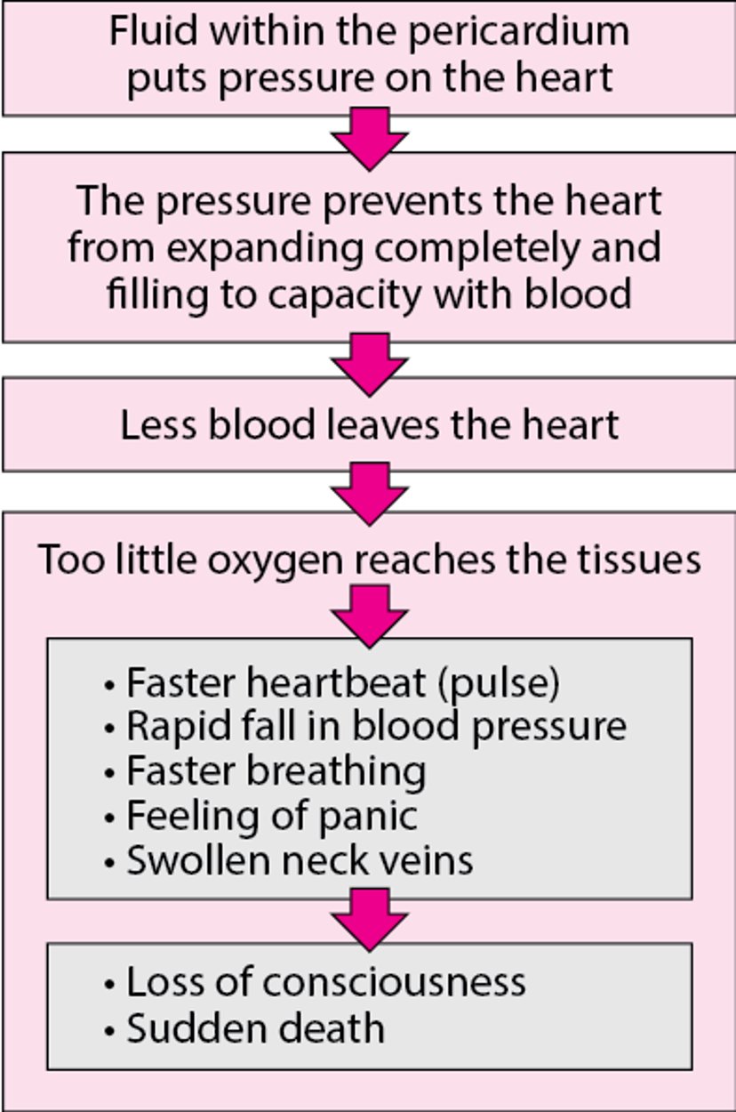
In cardiac tamponade, fluid or blood accumulates between the 2 layers of the pericardium, which then tightly squeezes the heart. This pressure can prevent the heart from filling with blood. As a result, less blood is pumped to the body, sometimes causing shock (with blood pressure becoming dangerously low) and death.
The most common causes are rupture of an aortic aneurysm (a bulge in the wall of the aorta), advanced lung cancer, acute pericarditis (inflammation of the pericardium), a heart attack, and heart surgery.
Chest injuries can also cause cardiac tamponade. The most common such injuries are stab wounds. Blunt injuries that tear the wall of the heart can cause tamponade, but many people with such injuries die before they can be brought for medical treatment.
Cardiac Tamponade

Symptoms of Cardiac Tamponade
People with cardiac tamponade may feel light-headed or short of breath. They may faint. They may have low blood pressure and a rapid heart rate. The skin may be cool, sweaty, and bluish. The veins in the neck may appear swollen or distended.
Diagnosis of Cardiac Tamponade
A doctor's evaluation
Echocardiography
Prompt diagnosis and treatment are essential because cardiac tamponade can be rapidly fatal. The diagnosis is based on symptoms, examination results, and usually echocardiography.
Echocardiography (which uses ultrasound waves to produce an image of the heart) is the gold standard to confirm the diagnosis.
Treatment of Cardiac Tamponade
Removal of blood or fluid from around the heart
Cardiac tamponade is a medical emergency. Doctors treat it immediately by using a needle (sometimes guided by ultrasound) to remove the blood or fluid from around the heart (pericardiocentesis). This procedure relieves pressure on the heart and enables it to beat normally.


This illustration shows pericardiocentesis, which is a medical procedure that is performed when fluid accumulates in the sac surrounding the heart (pericardium). A pericardial effusion is the medical term for the presence of fluid within the pericardium. A needle (often guided by an x-ray or ultrasound) is inserted into the pericardium to relieve pressure on the heart and/or to collect fluid to help diagnose the cause of fluid accumulation (such as infection, inflammation, or cancer).
Pepermpron/stock.adobe.com
Sometimes pericardiocentesis fails to remove enough fluid. Then, doctors must make an incision into the chest wall (thoracotomy) and then the pericardium (pericardiotomy) to drain the fluid. They may also need to remove part of the pericardium (pericardiectomy).


