


Cardiac catheterization is the passage of a catheter through peripheral arteries or veins into cardiac chambers, the pulmonary artery, and coronary arteries and veins.
Cardiac catheterization can be used to perform various tests, including:
Intracardiac and intravascular pressure measurement (including ventricular filling pressures and pulmonary artery pressure)
Angiography
Detection and quantification of shunts
Measurement of cardiac output (CO)
Measurements of myocardial metabolism
Endomyocardial biopsy
Intravascular ultrasound (IVUS)
These tests define coronary artery anatomy, cardiac anatomy, cardiac function, and pulmonary arterial hemodynamics to establish diagnoses and help clinicians select treatment.
Cardiac catheterization is also the basis for therapeutic interventions (see Percutaneous coronary intervention in Treatment of Coronary Artery Disease, Treatment of Aortic Stenosis, Treatment of Mitral Regurgitation).
Diagnostic and therapeutic cardiac catheterization for congenital heart disease is not discussed in detail here.
Procedure for Cardiac Catheterization
Many but not all preprocedure protocols require patients to fast for 4 to 6 hours before cardiac catheterization. Most patients do not require overnight hospitalization unless a therapeutic intervention is also performed. The procedure is typically performed with local anesthesia at the access site and light or moderate intravenous systemic sedation that keeps patients comfortable but awake and able to participate throughout.
Left heart catheterization
Left heart catheterization is most commonly used to assess:
Coronary artery anatomy and presence of coronary artery disease
Left heart catheterization is also used to assess:
Aortic blood pressure
Aortic valve function
Left ventricular pressure and function
Mitral valve function
Systemic vascular resistance
Left heart catheterization is performed via femoral, subclavian, radial, or brachial artery puncture, with a catheter passed into the coronary artery ostia and/or across the aortic valve into the left ventricle (LV). Radial artery access is preferred for coronary angiography and intervention as it is more comfortable and carries a lower risk of hematoma or pseudoaneurysm or arteriovenous fistula formation when compared with femoral artery access (1).
Catheterization of the left atrium (LA) and LV is occasionally performed using transseptal perforation during right heart catheterization, particularly during electrophysiologic studies when access to the left atrium is needed and the foramen ovale is not patent.
In addition to coronary artery evaluation, hemodynamic left heart catheterization can be performed to obtain direct left atrial and left ventricular pressure measurements and to directly measure transaortic and transmitral pressure gradients.
Right heart catheterization
Right heart catheterization is commonly used to measure:
Right atrial pressure
Right ventricular pressure
Pulmonary artery pressure
Pulmonary artery occlusion pressure (PAOP—see figure )
The most frequent indications for right heart catheterization are to assess hemodynamics to guide therapy in shock of unknown etiology or type, to evaluate the need for cardiac transplantation or mechanical cardiac support (eg, a ventricular assist device) in patients with severe cardiogenic shock, for periprocedural risk stratification among people with severe cardiopulmonary comorbidities, and to diagnose pulmonary hypertension.
PAOP (also called pulmonary capillary wedge pressure) approximates left atrial and left ventricular end-diastolic pressure in most cases. In seriously ill patients, PAOP helps assess volume status (by way of left ventricular end-diastolic pressure as a proxy for left ventricular preload) and, with simultaneous measurements of cardiac output, can help guide therapy.
Right heart catheterization is also useful for assessing cardiac filling pressures, pulmonary vascular resistance, tricuspid or pulmonic valve function, intracardiac shunts, and right ventricular pressure.
Right heart pressure measurements may help in the diagnosis of cardiomyopathy, constrictive pericarditis, and cardiac tamponade when noninvasive testing is nondiagnostic, and it is an essential part of the assessment for cardiac transplantation or mechanical cardiac support (eg, use of a ventricular assist device).
The procedure is performed via femoral, subclavian, internal jugular, or antecubital vein puncture. A catheter is passed into the right atrium, through the tricuspid valve, into the right ventricle, and across the pulmonary valve into the pulmonary artery.
Selective catheterization of the coronary sinus can also be performed.
Hemodynamic assessment via right heart catheterization during exercise is increasingly being performed as part of the workup for dyspnea of uncertain etiology. The test can be performed at the same time as cardiopulmonary exercise testing, called invasive cardiopulmonary exercise testing. This test is considered the gold standard for diagnosis of heart failure with preserved ejection fraction (HFpEF) as a cardiac limitation to exercise but is available at relatively few centers (2). Invasive cardiopulmonary exercise testing should be considered in patients at intermediate pretest probability for HFpEF if the diagnosis is uncertain after an initial evaluation. An increase in the PAOP > 25 mm Hg confirms the diagnosis when patients have signs and symptoms of heart failure irrespective of left ventricular ejection fraction.
Diagram of the Cardiac Cycle, Showing Pressure Curves of the Cardiac Chambers, Heart Sounds, Jugular Pulse Wave, and the ECG
The phases of the cardiac cycle are atrial systole (a), isovolumetric contraction (b), maximal ejection (c), reduced ejection (d), protodiastolic phase (e), isovolumetric relaxation (f), rapid inflow (g), and diastasis, or slow LV filling (h). For illustrative purposes, time intervals between valvular events have been modified, and the z point has been prolonged. In the jugular venous pulse tracing, the A wave represents atrial contraction at end-diastole, C wave is the carotid artery impulse or bulging of the tricuspid valve into the right atrium in early systole, V wave is the increase in pressure and volume of right atrial filling in late systole and early diastole, X and X' descents represent movement of the lower part of the right atrium toward the ventricle with X' at end-systole, and the Y descent is tricuspid valve opening and ventricular filling in diastole. AO = aortic valve opening; AC = aortic valve closing; LV = left ventricle; LA = left atrium; RV = right ventricle; RA = right atrium; MO = mitral valve opening. |


Procedure references
1. Rao SV, O'Donoghue ML, Ruel M, et al. 2025 ACC/AHA/ACEP/NAEMSP/SCAI Guideline for the Management of Patients With Acute Coronary Syndromes: A Report of the American College of Cardiology/American Heart Association Joint Committee on Clinical Practice Guidelines. Circulation. 2025;151(13):e771-e862. doi:10.1161/CIR.0000000000001309
2. Rajagopalan N, Borlaug BA, Bailey AL, et al. Practical Guidance for Hemodynamic Assessment by Right Heart Catheterization in Management of Heart Failure. JACC Heart Fail. 2024;12(7):1141-1156. doi:10.1016/j.jchf.2024.03.020
Specific Tests During Cardiac Catheterization
Angiography
Injection of a radiopaque contrast agent into coronary or pulmonary arteries, the aorta, and cardiac chambers is useful in certain circumstances. Digital subtraction angiography, in which images of background structures are removed to improve visualization of the structures of interest, is used for nonmoving arteries and for chamber cineangiography.
Selective coronary angiography via left heart catheterization is used to evaluate coronary artery anatomy in various clinical situations, as in patients with suspected coronary atherosclerotic or congenital coronary artery disease, valvular disorders before valvular replacement, or unexplained heart failure.
Aortic angiography via left heart catheterization is used to assess aortic regurgitation, coarctation, patent ductus arteriosus, and dissection.

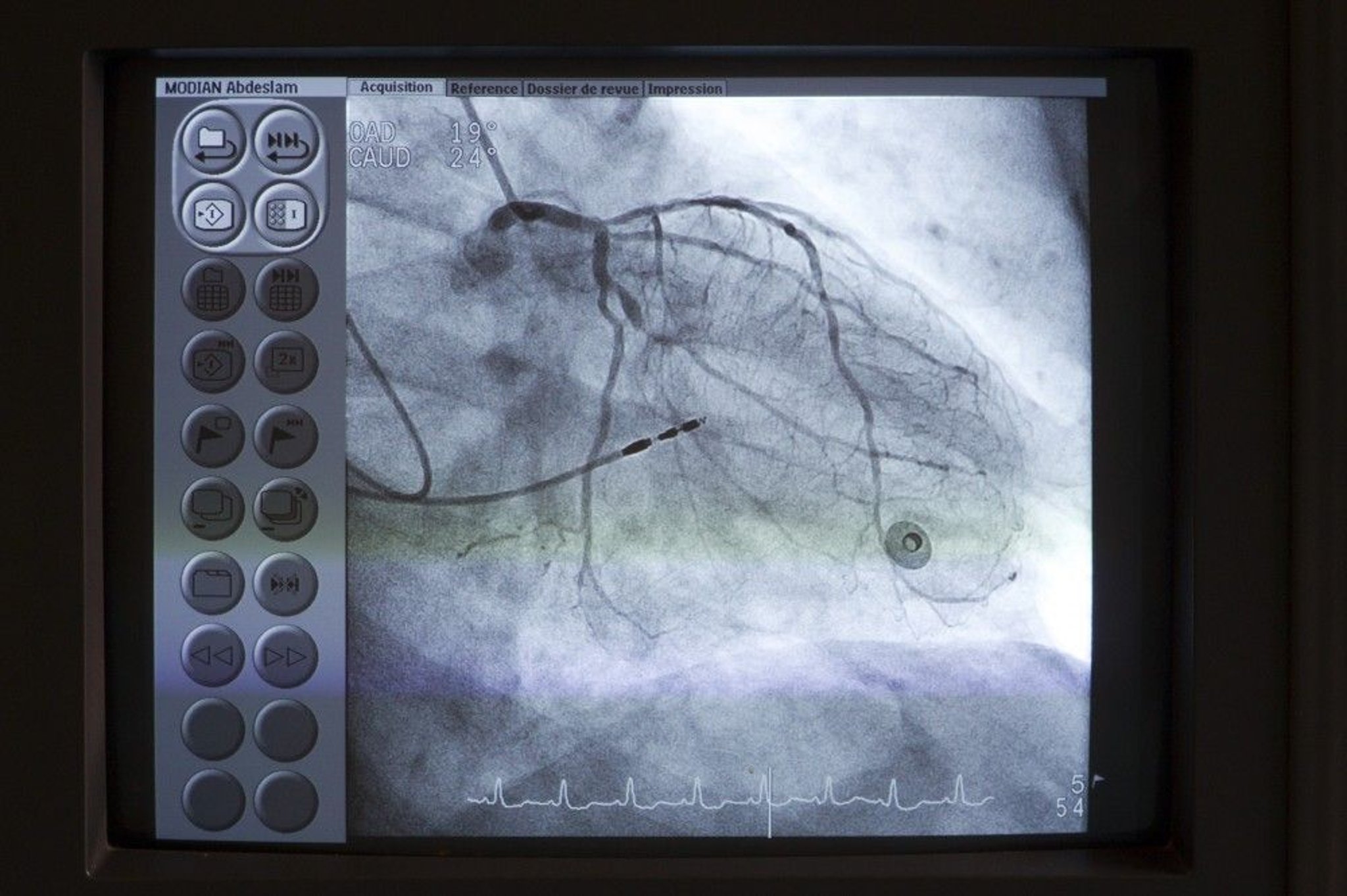
A catheter (upper left) is placed at the os of the left main coronary artery. Contrast is then injected into the coronary arteries under fluoroscopy to detect any anomalies such as stenoses.
Astier/BSIP/SCIENCE PHOTO LIBRARY
Pulmonary angiography via right heart catheterization can be used to diagnose pulmonary embolism. Intraluminal filling defects or arterial cutoffs are diagnostic. Radiopaque contrast agent is usually selectively injected into one or both pulmonary arteries and their segments. Computed tomographic pulmonary angiography (CTPA) has largely replaced right heart catheterization for diagnosis of acute pulmonary embolism. Pulmonary angiography via right heart catheterization remains commonly used to determine a management plan for suspected chronic thromboembolic disease. Pulmonary angiography can also be used evaluate pulmonary vascular abnormalities such as branch pulmonary artery stenosis.

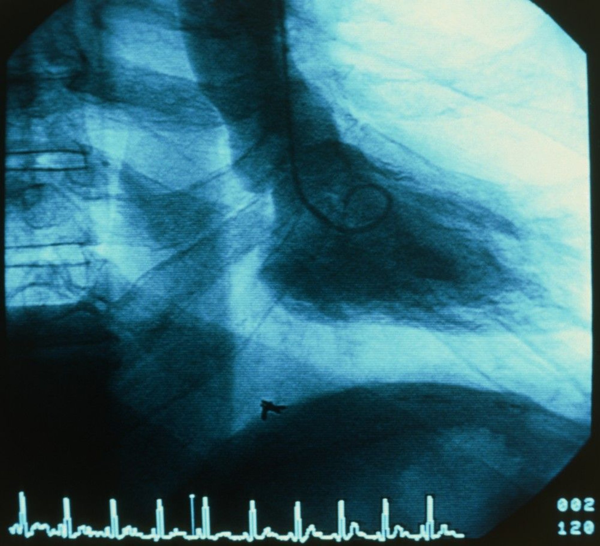
A catheter (center) is inserted into the left ventricle (LV). Contrast is then injected into the LV under fluoroscopy and the ejection fraction is visually estimated.
PETER MENZEL/SCIENCE PHOTO LIBRARY
Ventriculography, performed via right and/or left heart catheterization, is used to visualize ventricular wall motion and ventricular outflow tracts, including subvalvular, valvular, and supravalvular regions. It is also used to estimate severity of mitral valve regurgitation and determine its pathophysiology. After left ventricular mass and volume are determined from single planar or biplanar ventricular angiograms, end-systolic and end-diastolic volumes and ejection fraction can be calculated.
Coronary artery flow measurements
Coronary angiography shows the presence and degree of stenosis but not the functional significance of the lesion (ie, how much blood flows across the stenosis) or whether a specific lesion is likely to be the cause of symptoms.
Extremely thin guidewires with pressure sensors or Doppler flow sensors can be used to estimate coronary artery blood flow, which is expressed as fractional flow reserve (FFR). FFR is the ratio of maximal flow through the stenotic area to normal maximal flow obtained during hyperemia (most commonly with adenosine); an FFR of < 0.75 to 0.8 is considered abnormal (1). Other techniques of measuring coronary blood flow, which do not require hyperemia induced by adenosine, are collectively termed non-hyperemic pressure ratios (NHPR). They include instantaneous wave-free ratio (iFR) and diastolic hyperemia-free ratio (DFR). NHPR techniques measure gradients across a stenosis during a period in diastole; an NHPR of ≤ 0.89 is considered abnormal (2, 3, 4).
These flow estimates correlate well with the need for intervention and long-term outcome; patients with lesions with FFR > 0.8 or NHPR > 0.89 do not seem to benefit from placement of a stent. These flow measurements are most useful with intermediate lesions (40 to 69% stenosis) and with multiple lesions (to identify those that are clinically most significant), or with proximal lesion affecting large territories (5, 6, 7).
Noninvasive estimates of FFR can also be derived from data obtained during coronary CT angiography.
Intravascular ultrasound (IVUS)
Miniature ultrasound transducers on the end of coronary artery catheters can produce images of coronary vessel lumina and walls and delineate blood flow. Intravascular ultrasound is used to guide coronary angiography, including to guide optimal stent placement during percutaneous coronary intervention. It is also used to troubleshoot acute coronary complications and stent failure, to detect cardiac allograft vasculopathy after heart transplantation, to identify coronary artery dissections, and to guide endovascular therapy in aortic interventions (8, 9, 10).
Optical coherence tomography (OCT)
Optical coherence tomography is an optical analog of intracoronary ultrasound imaging that measures the amplitude of backscattered light to determine the temperature of coronary plaques and can help determine whether lesions are at high risk of future rupture (leading to acute coronary syndromes). Unlike in IVUS, injection of contrast is required to obtain images. The indications and appropriate use for OCT are uncertain.
Tests for cardiac shunts
Measuring blood oxygen content at successive levels in the heart and great vessels can help determine the presence, direction, and volume of central shunts. The maximal normal difference in oxygen content between structures is as follows:
The pulmonary artery and right ventricle: 0.5 mL/dL (0.5 vol%)
The right ventricle and right atrium: 0.9 mL/dL (0.9 vol%)
The right atrium and superior vena cava: 1.9 mL/dL (1.9 vol%)
If the blood oxygen content in a chamber exceeds that of the more proximal chamber by more than these values, a left-to-right shunt at that level is probable. Right-to-left shunts are strongly suspected when left arterial, left ventricular, or arterial oxygen saturation is low (≤ 92%) and does not improve when pure oxygen (fraction of inspired oxygen = 1.0) is given. Left heart or arterial desaturation plus increased oxygen content in blood samples drawn beyond the shunt site on the right side of circulation suggests a bidirectional shunt.
The overall ratio of pulmonary to systemic blood flow (Qp:Qs) can be also calculated using measured systemic arterial, mixed venous, and pulmonary arterial oxygen saturation, combined with an assumed or measured pulmonary venous oxygen saturation. This ratio can be used to estimate the direction and magnitude of shunting.
Measurement of cardiac output and flow
Cardiac output (CO) is the volume of blood ejected by the heart per minute (normal at rest: 4 to 8 L/minute). Techniques (see table ) used to calculate CO include:
Fick cardiac output technique
Indicator-dilution technique
Thermodilution technique
Cardiac Output Equations
Fick technique |
Numerator is oxygen consumption (O2 absorbed by lungs in mL/minute). |
Indicator-dilution technique |
Denominator is the sum of contrast agent concentrations (C) at each time interval (t). |
Thermodilution technique |
TB – TI is the difference between body and injectate temperatures; injectate is usually dextrose or saline. Denominator is the sum of changes in temperature at each time interval (t). |
SaO2= arterial oxygen saturation (%); SvO2= mixed venous oxygen saturation (%), measured in the pulmonary artery. |


With the Fick technique, CO is proportional to oxygen consumption divided by arteriovenous oxygen difference.
Dilution techniques rely on the assumption that after an indicator is injected into the circulation, it appears and disappears proportionately to CO.
Usually, CO is expressed in relation to body surface area (BSA) as the cardiac index (CI) in L/minute/m2 (ie, CI = CO/BSA—see table ). BSA can be estimated using DuBois height (ht)–weight (wt) equation:


Normal Values for Cardiac Index and Related Measurements
Measurement | Normal Value | SD |
|---|---|---|
Oxygen uptake | 143 mL/minute/m2* | 14.3 |
Arteriovenous oxygen difference | 4.1 dL | 0.6 |
Cardiac index | 3.5 L/minute/m2 | 0.7 |
Stroke index | 46 mL/beat/m2 | 8.1 |
Total systemic resistance | 1130 dynes-second-cm-5 | 178 |
Total pulmonary resistance | 205 dynes-second-cm-5 | 51 |
Pulmonary arteriolar resistance | 67 dynes-second-cm-5 | 23 |
* Varies with body mass index. | ||
SD = standard deviation. | ||
Adapted from Barratt-Boyes BG, Wood EH. Cardiac output and related measurements and pressure values in the right heart and associated vessels, together with an analysis of the hemodynamic response to the inhalation of high oxygen mixtures in healthy subjects. J Lab Clin Med. 1958;51(1):72–90. | ||

Endomyocardial biopsy
Endomyocardial biopsy helps assess transplant rejection and myocardial disorders due to infection, inflammation, or infiltrative diseases. The biopsy catheter (bioptome) can be passed into either ventricle, usually the right. Three to 5 samples of myocardial tissue are removed from the septal endocardium. The main complication of endomyocardial biopsy, cardiac perforation, occurs in 0.2 to 0.7% of patients (11, 12); it may cause hemopericardium leading to cardiac tamponade. Injury to the tricuspid valve and supporting chordae may also occur and can lead to tricuspid regurgitation.
Pulmonary vasodilator testing
During a cardiac catheterization to evaluate for pulmonary hypertension, pulmonary artery pressure, pulmonary vascular resistance, and cardiac output are measured at baseline, and then after oxygen and/or nitric oxide administered. The response (or lack thereof) to pulmonary vasodilators can guide pulmonary hypertension therapy.
Tests during cardiac catheterization references
1. Bangalore S, Fearon WF, Fugar S, et al. Evidence-Based Practices in the Cardiac Catheterization Laboratory: Invasive Epicardial Coronary Physiologic Assessment: A Scientific Statement From the American Heart Association. Circulation. Published online November 20, 2025. doi:10.1161/CIR.0000000000001389
2. Gotberg M, Christiansen EH, Gudmundsdottir IJ, et al. Instantaneous wave-free ratio versus fractional flow reserve to guide PCI. New Engl J Med . 2017;376 (19):1813-1823. doi: 10.1056/NEJMoa1616540
3. Johnson NP, Li W, Chen X, et al. Diastolic pressure ratio: new approach and validation vs. the instantaneous wave-free ratio. Eur Heart J. 2019;40(31):2585–2594. doi:10.1093/eurheartj/ehz230
4. Storozhenko T, Collison D, Johnson NP, et al. Diagnostic Performance of Non-Hyperemic Pressure Ratios Versus Fractional Flow Reserve Stratified by Coronary Artery: A Systematic Review and Meta-Analysis of Individual Patient Data. J Am Heart Assoc. 2026;15(2):e040916. doi:10.1161/JAHA.124.040916
5. Davies JE, Sen S, Dehbi HM, et al. Use of the Instantaneous Wave-free Ratio or Fractional Flow Reserve in PCI. N Engl J Med. 2017;376(19):1824-1834. doi:10.1056/NEJMoa1700445
6. Rajagopalan N, Borlaug BA, Bailey AL, et al. Practical Guidance for Hemodynamic Assessment by Right Heart Catheterization in Management of Heart Failure. JACC Heart Fail. 2024;12(7):1141-1156. doi:10.1016/j.jchf.2024.03.020
7. Toth GG, Johnson NP, Jeremias A, et al. Standardization of Fractional Flow Reserve Measurements. J Am Coll Cardiol. 2016;68(7):742-753. doi:10.1016/j.jacc.2016.05.067
8. Ugo F, Franzino M, Massaro G, et al. The Role of IVUS in Coronary Complications. Catheter Cardiovasc Interv. 2025;105(5):1171-1182. doi:10.1002/ccd.31433
9. Writing Committee Members, Isselbacher EM, Preventza O, et al. 2022 ACC/AHA Guideline for the Diagnosis and Management of Aortic Disease: A Report of the American Heart Association/American College of Cardiology Joint Committee on Clinical Practice Guidelines. J Am Coll Cardiol. 2022;80(24):e223-e393. doi:10.1016/j.jacc.2022.08.004
10. Writing Committee Members, Lawton JS, Tamis-Holland JE, et al. 2021 ACC/AHA/SCAI Guideline for Coronary Artery Revascularization: A Report of the American College of Cardiology/American Heart Association Joint Committee on Clinical Practice Guidelines. J Am Coll Cardiol. 2022;79(2):e21-e129. doi:10.1016/j.jacc.2021.09.006
11. Cooper LT, Baughman KL, Feldman AM, et al. The role of endomyocardial biopsy in the management of cardiovascular disease: a scientific statement from the American Heart Association, the American College of Cardiology, and the European Society of Cardiology. Endorsed by the Heart Failure Society of America and the Heart Failure Association of the European Society of Cardiology. J Am Coll Cardiol. 2007;50(19):1914-1931. doi:10.1016/j.jacc.2007.09.008
12. Shah Z, Vuddanda V, Rali AS, Pamulapati H, Masoomi R, Gupta K. National Trends and Procedural Complications from Endomyocardial Biopsy: Results from the National Inpatient Sample, 2007-2014. Cardiology. 2018;141(3):125-131. doi:10.1159/000493786
Contraindications to Cardiac Catheterization
Relative contraindications to cardiac catheterization include:
Fever
Radiopaque contrast agent allergies in patients who have not been appropriately premedicated
Systemic infection
Uncontrolled arrhythmia
Uncontrolled hypertension
Uncompensated heart failure
Relative contraindications balance the urgency of the procedure (eg, in an acute myocardial infarction vs an elective case) and the severity of the contraindicating disorder. Periprocedural management of anticoagulants or antiplatelet medications is individualized based on the type of procedure (ie, arterial vs venous access), the urgency of the procedure, the indication for the medication, and the patient's risk of bleeding. Catheterization laboratories frequently have policies for the periprocedural management of these medications.
Complications of Cardiac Catheterization
The incidence of major complications during or after diagnostic cardiac catheterization is approximately 1% (1, 2, 3).
Most complications are minor and can be easily treated. Serious complications of diagnostic catheterization (eg, cardiac arrest, anaphylactic reactions, shock, seizures, renal toxicity) are rare. Mortality rate after diagnostic catheterization is 0.01% to 0.7% (1, 2, 3), with not all deaths caused by the procedure. Myocardial infarction (< 0.011%) and stroke (0.03% to 0.17%) may result in significant morbidity.
In patients undergoing percutaneous coronary interventions (PCI), major complication rates are approximately 4.5%, generally higher than those for patients undergoing diagnostic catheterization (2). Reported mortality during PCI is 0.6 to 4.8% (higher in patients undergoing emergent intervention for ST-segment elevation myocardial infarction) (4). The approximate rate of periprocedural myocardial infarction is 2 to 18%, acute kidney injury is 4 to 7%, stroke is up to 1%, and major bleeding is 1 to 5% (5).
Patient factors that increase risk of complications include:
Increasing age
Contrast agent complications
Injection of radiopaque contrast agent produces a transient sense of warmth throughout the body in many patients. Tachycardia, a slight fall in systemic pressure, an increase in cardiac output, nausea, vomiting, and coughing may occur. Rarely, bradycardia occurs when a large amount of a contrast agent is injected; asking the patient to cough often restores normal rhythm by increasing coronary and cerebral perfusion and clearing contrast from coronary arteries.
More serious reactions (see also Radiographic Contrast Agents and Contrast Reactions) include:
Allergic-type contrast reactions
Contrast-induced acute kidney injury
Allergic-type reactions to contrast include urticaria and conjunctivitis, which usually respond to diphenhydramine 50 mg IV. Severe hypersensitivity reactions to iodinated radiopaque contrast, such as anaphylaxis and anaphylactoid reactions with bronchospasm, laryngeal edema, and dyspnea, are rare reactions, with an approximate frequency of 0.02 to 0.04% (6). One study of patients receiving intra-arterial contrast injection specifically found that 3.6% of patients had an immediate hypersensitivity reaction and 15% had a delayed reaction; however, the rate of severe reactions was only 0.3% (7). These reactions should be treated immediately with epinephrine. Patients with a history of allergic reaction to contrast, particularly after prior intra-arterial injection, may be premedicated with oral prednisone (or parenteral hydrocortisone if the study is urgent) and diphenhydramine.
Contrast-induced acute kidney injury is defined as impairment of renal function (a 25 to 50% increase in serum creatinine from baseline or a 0.3 to 0.5 mg/dL [23 to 44 micromole/L] increase in absolute value) within 2 to 7 days of IV contrast administration judged to be caused by contrast rather than alternative causes (8, 9). For patients at risk, using the lowest possible dose of low-osmolar or iso-osmolar contrast, discontinuing nephrotoxic medications, avoiding multiple contrast studies within a short period of time, and providing intravascular volume expansion before and after contrast exposure likely reduces this risk. Statins may reduce the risk of acute kidney injury. For some high-risk patients in whom the benefits of the procedure outweigh the risks, prophylactic dialysis may be performed.
Catheter-related complications
If the catheter tip contacts the ventricular endocardium, ventricular ectopy or arrhythmias commonly occur, but ventricular fibrillation is rare. If it occurs, direct current cardioversion (DC cardioversion) is administered immediately.
Disruption of an atherosclerotic plaque by the catheter can release a shower of atheroemboli. Emboli from the aorta may cause stroke or nephropathy. Emboli from proximal to distal coronary arteries may cause myocardial infarction.
Coronary artery dissection is also possible.
Access site complications
Access site complications include:
Bleeding
Hematoma
Pseudoaneurysm
Arteriovenous (AV) fistula
Limb ischemia
Bleeding from the access site may occur and usually resolves with compression. Mild bruises and small hematomas are common and do not require specific investigation or treatment.
A large or enlarging lump should be investigated using ultrasound to distinguish hematoma from pseudoaneurysm. A bruit at the site (with or without pain) suggests an AV fistula, which can be diagnosed using ultrasound. Hematomas usually resolve with time and do not require specific therapy. Pseudoaneurysms and AV fistulas usually resolve with compression; those that persist may require surgical repair.
Radial artery access carries a much lower risk of hematoma or pseudoaneurysm or AV fistula formation when compared with femoral artery access (10).
Complications references
1. Al-Hijji MA, Lennon RJ, Gulati R, et al. Safety and Risk of Major Complications With Diagnostic Cardiac Catheterization. Circ Cardiovasc Interv. 2019;12(7):e007791. doi:10.1161/CIRCINTERVENTIONS.119.007791
2. Dehmer GJ, Weaver D, Roe MT, et al. A contemporary view of diagnostic cardiac catheterization and percutaneous coronary intervention in the United States: a report from the CathPCI Registry of the National Cardiovascular Data Registry, 2010 through June 2011. J Am Coll Cardiol. 2012;60(20):2017-2031. doi:10.1016/j.jacc.2012.08.966
3. West R, Ellis G, Brooks N; Joint Audit Committee of the British Cardiac Society and Royal College of Physicians of London. Complications of diagnostic cardiac catheterisation: results from a confidential inquiry into cardiac catheter complications. Heart. 2006;92(6):810-814. doi:10.1136/hrt.2005.073890
4. Writing Committee Members, Harold JG, Bass TA, et al. ACCF/AHA/SCAI 2013 Update of the Clinical Competence Statement on Coronary Artery Interventional Procedures: a Report of the American College of Cardiology Foundation/American Heart Association/American College of Physicians Task Force on Clinical Competence and Training (Writing Committee to Revise the 2007 Clinical Competence Statement on Cardiac Interventional Procedures). Catheter Cardiovasc Interv. 2013;82(2):E69-E111. doi:10.1002/ccd.24985
5. Gaudino M, Andreotti F, Kimura T. Current concepts in coronary artery revascularisation. Lancet. 2023;401(10388):1611-1628. doi:10.1016/S0140-6736(23)00459-2
6. Broyles AD, Banerji A, Barmettler S, et al. Practical Guidance for the Evaluation and Management of Drug Hypersensitivity: Specific Drugs. J Allergy Clin Immunol Pract. 2020;8(9S):S16-S116. doi:10.1016/j.jaip.2020.08.006
7. Sohn KH, Kim GW, Lee SY, et al. Immediate and delayed hypersensitivity after intra-arterial injection of iodinated contrast media: a prospective study in patients with coronary angiography. Eur Radiol. 2019;29(10):5314-5321. doi:10.1007/s00330-019-06138-3
8. Mandurino-Mirizzi A, Munafò A, Crimi G. Contrast-Associated Acute Kidney Injury. J Clin Med. 2022;11(8):2167. doi:10.3390/jcm11082167
9. Mehran R, Dangas GD, Weisbord SD. Contrast-Associated Acute Kidney Injury. N Engl J Med. 2019;380(22):2146-2155. doi:10.1056/NEJMra1805256
10. Rao SV, O'Donoghue ML, Ruel M, et al. 2025 ACC/AHA/ACEP/NAEMSP/SCAI Guideline for the Management of Patients With Acute Coronary Syndromes: A Report of the American College of Cardiology/American Heart Association Joint Committee on Clinical Practice Guidelines. Circulation. 2025;151(13):e771-e862. doi:10.1161/CIR.0000000000001309
More Information
The following English-language resource may be useful. Please note that The Manual is not responsible for the content of this resource.
American College of Radiology (ACR) Manual on Contrast Media. 2025 ACR Committee on Drugs and Contrast Media provides a guide for safe and effective use of contrast media
Drug Information for the Topic


