Percutaneous coronary intervention (PCI) is percutaneous transluminal coronary angioplasty (PTCA) with or without stent insertion. Primary indications are treatment of:
Stable ischemic heart disease with angina pectoris refractory to medical treatment
Acute coronary syndromes (including unstable angina, non-ST-elevation myocardial infarction [NSTEMI], and ST-elevation myocardial infarction [STEMI]
PTCA and stent placement within 90 minutes of onset of pain is the optimal treatment of STEMI, or for any coronary syndrome with features of clinical instability (eg, refractory chest pain, new or worsening heart failure, hemodynamic instability, cardiogenic shock, or unstable arrhythmias). PCI is also indicated on a more delayed time frame for NSTEMI and some cases of unstable angina. Elective PCI may be appropriate for patients who have had a myocardial infarction (MI) and who have recurrent or inducible angina before hospital discharge and for patients who have angina and remain symptomatic despite medical treatment.


This coronary angiogram shows a patient's artery before intervention.
Stephen Gerard/SCIENCE PHOTO LIBRARY


This coronary angiogram shows a patient's heart after angioplasty.
Stephen Gerard/SCIENCE PHOTO LIBRARY

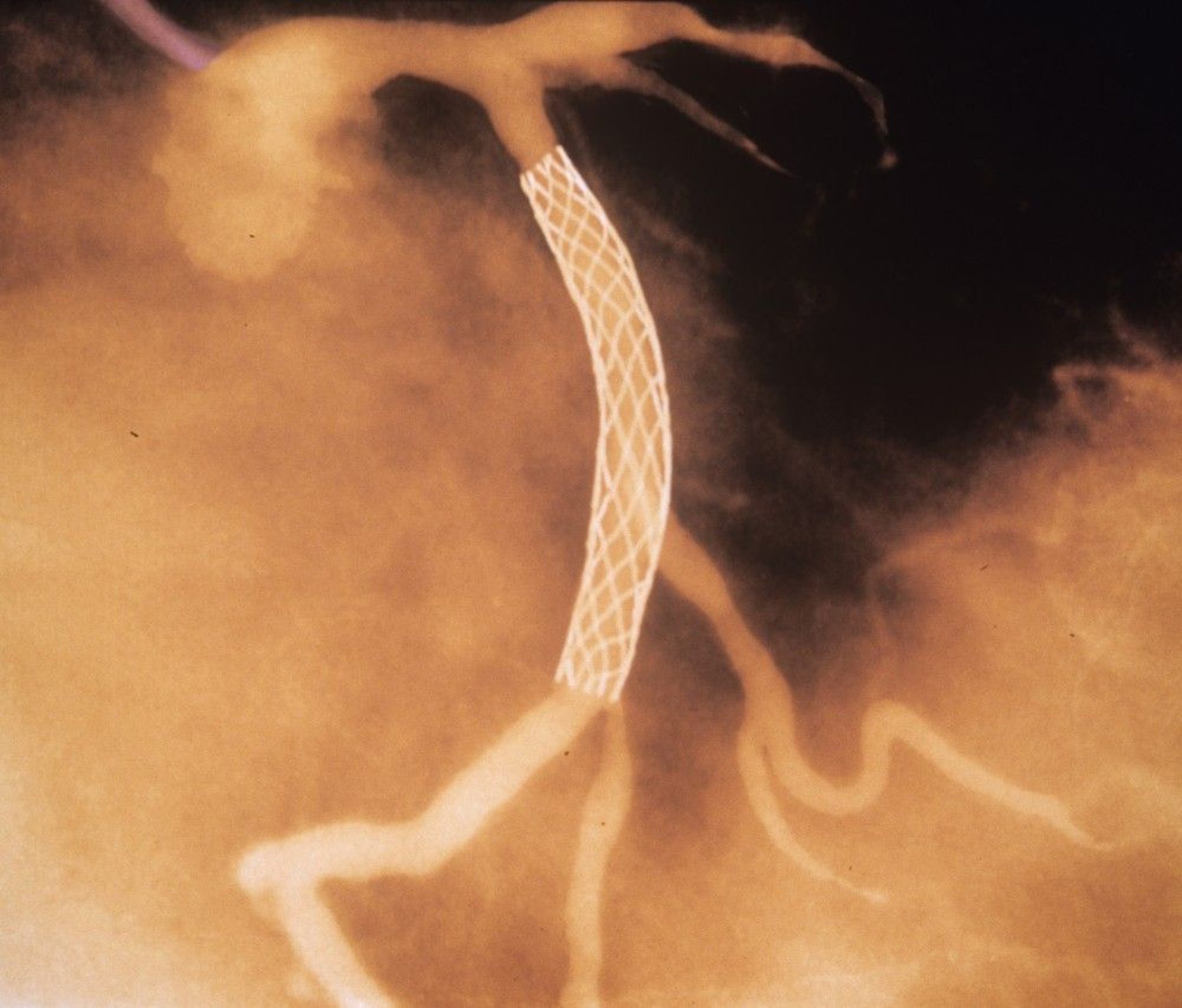
This colored coronary angiogram shows a stent (white mesh) after placement in a coronary artery.
GJLP/CNRI/SCIENCE PHOTO LIBRARY
Percutaneous transluminal angioplasty (PTA) is also used to treat peripheral arterial disease.
Procedure for PCI
PTCA is performed via percutaneous femoral, radial, or brachial artery puncture. The radial approach is preferred as it reduces patient discomfort, improves time to ambulation, and reduces the incidence of some complications (eg, bleeding, pseudoaneurysm formation) (1, 2).
A guiding catheter is inserted into a large peripheral artery and threaded to the appropriate coronary ostium. A balloon-tipped catheter, guided by fluoroscopy or intravascular ultrasound, is aligned within the stenosis, then inflated to disrupt the atherosclerotic plaque and thrombus and dilate the artery. A stent is usually placed. Angiography is repeated after the procedure to document any changes.
PCI procedure references
1. Rao SV, O'Donoghue ML, Ruel M, et al. 2025 ACC/AHA/ACEP/NAEMSP/SCAI Guideline for the Management of Patients With Acute Coronary Syndromes: A Report of the American College of Cardiology/American Heart Association Joint Committee on Clinical Practice Guidelines. Circulation. 2025;151(13):e771-e862. doi:10.1161/CIR.0000000000001309
2. Writing Committee Members, Lawton JS, Tamis-Holland JE, et al. 2021 ACC/AHA/SCAI Guideline for Coronary Artery Revascularization: A Report of the American College of Cardiology/American Heart Association Joint Committee on Clinical Practice Guidelines. J Am Coll Cardiol. 2022;79(2):e21-e129. doi:10.1016/j.jacc.2021.09.006
Stents for PCI
Stents for coronary arteries are expandable wire mesh cylinders that help hold stenotic areas open, improve luminal gain, and prevent abrupt vessel closure during PTCA.
Stents may be used for treatment of acute myocardial infarction, ostial or left main disease, chronic total occlusions, and bifurcation lesions.
Types of stents
Bare-metal stents (BMS) are made of nickel-titanium alloy, and their use is limited (1). Drug-eluting stents (DES) have medications (eg, first-generation: sirolimus, paclitaxel; second-generation: everolimus, ridaforolimus, zotarolimus) bonded to the metal that limit neointimal proliferation to reduce the risk of restenosis. Third-generation stents may be coated with biodegradable polymers or be polymer free. Third-generation stents may improve endothelialization. Completely biodegradable stents are being developed, and initial short-term outcomes have shown promise, but their use is currently limited to clinical trials (2).
Stent-free approaches using drug-eluting balloons may also be used for some small vessel lesions.
Stent references
1. Colombo A, Giannini F, Briguori C. Should We Still Have Bare-Metal Stents Available in Our Catheterization Laboratory? J Am Coll Cardiol. 2017;70(5):607-619. doi:10.1016/j.jacc.2017.05.057
2. Iglesias JF, Muller O, Heg D, et al. Biodegradable polymer sirolimus-eluting stents versus durable polymer everolimus-eluting stents in patients with ST-segment elevation myocardial infarction (BIOSTEMI): a single-blind, prospective, randomised superiority trial. Lancet 2019;394(10205):1243-1253. doi:10.1016/S0140-6736(19)31877-X
Anticoagulation and Antiplatelet Therapy
Various anticoagulation and antiplatelet regimens are used during and after PCI to reduce the incidence of thrombosis at the site of balloon dilation and stent placement.
Anticoagulation is usually initiated with unfractionated heparin (1). Enoxaparin and bivalirudin are alternatives. Bivalirudin or argatroban should be used in place of unfractionated heparin in patients with heparin-induced thrombocytopenia. In patients undergoing PCI, dual antiplatelet treatment with P2Y12 inhibitors (clopidogrel, prasugrel, ticagrelor) and aspirin is started at the time of the procedure. In patients with unstable non-ST-segment elevation myocardial infarction, glycoprotein IIb/IIIa inhibitors (abciximab, eptifibatide, tirofiban) may be added periprocedurally.
P2Y12 inhibitors (often in combination with aspirin) are usually continued for at least 6 to 12 months after PCI to decrease the risk of in-stent thrombosis until endothelialization of the stent has occurred. Dual antiplatelet treatment may be of shorter duration (1 or 3 months) when newer generation stents are used in certain clinical scenarios (eg, in patients with a high risk of bleeding and/or low risk of recurrent ischemia); however, the 12-month duration is still the default recommendation in patients with ACS (2). If dual antiplatelet therapy is continued for 12 months, prasugrel or ticagrelor may be de-escalated to clopidogrel, a less potent P2Y12 inhibitor that confers less bleeding risk.
For patients who require anticoagulation for another reason (eg, atrial fibrillation), direct-acting oral anticoagulants (apixaban, dabigatran, edoxaban, or rivaroxaban) are preferred long-term over warfarin, unless there is a contraindication to them (3, 4). For most patients requiring anticoagulation, triple therapy with oral anticoagulation, a P2Y12 inhibitor, and aspirin is stopped within 1 week to 1 month after intervention, and patients are continued on oral anticoagulation and a P2Y12 inhibitor for 6 months to 1 year (1). Calcium channel blockers and nitrates may also be given to reduce risk of coronary spasm.
Anticoagulation references
1. Rao SV, O'Donoghue ML, Ruel M, et al. 2025 ACC/AHA/ACEP/NAEMSP/SCAI Guideline for the Management of Patients With Acute Coronary Syndromes: A Report of the American College of Cardiology/American Heart Association Joint Committee on Clinical Practice Guidelines. Circulation. 2025;151(13):e771-e862. doi:10.1161/CIR.0000000000001309
2. Byrne RA, Rossello X, Coughlan JJ, et al. 2023 ESC Guidelines for the management of acute coronary syndromes. Eur Heart J. 2023;44(38):3720-3826. doi:10.1093/eurheartj/ehad191
3. Berwanger O, Wojdyla DM, Fanaroff AC, et al. Antithrombotic Strategies in Atrial Fibrillation After ACS and/or PCI: A 4-Way Comparison From AUGUSTUS. J Am Coll Cardiol. 2024;84(10):875-885. doi:10.1016/j.jacc.2024.06.022
4. Rodriguez F, Harrington RA. Management of Antithrombotic Therapy after Acute Coronary Syndromes. N Engl J Med. 2021;384(5):452-460. doi:10.1056/NEJMra1607714
Contraindications to PCI
Relative contraindications to PCI include:
Coagulopathy or other condition leading to high bleeding risk
Hypercoagulable states
Advanced kidney disease or acute kidney injury
Limited life expectancy (< 1 to 2 years)
Advanced dementia
Patient preference
A single diseased vessel providing all perfusion to the myocardium
Critical left main coronary stenosis without collateral flow from a native vessel or previous bypass graft to the left anterior descending artery
Diffusely diseased vessels without focal stenoses
Lack of cardiac surgical support
Stenosis < 50%
Total occlusion of a coronary artery
Although lack of cardiac surgical support is sometimes considered an absolute contraindication to PCI, many experts advocate that when revascularization is required urgently in STEMI, experienced operators in approved catheterization laboratories should proceed with PCI even if surgical backup is not available (1).
Although bypass is typically preferred for patients with critical left main coronary stenosis without collateral flow from either a native vessel or previous bypass graft (2), PCI is often and increasingly being used in this scenario in selected patients.
Contradictions references
1. Grines CL, Box LC, Mamas MA, et al. SCAI Expert Consensus Statement on Percutaneous Coronary Intervention Without On-Site Surgical Backup. J Soc Cardiovasc Angiogr Interv. 2023;2(2):100560. doi:10.1016/j.jscai.2022.100560
2. Holm NR, Mäkikallio T, Lindsay MM, et al. Percutaneous coronary angioplasty versus coronary artery bypass grafting in the treatment of unprotected left main stenosis: updated 5-year outcomes from the randomised, non-inferiority NOBLE trial. Lancet. 2020;395(10219):191-199. doi:10.1016/S0140-6736(19)32972-1
Complications of PCI
The main complications of PTCA with or without stent placement are:
Arterial dissection
Bleeding caused by adjunctive anticoagulation
Restenosis
Standard complications of cardiac catheterization and coronary angiography
Thrombosis and distal embolization
The overall in-hospital mortality rate after PCI is 0.5 to 1.9% (1, 2). Some, but not all, studies show higher overall long-term (5 to 10 year) mortality after PCI than coronary artery bypass grafting (CABG), especially in patients with complex or multivessel disease, although mortality rates are equivalent with left main disease and favor PCI in patients with diabetes (3, 4, 5). Risk of stroke is lower with PCI than with CABG, whereas risk of periprocedural myocardial infarction and need for a repeat revascularization procedure are generally higher with PCI (6, 7, 8).
PCI has a high risk of contrast-induced nephropathy relative to many other angiographic procedures (due to increased contrast load, urgency, severity of patient illness, and procedural time) (9, 10); this risk can be reduced by preprocedural hydration, and in patients with preexisting chronic kidney disease, possibly by use of a nonionic contrast agent or hemofiltration. Best practice also includes preprocedural identification of patients at risk of contrast-induced nephropathy and articulation of a maximal "desirable" volume of contrast to be targeted for administration.
Thrombosis
Stent thrombosis causes complete blockage and may occur at any time:
Acutely (immediately during or after the procedure)
Subacutely (within 30 days)
Late (> 30 days)
Very late ( > 1 year)
Stent thrombosis may be due to inadequate stent expansion or incomplete stent apposition at the time of the procedure, premature discontinuation of dual antiplatelet therapy (eg, due to nonadherence, need for noncardiac surgery), or both. Rarely, the stent may break up an intracoronary clot (ie, as may be present in acute myocardial infarction), which may embolize distally and cause myocardial infarction. Use of protection strategies (eg, temporarily blocking blood flow within the artery using a balloon and then aspirating the emboli, deploying a small filter distal to the site of PCI to capture emboli) may improve outcome in PCI performed on a previous saphenous vein graft but is not commonly done (11).
Use of stents has almost eliminated the need for emergency coronary artery bypass grafting following PCI; the rate of acute (0 to 24 hours) and subacute (24 hours to 30 days) thrombosis is < 0.5 to 1.3% and is higher in patients with ACS versus those undergoing PCI for stable ischemic heart disease (12, 13). With current drug-eluting stents and dual antiplatelet therapy, the risk of late (> 30 days) stent thrombosis is approximately 0.1% per year, affecting up to 2.5% of PCI patients overall (14).
With balloon angioplasty alone, risk of acute thrombosis is between 1 and 2% (15).
Restenosis
Restenosis is typically due to collagen deposition and thus does not occur until several weeks after the procedure or later; it may cause partial or, less commonly, complete vessel blockage.
With current drug-eluting stents and antiplatelet therapies, the rate of restenosis requiring revascularization is approximately 0.5 to 1% per year up to 10 years after PCI (14).
With balloon angioplasty alone, the risk of restenosis within months of PCI is approximately 20 to 30% (16).
Arterial dissection
The reported rate of catheter-induced coronary artery dissection during PCI varies widely, from < 0.1% to approximately 2% in several large series (17, 18, 19). This complication carries an increased risk of mortality. Insertion of another stent ("bailout stenting") often reopens the dissected segment; emergency CABG is a backup strategy.
Stroke
Complications references
1. Castro-Dominguez YS, Wang Y, Minges KE, et al. Predicting In-Hospital Mortality in Patients Undergoing Percutaneous Coronary Intervention. J Am Coll Cardiol. 2021;78(3):216-229. doi:10.1016/j.jacc.2021.04.067
2. Strepkos D, Alexandrou M, Mutlu D, et al. Death Following Percutaneous Coronary Intervention: Data From the PROGRESS-COMPLICATIONS Registry. Am J Cardiol. 2026:Feb 13:266:35-37. doi:10.1016/j.amjcard.2026.02.026
3. Fearon WF, Zimmermann FM, Ding VY, et al. Outcomes after fractional flow reserve-guided percutaneous coronary intervention versus coronary artery bypass grafting (FAME 3): 5-year follow-up of a multicentre, open-label, randomised trial. Lancet. 2025;405(10488):1481-1490. doi:10.1016/S0140-6736(25)00505-7
4. Head SJ, Milojevic M, Daemen J, et al. Mortality after coronary artery bypass grafting versus percutaneous coronary intervention with stenting for coronary artery disease: a pooled analysis of individual patient data. Lancet. 2018;391(10124):939-948. doi:10.1016/S0140-6736(18)30423-9
5. Kawczynski MJ, Gabrio A, Maessen JG, et al. Percutaneous coronary intervention with drug-eluting stents versus coronary bypass surgery for coronary artery disease: A Bayesian perspective. J Thorac Cardiovasc Surg. 2025;170(2):558-565.e10. doi:10.1016/j.jtcvs.2024.08.017
6. Head SJ, Milojevic M, Daemen J, et al. Stroke Rates Following Surgical Versus Percutaneous Coronary Revascularization. J Am Coll Cardiol. 2018;72(4):386-398. doi:10.1016/j.jacc.2018.04.071
7. Lee PH, Park H, Lee JS, Lee SW, Lee CW. Meta-Analysis Comparing the Risk of Myocardial Infarction Following Coronary Artery Bypass Grafting Versus Percutaneous Coronary Intervention in Patients With Multivessel or Left Main Coronary Artery Disease. Am J Cardiol. 2019;124(6):842-850. doi:10.1016/j.amjcard.2019.06.009
8. Palmerini T, Biondi-Zoccai G, Reggiani LB, et al. Risk of stroke with coronary artery bypass graft surgery compared with percutaneous coronary intervention. J Am Coll Cardiol. 2012;60(9):798-805. doi:10.1016/j.jacc.2011.10.912
9. Bagai J, Beavers CJ, Boudoulas KD. SCAI (Society for Cardiovascular Angiography and Interventions. Quality Initiatives for Prevention of Contrast-Induced Acute Kidney Injury. January 23, 2025. Accessed March 10, 2026.
10. Mehran R, Dangas GD, Weisbord SD. Contrast-Associated Acute Kidney Injury. N Engl J Med. 2019;380(22):2146-2155. doi:10.1056/NEJMra1805256
11. Writing Committee Members, Lawton JS, Tamis-Holland JE, et al. 2021 ACC/AHA/SCAI Guideline for Coronary Artery Revascularization: A Report of the American College of Cardiology/American Heart Association Joint Committee on Clinical Practice Guidelines. J Am Coll Cardiol. 2022;79(2):e21-e129. doi:10.1016/j.jacc.2021.09.006
12. Chau KH, Kirtane AJ, Easterwood RM, et al. Stent Thrombosis Risk Over Time on the Basis of Clinical Presentation and Platelet Reactivity: Analysis From ADAPT-DES. JACC Cardiovasc Interv. 2021;14(4):417-427. doi:10.1016/j.jcin.2020.12.005
13. Moreno R, Džavík V, Cairns J, et al. Stent thrombosis in the setting of ST-segment elevation acute myocardial infarction in the contemporary practice: results from the TOTAL randomized trial. Coron Artery Dis. 2025;36(2):126-138. doi:10.1097/MCA.0000000000001456
14. Gaudino M, Andreotti F, Kimura T. Current concepts in coronary artery revascularisation. Lancet. 2023;401(10388):1611-1628. doi:10.1016/S0140-6736(23)00459-2
15. Dangas G, Aymong ED, Mehran R, et al. Predictors of and outcomes of early thrombosis following balloon angioplasty versus primary stenting in acute myocardial infarction and usefulness of abciximab (the CADILLAC trial). Am J Cardiol. 2004;94(8):983-988. doi:10.1016/j.amjcard.2004.06.050
16. Byrne RA, Stone GW, Ormiston J, Kastrati A. Coronary balloon angioplasty, stents, and scaffolds. Lancet. 2017;390(10096):781-792. doi:10.1016/S0140-6736(17)31927-X
17. Hiraide T, Sawano M, Shiraishi Y, et al. Impact of catheter-induced iatrogenic coronary artery dissection with or without postprocedural flow impairment: A report from a Japanese multicenter percutaneous coronary intervention registry. PLoS One. 2018;13(9):e0204333. doi:10.1371/journal.pone.0204333
18. Kuno T, Ohata T, Nakamaru R, et al. Long-term outcomes of periprocedural coronary dissection and perforation for patients undergoing percutaneous coronary intervention in a Japanese multicenter registry. Sci Rep. 2023;13(1):20318. doi:10.1038/s41598-023-47444-7
19. Ramasamy A, Bajaj R, Jones DA, et al. Iatrogenic catheter-induced ostial coronary artery dissections: Prevalence, management, and mortality from a cohort of 55,968 patients over 10 years. Catheter Cardiovasc Interv. 2021;98(4):649-655. doi:10.1002/ccd.29382
Drug Information for the Topic