


An evaluation of the ankle includes a physical examination and sometimes arthrocentesis (see How To Do Ankle Arthrocentesis).
(See also Evaluation of the Patient With Joint Symptoms and Overview of Foot and Ankle Disorders.)
Physical Examination of the Ankle
The patient is observed walking, but only if there is no serious injury that could be aggravated or complicated by weight bearing.
The ankle is inspected for deformities, swelling, skin discoloration, muscle atrophy, and asymmetry with the opposite side. The lower leg muscles are inspected for atrophy.


Sensation to light touch is tested, at minimum, on the top of the first webbed space and the side of the foot. The dorsalis pedis pulse is palpated over the anterior foot, and the posterior tibial pulse is palpated behind the medial malleolus.
The ankle is gently felt for warmth and to detect subtle swelling. Comparison to the unaffected side is useful. Palpation for tenderness is done over the bones and then the major ligaments. Touching only the bone, and then only the ligament, can help distinguish bony from ligamentous injury.
Laterally, palpation includes the tip of the lateral malleolus, the fibula, and the three lateral ligaments: anterior talofibular, posterior talofibular, and fibulocalcaneal. Because an inversion injury of the ankle can fracture the proximal fibula, the proximal fibula is palpated. The base of the 5th metatarsal is also palpated.
Bones of the Foot and Ankle

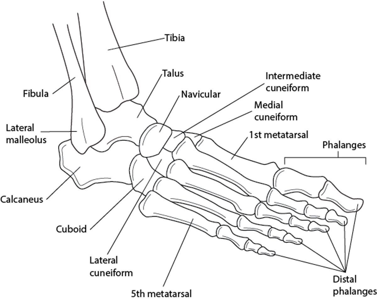
The dome of the talus is palpated if ankle swelling is severe and egg-shaped after an ankle injury.
Medially, palpation includes the tip of the medial malleolus, the tibia, navicular bone, and the medial deltoid ligament complex.
Passive range of motion is tested for dorsiflexion, plantar flexion, eversion (while securing the heel), and inversion (by rotating the heel inward). Active range of motion is tested for dorsiflexion, plantar flexion, and eversion.
After injury, if the patient’s symptoms permit, provocative testing for damage to ligaments and tendons may be done. Instability after ankle sprains, particularly lateral sprains, is assessed by the anterior drawer test. For this test, the examiner stabilizes the patient's lower leg with one hand, puts the other hand under the patient's foot and cups the heel, and pulls the heel anteriorly. If the ligaments are intact, there should be no laxity with anterior stress. To check for tears of the Achilles tendon, the Thompson test is done. For this test, the examiner squeezes the calf muscles while the patient is prone. Absence of the normal plantar flexion with this maneuver suggests a complete or functionally significant tear.


