

Intracranial tumors may involve the brain or other structures (eg, cranial nerves, meninges). The tumors usually develop during early or middle adulthood but may develop at any age; they are becoming more common among older adults. Brain tumors are present in approximately 20 to 30/100,000 adults in the United States (there are fewer in children); mortality from brain tumors is approximately 5 to 6/100,000 adults (1).
Some tumors are benign, but because the cranial vault allows no room for expansion, even benign tumors can cause serious neurologic dysfunction or death.
(See also Overview of Central Nervous System Tumors in Children.)
Reference
1. Ostrom QT, Gittleman H, Fulop J, et al: CBTRUS statistical report: Primary brain and central nervous system tumors diagnosed in the United States in 2008-2012. Neuro Oncol 17 Suppl 4(Suppl 4):iv1-iv62, 2015. doi: 10.1093/neuonc/nov189
Classification of Intracranial Tumors
There are 2 types of brain tumors:
Primary brain tumors: Originate in the brain in the brain parenchyma (eg, gliomas, which include astrocytomas, oligodendrogliomas, and ependymomas; medulloblastomas; primary central nervous system [CNS] lymphomas) or in extraneural structures (eg, meningiomas, vestibular schwannomas)
Secondary brain tumors (brain metastases): Originate in tissues outside the brain and spread to the brain
Brain metastases are about 10 times more common than primary tumors (1).
Pearls & Pitfalls
|

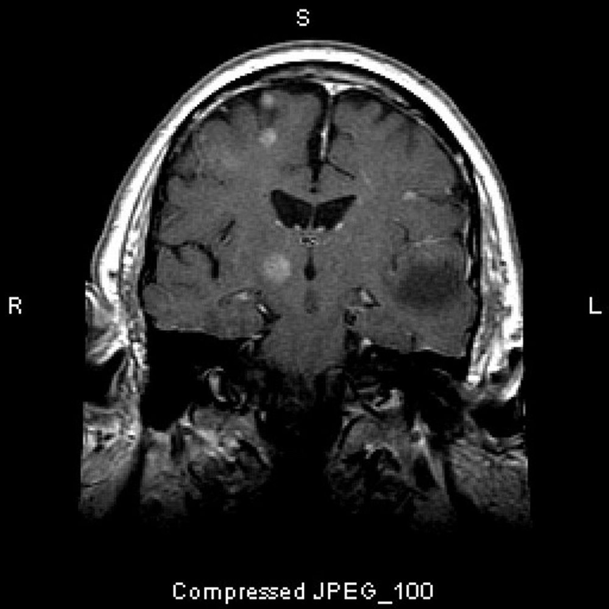
This MRI scan shows multiple brain lesions, representing metastatic tumors. Over 80% of brain metastases are multiple.
Image courtesy of William R. Shapiro, MD.
Type of tumor varies somewhat by site (see table ) and patient age (see table ).
Common Localizing Manifestations of Primary Brain Tumors
Tumor Site | Findings | Common Primary Tumor Types* |
|---|---|---|
Anterior corpus callosum | Cognitive impairment | Astrocytoma (including low-grade gliomas) |
Basal ganglia | Hemiparesis (contralateral), movement disorders | Astrocytoma (including low-grade gliomas) Primary CNS lymphoma |
Brain stem | Unilateral or bilateral motor or sensory loss, cranial nerve deficits (eg, gaze palsies, hearing loss, vertigo, palatal paresis, facial weakness), ataxia, intention tremor, nystagmus | Astrocytoma (including juvenile pilocytic astrocytoma) Diffuse pontine glioma |
Cerebellopontine angle | Tinnitus and hearing loss (both ipsilateral), vertigo, loss of vestibular response to caloric stimulation If tumor is large, ataxia, loss of facial sensation and facial weakness (both ipsilateral), possibly other cranial nerve or brain stem deficits | |
Cerebellum | Ataxia, nystagmus, tremor, hydrocephalus with suddenly increased intracranial pressure | Astrocytoma (including juvenile pilocytic astrocytoma) |
2nd cranial (optic) nerve | Loss of vision | Astrocytoma (including pilocytic astrocytomas and lower-grade gliomas; optic nerve location most common in neurofibromatosis) |
5th cranial (trigeminal) nerve | Facial pain, loss of sensation, jaw weakness | Meningioma Schwannoma |
Frontal lobe | Generalized or focal (contralateral) seizures, gait disorders, urinary urgency or incontinence, impaired attention and cognition and apathy (particularly if tumor is bilateral), hemiparesis Expressive aphasia if tumor is in dominant hemisphere Anosmia if tumor is at base of lobe | Astrocytoma Glioblastoma Oligodendroglioma |
Hypothalamus | Eating and drinking disorders (eg, polydipsia), precocious puberty (especially in boys), hypothermia | Astrocytoma |
Occipital lobe | Generalized seizures with visual aura, visual hallucinations, hemianopia or quadrantanopia (contralateral) | Astrocytoma Glioblastoma Oligodendroglioma |
Parietal lobe | Deficits in position sensation and in 2-point discrimination (contralateral), anosognosia (no recognition of bodily defects), denial of illness, hemianopia (contralateral), generalized or focal seizures, inability to perceive (extinguishing of) a contralateral stimulus when stimuli are applied to both sides of the body (called double simultaneous stimulation) Receptive aphasia if tumor is in dominant hemisphere | Astrocytoma Glioblastoma Oligodendroglioma |
Paresis of upward gaze, ptosis, loss of pupillary light and accommodation reflexes, sometimes hydrocephalus with suddenly increased intracranial pressure | Germ cell tumor Pineocytoma (rare) | |
Pituitary or suprasellar region | Endocrinopathies, monocular visual loss, headache without increased intracranial pressure, bitemporal hemianopia | Craniopharyngioma Pituitary carcinoma (rare) |
Temporal lobe | Complex partial seizures, generalized seizures with or without aura, hemianopia (contralateral), mixed expressive and receptive aphasia or anomia | Astrocytoma Glioblastoma Oligodendroglioma |
Thalamus | Sensory impairment (contralateral) | Astrocytoma |
* Similar manifestations may result from brain parenchymal metastases or from tumors around the dura (eg, metastatic tumors; meningeal tumors such as meningiomas, sarcomas, or gliomas) or skull lesions (eg, granulomas, hemangiomas, osteitis deformans, osteomas, xanthomas) that compress the underlying brain. | ||
CNS = central nervous system. | ||
Common Brain Tumors by Age
Age Group | Primary | Metastases |
|---|---|---|
Children | Cerebellar astrocytomas, juvenile pilocytic astrocytomas, and medulloblastomas Primitive neuroectodermal tumors and neurocytoma Gliomas of the brain stem or optic nerve Germinomas Congenital tumors* | Neuroblastoma (usually epidural) Leukemia (meningeal) |
Adults | Schwannomas Primary central nervous system lymphomas Gliomas of the cerebral hemispheres, particularly glioblastoma, anaplastic astrocytoma, low-grade astrocytoma, pilocytic astrocytoma, and oligodendroglioma | Adenocarcinoma of the lung, breast, colon, thyroid, or kidneys Malignant melanoma Metastatic lymphoma |
* Congenital tumors include craniopharyngiomas, chordomas, germinomas, teratomas, dermoid cysts, angiomas, and hemangioblastomas. | ||
Classification reference
1. Lin X, DeAngelis LM: Treatment of brain metastases. J Clin Oncol 33(30):3475-3484, 2015. doi: 10.1200/JCO.2015.60.9503
Pathophysiology of Intracranial Tumors
Neurologic dysfunction may result from the following:
Invasion and destruction of brain tissue by the tumor
Direct compression of adjacent tissue by the tumor
Increased intracranial pressure (because the tumor occupies space within the skull)
Bleeding within or outside the tumor
Cerebral edema
Obstruction of dural venous sinuses (especially by bone or extradural metastatic tumors)
Obstruction of cerebrospinal fluid (CSF) drainage (occurring early with third-ventricle or posterior fossa tumors)
Obstruction of CSF absorption (eg, when leukemia or carcinoma involves the meninges)
Obstruction of arterial flow
Rarely, paraneoplastic syndromes
A malignant tumor can develop new internal blood vessels, which can bleed or become occluded, resulting in necrosis and neurologic dysfunction that mimics stroke. Bleeding as a complication of metastatic tumors is most likely to occur in patients with melanoma, renal cell carcinoma, choriocarcinoma, or thyroid, lung, or breast cancer.
Benign tumors grow slowly. They may become quite large before causing symptoms, partly because often there is no cerebral edema. Malignant primary tumors grow rapidly but rarely spread beyond the CNS. Death results from local tumor growth and/or tumor-related hemorrhage and thus can result from benign as well as malignant tumors.
Symptoms and Signs of Intracranial Tumors
Symptoms caused by primary tumors and metastatic tumors are the same. Many symptoms result from increased intracranial pressure:
Headache
Deterioration in mental status
Focal brain dysfunction
Headache is the most common symptom. Headache may be most intense when patients awake from deep nonrapid eye movement (non-REM) sleep (usually several hours after falling asleep) because hypoventilation, which increases cerebral blood flow and thus intracranial pressure, is usually maximal during non-REM sleep. Headache is also progressive and may be worsened by recumbency or the Valsalva maneuver. When intracranial pressure is very high, the headache may be accompanied by vomiting, sometimes with little nausea preceding it.
Papilledema develops in approximately 25 to 35% of patients with a brain tumor (1) but may be absent even when intracranial pressure is increased. In infants and very young children, increased intracranial pressure may enlarge the head. If intracranial pressure increases sufficiently, brain herniation occurs.
Deterioration in mental status is the second most common symptom. Manifestations include drowsiness, lethargy, personality changes, disordered conduct, and impaired cognition, particularly with malignant brain tumors. Airway reflexes may be impaired.
Focal brain dysfunction causes some symptoms. Focal neurologic deficits, endocrine dysfunction, or focal seizures (sometimes with secondary generalization) may develop depending on the tumor’s location (see table ). Focal deficits often suggest the tumor’s location. However, sometimes focal deficits do not correspond to the tumor’s location. Such deficits, called false localizing signs, include the following:
Unilateral or bilateral lateral rectus palsy (with paresis of eye abduction) due to increased intracranial pressure compressing the 6th cranial nerve
Ipsilateral hemiplegia due to compression of the contralateral cerebral peduncle against the tentorium (Kernohan notch)
Ipsilateral visual field defect due to ischemia in the contralateral occipital lobe
Generalized seizures may occur, more often with primary than metastatic brain tumors. Impaired consciousness can result from herniation, brain stem dysfunction, or diffuse bilateral cortical dysfunction.
Some tumors cause meningeal inflammation, resulting in subacute or chronic meningitis.
Symptoms and signs reference
1. Serova N, Eliseeva N, Shifrin: Papilloedema in patients with brain tumour. Neuro-ophthalmology 33(3):100-105, 2009. doi.org/10.1080/01658100902930545
Diagnosis of Intracranial Tumors
T1-weighted MRI with gadolinium or CT with contrast
Sometimes biopsy
Early-stage brain tumors are often misdiagnosed. A brain tumor should be considered in patients with any of the following:
Progressive focal or global deficits of brain function
New-onset seizures
Persistent, unexplained, recent-onset headaches, particularly if worsened by sleep
Evidence of increased intracranial pressure (eg, papilledema, unexplained vomiting)
Pituitary or hypothalamic endocrinopathy
Similar findings can result from other intracranial masses (eg, abscess, aneurysm, arteriovenous malformation, intracerebral hemorrhage, subdural hematoma, granuloma, parasitic cysts such as neurocysticercosis) or ischemic stroke.
A complete neurologic examination, neuroimaging, and chest radiographs (for a source of metastases) should be performed. T1-weighted MRI with gadolinium is the study of choice. CT with contrast agent is an alternative. MRI usually detects low-grade astrocytomas and oligodendrogliomas earlier than CT and shows brain structures near bone (eg, the posterior fossa) more clearly. If whole-brain imaging does not show sufficient detail in the target area (eg, sella turcica, cerebellopontine angle, optic nerve), closely spaced images or other special views of the area are obtained. If neuroimaging is normal but increased intracranial pressure is suspected, idiopathic intracranial hypertension should be considered and lumbar puncture done.
Clues to the type of tumor, mainly clinically suspected location (see table ) and pattern of enhancement on MRI, may be inconclusive; brain biopsy, sometimes excisional biopsy, may be required.
Specialized tests (eg, molecular and genetic tumor markers in blood and cerebrospinal fluid [CSF]) can help in some cases. In patients with end-stage HIV infection, Epstein-Barr virus titers in CSF typically increase as CNS lymphoma develops.
Treatment of Intracranial Tumors
Airway protection
Dexamethasone for increased intracranial pressureDexamethasone for increased intracranial pressure
Mannitol for herniationMannitol for herniation
Antiseizure medications for seizures
Definitive therapy with excision, radiation therapy, systemic cancer therapy (eg, chemotherapy, targeted therapy, immunotherapy), or a combination
Patients in a coma or with impaired airway reflexes require endotracheal intubation.
Brain herniation due to tumors is treated with hyperosmotic therapy (mannitol 25 to 100 g infused IV or 23.4 % hypertonic saline 20 mL infused through a central line and a corticosteroid [eg, dexamethasone 16 mg IV, followed by 4 mg orally or IV every 6 hours]) plus endotracheal intubation. Hyperventilation to a carbon dioxide partial pressure (PCO2) of 26 to 30 mm Hg can help decrease intracranial pressure temporarily in emergencies. Mass lesions should be surgically decompressed as soon as possible.due to tumors is treated with hyperosmotic therapy (mannitol 25 to 100 g infused IV or 23.4 % hypertonic saline 20 mL infused through a central line and a corticosteroid [eg, dexamethasone 16 mg IV, followed by 4 mg orally or IV every 6 hours]) plus endotracheal intubation. Hyperventilation to a carbon dioxide partial pressure (PCO2) of 26 to 30 mm Hg can help decrease intracranial pressure temporarily in emergencies. Mass lesions should be surgically decompressed as soon as possible.
Increased intracranial pressure due to tumors but without herniation can be treated with corticosteroids (eg, dexamethasone 4 mg orally every 6 to 12 hours or prednisone 30 to 40 mg orally twice a day).Increased intracranial pressure due to tumors but without herniation can be treated with corticosteroids (eg, dexamethasone 4 mg orally every 6 to 12 hours or prednisone 30 to 40 mg orally twice a day).
Treatment of the brain tumor depends on pathology and location. Surgical excision should be used for diagnosis (excisional biopsy) and symptom relief. It may cure benign tumors. For tumors infiltrating the brain parenchyma, treatment is multimodal. Radiation therapy is required, and chemotherapy, targeted therapy, and/or immunotherapy appears to benefit some patients.
Treatment of metastatic tumors includes radiation therapy, which can be delivered as whole-brain or conformal stereotactic radiosurgery (1). For patients with a single metastasis, surgical excision of the tumor before radiation therapy improves outcome.
End-of-life issues
If patients have an incurable tumor, end-of-life issues should be discussed, and palliative care consultation should be considered.
Cranial Radiation Therapy and Neurotoxicity
Radiation therapy may be directed at the whole head for diffuse or multicentric tumors or locally for well-demarcated tumors (1).
There are 2 types of localized brain radiation therapy; both aim to spare normal brain tissue:
Conformal: Using CT to create a 3-dimensional map of the tumor facilitates precise targeting of the tumor
Stereotactic: Using gamma knife or proton beam therapy to deliver multiple focused beams of high energy to the tumor
Gliomas are treated with conformal radiation therapy; a stereotactically directed gamma knife or proton beam therapy is useful for metastases. Current recommendations are to treat ≤ 4 metastatic lesions with stereotactic or other focal radiation interventions and to treat > 4 lesions with whole-brain radiation therapy (2, 3); however, more recent data may support stereotactic surgery for up to 10 metastatic lesions (4, 5). Giving radiation in smaller fractionated daily doses tends to maximize efficacy while minimizing neurotoxicity and damage to normal CNS tissue (see Radiation Exposure and Contamination).
Degree of neurotoxicity depends on
Cumulative radiation dose
Individual dose size
Duration of therapy
Volume of tissue irradiated
Individual susceptibility
Because susceptibility varies, prediction of radiation neurotoxicity is imprecise. Symptoms can develop in the first few days (acute) or months of treatment (early-delayed) or several months to years after treatment (late-delayed). Rarely, radiation causes gliomas, meningiomas, or peripheral nerve sheath tumors years after therapy.
Acute radiation neurotoxicity
Typically, acute neurotoxicity involves headache, nausea, vomiting, somnolence, and sometimes worsening focal neurologic signs in children and adults.
Acute neurotoxicity largely results from transient swelling and edema; thus, it is particularly likely if intracranial pressure is already high. Using corticosteroids to lower intracranial pressure can prevent or treat acute toxicity. Acute toxicity lessens with subsequent treatments.
Early-delayed neurotoxicity
In children or adults, early-delayed neurotoxicity can cause encephalopathy, which must be distinguished by MRI or CT from worsening or recurrent brain tumor. It may occur in children who have received prophylactic whole-brain radiation therapy for leukemia; they may develop somnolence, which lessens spontaneously over several days to weeks, possibly more rapidly if corticosteroids are used.
After radiation therapy to the neck or upper thorax, early-delayed neurotoxicity can result in a myelopathy, characterized by spinal symptoms such as Lhermitte sign (an electric shocklike sensation radiating down the back and into the legs when the neck is flexed). This early-delayed myelopathy typically resolves spontaneously.
Late-delayed neurotoxicity
After diffuse or whole-brain radiation therapy, many children and adults develop late-delayed neurotoxicity if they survive long enough. The most common cause in children is diffuse therapy given to prevent leukemia or to treat medulloblastoma. After diffuse therapy, the most common symptom is progressive dementia; adults may also develop an unsteady gait and focal neurologic symptoms. MRI or CT can show cerebral atrophy and often white matter loss.
After localized therapy, neurotoxicity more often involves focal neurologic deficits.
MRI or CT shows a mass that may be enhanced by contrast agent and that may be difficult to distinguish from recurrence of the primary tumor. Excisional biopsy of the mass is diagnostic and often ameliorates symptoms.
Late-delayed myelopathy can develop after radiation therapy for extraspinal tumors (eg, due to Hodgkin lymphoma). It is characterized by progressive paresis and sensory loss, often as a Brown-Séquard syndrome (ipsilateral paresis and proprioceptive sensory loss, with contralateral loss of pain and temperature sensation). Most patients eventually become paraplegic.
Treatment references
1. Gondi V, Bauman G, Bradfield L, et al: Radiation therapy for brain metastases: An ASTRO clinical practice guidelines. Pract Radiation Oncol 12(4):265–282, 2022. https://doi.org/10.1016/j.prro.2022.02.003
2. Gaspar L, Prabhu R, Hdeib A, et al: Congress of Neurological Surgeons systematic review and evidence-based guidelines on the role of whole brain radiation therapy in adults with newly diagnosed metastatic brain tumors. Neurosurgery 84 (3):E159–E162, 2019. doi: 10.1093/neuros/nyy541
3. Vogelbaum MA, Brown PD, Messersmith H, et al: Treatment for brain metastases: ASCO-SNO-Astro guideline. J Clin Oncol 40 (5):492–516, 2022. doi: 10.1200/JCO.21.02314
4. Long GV, Atkinson V, Lo S, et al: Combination nivolumab and ipilimumab or nivolumab alone in melanoma brain metastases: A multicentre randomised phase 2 study. : Combination nivolumab and ipilimumab or nivolumab alone in melanoma brain metastases: A multicentre randomised phase 2 study.Lancet Oncol 19 (5):672–681, 2018. doi: 10.1016/S1470-2045(18)30139-6 Epub 2018 Mar 27.
5. Moss NS, Tosi U, Santomasso BD, et al: Multifocal and pathologically-confirmed brain metastasis complete response to trastuzumab deruxtecan. : Multifocal and pathologically-confirmed brain metastasis complete response to trastuzumab deruxtecan.CNS Oncol 11 (3):CNS90, 2022. doi: 10.2217/cns-2022-0010
Drug Information for the Topic


