


A variety of tests can be done to confirm an eye problem or to determine the extent or severity of an eye disorder. Each eye is tested separately.
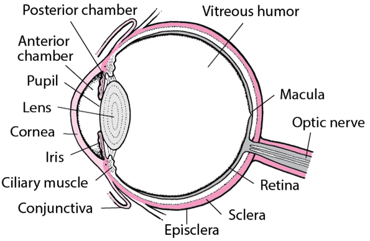
An Inside Look at the Eye

Angiography
In general, angiography involves injecting dye into blood vessels to make them more visible on imaging tests. Angiography of the eye, however, uses dye to make blood vessels more visible when doctors directly examine or photograph them.
Fluorescein angiography allows a doctor to clearly see the blood vessels at the back of the eye. A fluorescent dye, which is visible in blue light, is injected into a vein in the person's arm. The dye circulates through the person's bloodstream, including the blood vessels in the retina. Shortly after the dye is injected, a rapid sequence of photographs is taken of the retina, choroid, optic disk, iris, or a combination. The dye inside the blood vessels fluoresces, making the vessels stand out.
Fluorescein angiography is particularly useful in the diagnosis of macular degeneration, blocked retinal blood vessels, and diabetic retinopathy. This type of angiography is also used to assess people who may need laser procedures done on the retina.
Indocyanine green angiography allows doctors to see the blood vessels of the retina and choroid. As in fluorescein angiography, a fluorescent dye is injected into a vein. This type of angiography gives doctors more detail of the blood vessels of the choroid than fluorescein angiography. Indocyanine green angiography is used to show macular degeneration and detect the development of new blood vessels in the eye.
Electroretinography
Electroretinography allows a doctor to examine the function of the light-sensing cells (photoreceptors) in the retina by measuring the response of the retina to flashes of light. Eye drops numb the eye and dilate the pupil. A recording electrode in the form of a contact lens is then placed on the cornea, and another electrode is placed on the skin of the face nearby. The eyes are then propped open. The room is darkened, and the person stares at a flashing light. The electrical activity generated by the retina in response to the flashes of light is recorded by the electrodes.
Electroretinography is particularly useful for evaluating diseases, such as retinitis pigmentosa, in which the photoreceptors are affected.
Ultrasound
The eye can be examined by ultrasound. A probe is placed gently against the closed eyelid and painlessly bounces sound waves off the eyeball. The reflected sound waves produce a two-dimensional image of the inside of the eye.
Ultrasound is useful when an ophthalmoscope or slit lamp cannot view the retina because the inside of the eye is cloudy or something is blocking the line of sight. Ultrasound can be used to determine the nature of abnormal structures, such as a tumor or retinal detachment. Ultrasound can also be used to examine blood vessels supplying the eye (Doppler ultrasound) and to determine the thickness of the cornea (pachymetry).
Pachymetry
Pachymetry (measuring the thickness of the cornea) is very important in refractive eye surgery, such as laser in situ keratomileusis (LASIK).
Pachymetry is usually done by using ultrasound. For ultrasound pachymetry, the eye is numbed with drops, and an ultrasound probe is placed gently onto the surface of the cornea. In contrast, optical pachymetry does not require numbing eye drops because the instruments do not touch the eye.
Optical Coherence Tomography
Optical coherence tomography (OCT) provides high-resolution images of structures at the back of the eye, such as the optic nerve, retina, choroid, and vitreous humor. OCT can be used to identify swelling of the retina. OCT is similar to ultrasound but uses light instead of sound.
Doctors use OCT to view disorders of the retina, including macular degeneration, disorders that can cause new blood vessels to develop in the eye, and glaucoma.
Computed Tomography (CT) and Magnetic Resonance Imaging (MRI)
Computed tomography and magnetic resonance imaging can be used to provide detailed information about the structures inside the eye and the bony structure that surrounds the eye (the orbit). These techniques are used to evaluate eye injuries such as a suspected foreign object in the eye, tumors of the orbit and optic nerve, and optic neuritis.
Drug Information for the Topic


