


Tumors, both noncancerous and cancerous, within the bile ducts or gallbladder are rare.
Ultrasound, magnetic resonance imaging (MRI), or magnetic resonance cholangiopancreatography (MRCP) can usually detect a tumor in the bile ducts or gallbladder.
These cancers are usually fatal, but symptoms can be treated.
Bile is a fluid that is produced by the liver and aids in digestion. Bile is transported through small tubes (bile ducts) that carry bile through the liver and then from the liver to the gallbladder and to the small intestine. The gallbladder is a small, pear-shaped sac located beneath the liver that stores bile and releases it when needed, such as when people eat. (See also Overview of Gallbladder and Bile Duct Disorders and figure .)
Cancer of the bile ducts (cholangiocarcinoma) is rare. It can originate anywhere in the bile ducts, particularly in the bile ducts located just outside of the liver. Having primary sclerosing cholangitis, liver flukes, viral hepatitis, cirrhosis, consuming alcohol, or having a cyst in the bile duct (choledochal cyst) all increase the risk of developing this cancer.
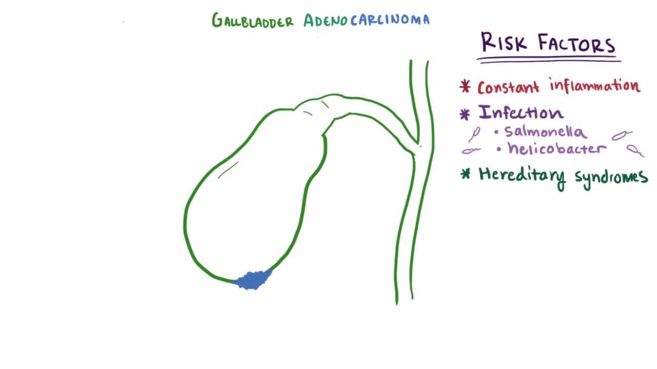
Cancer of the gallbladder is also rare. Nearly everyone with gallbladder cancer has gallstones. Many people live only a few months after this cancer develops. This cancer is more common among older adults, women, people with gallstones, American Indians, and probably in people with a extensive scarring of the gallbladder, which can occur in severe chronic cholecystitis.

Gallbladder polyps, which are noncancerous (benign) outgrowths of tissue, may develop in the gallbladder. They rarely cause symptoms or require treatment. They are found in about 5% of people during ultrasound. Surgery may be needed to remove larger polyps.
Symptoms of Bile Duct and Gallbladder Tumors
Early symptoms include the following:
Worsening jaundice (yellowish discoloration of the skin and whites of the eyes)
Abdominal discomfort
Loss of appetite
Weight loss
Itchiness
Sometimes cancers and noncancerous tumors can block the flow of bile (although most blockages are caused by gallstones). Even less often, cancer can spread (metastasize) from elsewhere in the body to adjacent structures or nearby lymph nodes, causing blockage.
Symptoms of bile duct cancer gradually worsen. Abdominal pain may become increasingly severe and constant. The pain is usually caused by blockage of the bile ducts. The stool may become pale. People feel tired and uncomfortable. They may feel a mass in their abdomen.
Symptoms of gallbladder cancer, if present, may include pain, weight loss, a mass in the abdomen, or jaundice.
Most gallbladder polyps cause no symptoms.


This photo shows yellowing of the eyes and skin (jaundice).
DR P. MARAZZI/SCIENCE PHOTO LIBRARY
Diagnosis of Bile Duct and Gallbladder Tumors
Sometimes blood tests
Ultrasound, followed by magnetic resonance cholangiopancreatography (MRCP) or CT cholangiography
Sometimes endoscopic retrograde cholangiopancreatography (ERCP) or taking a tissue sample (biopsy)
Doctors suspect bile duct or gallbladder cancer when a bile duct is blocked and no other cause is identified. Bile duct cancer is suspected especially in people with primary sclerosing cholangitis (PSC). If people have PSC, blood tests to measure substances secreted by tumors (tumor markers) are done periodically to check for this cancer.
The diagnosis is confirmed by imaging tests. Usually, ultrasound is done first. Sometimes computed tomography (CT) or magnetic resonance imaging (MRI) are done to provide additional information, especially if gallbladder cancer is suspected. Magnetic resonance cholangiopancreatography (MRCP) or CT cholangiography (CT of the bile ducts done after a radiopaque contrast agent is injected into a vein) is usually the next step.
If results of the initial imaging tests are unclear, or if bile duct cancer is suspected endoscopic retrograde cholangiopancreatography (ERCP) is done. In this procedure, a viewing tube (endoscope) is inserted through the mouth and into the small intestine. A thin tube (catheter) is inserted through the endoscope, and a radiopaque contrast agent, which is visible on x-rays, is injected through the catheter into the bile ducts. Then x-rays are taken to detect any abnormalities. This procedure enables doctors to obtain images and a tissue sample for examination under a microscope (see figure ).
If these tests suggest a tumor but are not conclusive, doctors may take a tissue sample by inserting a thin needle through the skin into the area thought to be abnormal. Ultrasound or computed tomography (CT) is used to guide the needle.
To determine how extensive the cancer is, doctors may be able to use a CT scan, but often have to do surgery to directly examine the area (a procedure called a diagnostic laparoscopy or an open laparotomy).
Treatment of Bile Duct and Gallbladder Tumors
Sometimes surgery to remove tumor
Insertion of stents into blocked bile ducts
Most bile duct and gallbladder cancers are fatal, but treatment can help control symptoms.
Surgery to remove a cancer of the bile duct may be done, but usually the tumor cannot be completely removed. Chemotherapy, either before or after surgery, may be used to treat or shrink areas of cancer that were not removed. If tumors have spread from other parts of the body (metastasized), chemotherapy may provide some symptom relief but does not dramatically improve survival.
If surgery to remove the bile duct cancer is not possible, stents may be passed through an endoscope (a viewing tube) and placed in bile ducts that are blocked by the cancer. These stents allow bile to flow past the blockage and can improve jaundice, and prevent recurrent infections. If bile duct cancer is limited to the bottom of the liver (where the bile ducts outside of the liver meet the bile ducts inside the liver), a liver transplant may be a curative option.
Very early gallbladder cancer that is found during surgery for gallstones can often be cured by removing the gallbladder. Treatment of other gallbladder cancers may involve surgery on the gallbladder, liver, and surrounding lymph nodes, as well as chemotherapy.
Large gallbladder polyps are treated by removing the gallbladder.
More Information
The following English-language resources may be useful. Please note that The Manual is not responsible for the content of these resources.
International Foundation for Gastrointestinal Disorders (IFFGD): A resource that helps people with gastrointestinal disorders manage their health.
National Institute of Diabetes and Digestive and Kidney Diseases (NIDDK): Comprehensive information on how the digestive system works and links to related topics, such as research and treatment options.


