Worldwide, an estimated 1.57 billion people (20.3% of the world's population) had hearing loss in 2019, with approximately 403 million having moderate or greater severity (1). In the United States, an estimated 72.9 million people (22.2% of the population) had hearing loss in 2019 (2).
Approximately 2 to 3 out of every 1,000 children in the United States are born with a detectable level of hearing loss in one or both ears (3). During childhood and adolescence, the prevalence of moderate to severe hearing loss increases to 6 out of 1,000 (4). Adolescents are at risk from excessive exposure to noise, head trauma, or both. It is estimated that approximately 104 million people in the United States are exposed to levels of noise on a continuous basis that can cause hearing loss (5). Overall, an estimated 37.9 million Americans across all ages experience bilateral hearing loss (11.6% of the population) with 24.9 million experiencing mild hearing loss and 13.0 million experiencing moderate or worse impairment (6).
Older adults typically experience a progressive decrease in hearing (presbycusis), which is associated with a combination of aging, noise exposure, and genetic factors. Approximately 5% of adults ages 45 to 54 have disabling hearing loss (3). The rate increases to 10% for adults aged 55 to 64 years. Approximately 22% of those ages 65 to 74 and 55% of those who are 75 years and older have disabling hearing loss.
Hearing deficits in early childhood can result in lifelong impairments in receptive and expressive language skills. The severity of the impairment is determined by:
The age at which the hearing loss occurred
The nature of the loss (its duration, the frequencies affected, and the degree)
The susceptibilities of the individual child (eg, coexisting visual impairment, intellectual disability, primary language deficits, inadequate linguistic environment)
Children who have other sensory, linguistic, or cognitive deficiencies are affected most severely.
(See also Sudden Hearing Loss.)
General references
1. GBD 2019 Hearing Loss Collaborators. Hearing loss prevalence and years lived with disability, 1990-2019: findings from the Global Burden of Disease Study 2019. Lancet. 2021;397(10278):996-1009. doi:10.1016/S0140-6736(21)00516-X
2. Haile LM, Orji AU, Reavis KM, et al. Hearing Loss Prevalence, Years Lived With Disability, and Hearing Aid Use in the United States From 1990 to 2019: Findings From the Global Burden of Disease Study. Ear Hear. 2024;45(1):257-267. doi:10.1097/AUD.0000000000001420
3. National Institutes of Health: National Institute on Deafness and Other Communication Disorders: Quick Statistics About Hearing, Balance and Dizziness. Updated March 4, 2024. Accessed July 9, 2026.
4. Zablotsky B, Black LI: Prevalence of Children Aged 3-17 Years With Developmental Disabilities, by Urbanicity: United States, 2015-2018. Natl Health Stat Report (139):1–7, 2020.
5. Hammer MS, Swinburn TK, Neitzel RL. Environmental noise pollution in the United States: developing an effective public health response. Environ Health Perspect. 2014;122(2):115-119. doi:10.1289/ehp.1307272
6. Rein DB, Franco C, Reed NS, et al. The prevalence of bilateral hearing loss in the United States in 2019: a small area estimation modelling approach for obtaining national, state, and county level estimates by demographic subgroup. Lancet Reg Health Am. 2024;30:100670. Published 2024 Jan 24. doi:10.1016/j.lana.2023.100670
Pathophysiology of Hearing Loss
Hearing loss can be classified as conductive, sensorineural, or both (mixed hearing loss) (1).
Conductive hearing loss occurs secondary to lesions in the external auditory canal, tympanic membrane (TM), or middle ear. These lesions prevent sound from being effectively conducted to the inner ear.
Sensorineural hearing loss is caused by lesions of either the inner ear (sensory) or the auditory (8th) cranial nerve (neural). These lesions cause an inability to convert the mechanical vibrations of sounds into electric nerve signals that are transmitted via the auditory nerve to the brain. This is commonly caused by pathology within the cochlear outer hair cells (eg, presbycusis), but depending on the cause, pathology can affect multiple sites along the neural pathway. Auditory testing can be helpful in some cases to localize the site of damage (see Congenital Causes of Hearing Loss and Some Causes of Acquired Hearing Loss).
Auditory neuropathy spectrum disorder is a type of sensorineural hearing loss, when sound can be detected but the signal is not sent correctly to the brain, and is thought to be due to an abnormality of the inner hair cells or the neurons that innervate them within the cochlea (2).
Mixed hearing loss may be caused by severe head injury with or without fracture of the skull or temporal bone, by chronic infection, or by one of many genetic disorders (3). It may also occur when a transient conductive hearing loss, commonly due to otitis media, is superimposed on a sensorineural hearing loss.
Differences Between Sensory and Neural Hearing Losses
Test | Sensory Hearing Loss | Neural Hearing Loss |
|---|---|---|
Speech discrimination | Moderate decrease | Severe decrease |
Discrimination with increasing sound intensity | Usually improves up to a point, depending on the severity and distribution of loss of sensory elements | Deteriorates |
Recruitment in which the perception of sound is exaggerated, especially at louder sound levels | Present | Absent |
Acoustic reflex decay in which the acoustic reflex response is reduced over time during a measurement | Absent or mild | Present |
Waveforms in auditory brain stem responses | Well formed with normal latencies for mild to moderate hearing losses; reduced for more severe losses | Absent or with abnormally long latencies |
Otoacoustic emissions | Absent | Present |
Pathophysiology references
1. U.S. Centers for Disease Control and Prevention. Hearing Loss in Children: Types of Hearing loss. Published May 15, 2024. Accessed July 9, 2026.
2. Pham NS. The management of pediatric hearing loss caused by auditory neuropathy spectrum disorder. Curr Opin Otolaryngol Head Neck Surg. 2017;25(5):396–399. doi: 10.1097/MOO.0000000000000390
3. Shearer AE, Hildebrand MS, Odell AM, et al. Genetic hearing loss overview. 1999 Feb 14 [Updated 2025 Apr 3]. In: Adam MP, Bick S, Mirzaa GM, et al., eds. GeneReviews® [Internet]. Seattle (WA): University of Washington, Seattle; 1993-2026.
Etiology of Hearing Loss


Hearing loss can be:
Congenital (see table ) or acquired (see table )
Progressive or sudden
Temporary or permanent
Unilateral or bilateral
Mild or profound
Congenital Causes of Hearing Loss*
Anatomic Area Affected | Etiology† |
|---|---|
Conductive | |
External and middle ear | Genetic Developmental (eg, ossicular fixation) Idiopathic (unknown) malformation Drug-induced malformation (eg, with thalidomide) |
Sensory | |
Inner ear | Genetic Idiopathic (unknown) malformation Congenital infection (eg, rubella, cytomegalovirus infection, toxoplasmosis, syphilis) Severe hyperbilirubinemia (eg, as a result of Rh incompatibility) Anoxia Maternal ingestion of ototoxic medications (eg, such as aminoglycosides for tuberculosis or severe infection) Drug-induced malformation (eg, with thalidomide) |
Neural | |
Central nervous system | Anoxia Idiopathic (unknown) malformation Genetic Congenital infection (eg, cytomegalovirus infection, toxoplasmosis, syphilis, rubella) Acoustic neuroma (also called vestibular schwannoma, as a result of neurofibromatosis [type 2]) Severe hyperbilirubinemia (eg, as a result of Rh incompatibility) |
* A number of congenital hearing losses may be mixed losses (a combination of conductive and sensory with or without a neural component). | |
† Causes are listed in approximate order of greatest frequency first. | |
The most common causes of hearing loss overall are the following:
Cerumen accumulation
Noise
Aging (presbycusis)
Infections (particularly among children and young adults)
Some Causes of Acquired Hearing Loss
Cause* | Suggestive Findings | Diagnostic Approach† |
|---|---|---|
External ear (conductive loss) | ||
Obstruction (eg, caused by cerumen, a foreign body, otitis externa, or, rarely, tumor) | Visible during examination | Otoscopy |
Middle ear (conductive loss) | ||
Hearing loss that may fluctuate Sometimes also dizziness, pain, or fullness in the ear Usually abnormal-looking TM Often a history of acute otitis media or other causative event | Otoscopy Tympanogram | |
Chronic ear discharge Usually visible perforation Granulation tissue or polyp in the canal Sometimes cholesteatoma | Otoscopy For cholesteatoma, CT or MRI | |
Apparent by history Often visible perforation of the TM, blood in the canal or behind the TM (if intact) | Otoscopy | |
Family history Age at onset in 20s to 30s Slowly progressive | Tympanogram | |
Tumors (benign and malignant) | Unilateral loss Often lesion visible during otoscopy | CT or MRI |
Inner ear (sensory loss) | ||
Genetic disorders (eg, connexin 26 mutation, Waardenburg syndrome, Usher syndrome, Pendred syndrome, OTOF-related hearing loss) | Sometimes a positive family history (but usually negative) Consanguinity Connexin 26 mutations account for the vast majority of nonsyndromic hearing loss cases and should be screened for initially Sometimes a white forelock of hair or different colored eyes suggests Waardenburg syndrome Loss of both vision and hearing can suggest Usher syndrome | Genetic testing CT and/or MRI |
Autoimmune inner ear disease | Bilateral fluctuating or progressive hearing loss | Serologic testing (to exclude systemic rheumatic and other autoimmune disorders) |
Usually apparent by history | History and physical examination | |
> 55 years in men, > 65 years in women Progressive, bilateral loss Normal neurologic examination | History and physical examination | |
Ototoxic medications (eg, aspirin, aminoglycosides, vancomycin, cisplatin, furosemide, ethacrynic acid, quinine) | History of use Bilateral loss Variable vestibular symptoms Renal failure | History and physical examination Blood tests to measure medication levels |
Infections (eg, meningitis, purulent labyrinthitis) | Obvious history of infection Symptoms that begin during or shortly after an infection | History and physical examination Specific tests to identify cause of infection (eg, lumbar puncture for CSF cytology, electrolytes) |
Systemic rheumatic disorders (eg, rheumatoid arthritis, systemic lupus erythematosus) | Joint inflammation, rash Sometimes a sudden change in vision or eye irritation Often known history of the disorder | Serologic testing |
Episodes of unilateral, fluctuating hearing loss accompanied by aural fullness, tinnitus, and vertigo | Gadolinium-enhanced MRI to exclude tumor | |
Barotrauma (with perilymphatic fistula)‡ | History of abrupt pressure change (eg, scuba diving, rapid descent in airplane) or a blow to the ear canal Sometimes severe ear pain or vertigo | Tympanometry and balance function tests CT of temporal bone Surgical exploration if vertigo persists |
Head trauma (with basilar skull fracture or cochlear concussion)‡ | History of significant injury Possibly vestibular symptoms, facial weakness Sometimes blood behind the TM, CSF leak, ecchymosis over the mastoid | CT or MRI |
Auditory neuropathy‡ | Good sound detection, but poor word understanding | Auditory testing (auditory brain stem response [ABR], otoacoustic emissions) MRI |
Central nervous system (neural loss) | ||
Tumors of the cerebellopontine angle (eg, vestibular schwannoma, meningioma) | Unilateral hearing loss, often with tinnitus Vestibular abnormalities Sometimes facial or trigeminal nerve deficits | Gadolinium-enhanced MRI |
Demyelinating disease (eg, multiple sclerosis) | Unilateral loss Multifocal neurologic deficits Waxing and waning symptoms | MRI of the brain Sometimes lumbar puncture |
* Each group is listed in approximate order of frequency. | ||
† All patients should have otoscopy and audiologic testing. | ||
‡ Mixed conductive and sensorineural loss may also be present. | ||
CSF = cerebrospinal fluid; TM = tympanic membrane. | ||
Cerumen (earwax) accumulation is the most common cause of treatable conductive hearing loss, especially in older patients (1). Foreign bodies obstructing the canal are a well-recognized cause of hearing loss, particularly in children. Such hearing impairment can result both because of the presence of a foreign body and because of any damage inadvertently caused iatrogenically during removal of the foreign body.
Noise can cause sudden or gradual sensorineural hearing loss. In acoustic trauma, hearing loss results from exposure to a single, extreme noise (eg, a nearby gunshot or explosion); some patients develop tinnitus as well. The loss is usually temporary (unless there is also blast damage, which may destroy the tympanic membrane, ossicles, or both). In noise-induced hearing loss, the loss develops over time because of chronic exposure to noise > 85 decibels (dB—see ). Even before hearing loss can be documented, noise exposure can damage auditory neurons and their synapses on hair cells; this damage is referred to as "hidden hearing loss" or "synaptopathy," and patients may notice difficulty hearing in noisy environments and have accelerated age-related hearing loss (2). Although people vary somewhat in susceptibility to noise-induced hearing loss, nearly everyone loses some hearing if they are exposed to sufficiently intense noise for an adequate time. Repeated exposure to loud noise ultimately results in loss of hair cells in the organ of Corti. Hearing loss typically occurs first at 4 kHz and gradually spreads to the lower and higher frequencies as exposure continues. In contrast to most other causes of sensorineural hearing loss, noise-induced hearing loss may be less severe at 8 kHz than at 4 kHz.
Aging, together with noise exposure and genetic factors, is a common risk factor for progressive decrease in hearing. Age-related hearing loss is termed presbycusis. Presbycusis is due to a combination of sensory cell (hair cell) and neuronal loss. Research also strongly suggests that early noise exposure accelerates age-related hearing loss (3). Higher frequencies are more affected than lower frequencies in age-related hearing loss.
Acute otitis media (AOM) is a common cause of transient mild to moderate hearing loss (mainly in children) and typically resolves without permanent sequelae. However, recurrent or untreated AOM can progress to chronic suppurative otitis media (CSOM), characterized by a persistent tympanic membrane perforation and chronic middle ear infection, which is a leading cause of preventable permanent hearing loss worldwide. CSOM may predispose to the development of cholesteatoma and cause irreversible hearing loss. In rare cases, purulent labyrinthitis, a complication of either AOM or CSOM, can cause irreversible sensorineural hearing loss.
Otitis media with effusion (OME) occurs in several ways. Almost all episodes of AOM are followed by a period of 2 to 4 weeks of OME. OME can also be caused by eustachian tube dysfunction (eg, resulting from cleft palate, benign or malignant tumors of the nasopharynx, or barotrauma due to rapid changes in external air pressure as occur during descent from high altitudes or rapid ascent while scuba diving).
Systemic rheumatic and other autoimmune disorders can cause sensorineural hearing loss at any age and can cause other symptoms and signs as well.
Autoimmune inner ear disease causes inflammation in the inner ear that results in a fluctuating or progressive bilateral hearing loss.
Ototoxic medications can cause sensorineural hearing loss, and many also have vestibular toxicity.
Sound Levels
Sound intensity and pressure (the physical correlates of loudness) are measured in decibels (dB). A dB is a unitless figure that compares 2 values and is defined as the logarithm of the ratio of a measured value to a reference value, multiplied by a constant: dB = k log (Vmeasured/Vref) By convention, the reference value for sound pressure level (SPL) is taken as the quietest 1000-Hz sound detectable by young, healthy human ears.* The sound may be measured in terms of pressure (N/m2) or intensity (watts/m2). Because sound intensity equals the square of sound pressure, the constant (k) for SPL is 20; for sound intensity, 10. Thus, each 20-dB increase represents a 10-fold increase in SPL but a 100-fold increase in sound intensity. The dB values in the table below give only a rough idea of the risk of hearing loss. Some of them are dB SPL values (referenced to N/m2), whereas others represent peak dB or dB on the A-scale (a scale that emphasizes the frequencies that are most hazardous to human hearing). | |
Db | Example |
0 | Faintest sound heard by human ear |
30 | Whisper, quiet library |
60 | Normal conversation, sewing machine, typewriter |
90 | Lawnmower, shop tools, truck traffic (90 dB for 8 hours a day is the maximum exposure without protection†) |
100 | Chain saw, pneumatic drill, snowmobile (2 hours a day is the maximum exposure without protection) |
115 | Sandblasting, loud concert, automobile horn (15 minutes a day is the maximum exposure without protection) |
140 | Gun muzzle blast, jet engine (noise causes pain and even brief exposure injures unprotected ears; injury may occur even with hearing protectors) |
180 | Rocket launching pad |
* In audiometric testing, because human ears respond differently at different frequencies, the reference value changes for each frequency tested. Threshold values reported on audiograms take this into account; the normal threshold is always 0 dB, regardless of the actual sound pressure level (SPL). | |
† This is the mandatory federal standard, but protection is recommended for more than brief exposure to sound levels > 85 dB. | |
Etiology references
1. Tolan MM, Choi JS, Tibesar MT, Adams ME. Cerumen impaction: Prevalence and associated factors in the United States population. Laryngoscope Investig Otolaryngol. 2024;9(2):e1228, 2024. doi:10.1002/lio2.1228
2. Liberman MC, Kujawa SG. Cochlear synaptopathy in acquired sensorineural hearing loss: manifestations and mechanisms. Hear Res. 2017;349:138-147. doi: 10.1016/j.heares.2017.01.003
3. Kujawa SG, Liberman MC. Acceleration of age-related hearing loss by early noise exposure: evidence of a misspent youth. J Neurosci. 2006;26(7):2115–2123. doi:10.1523/JNEUROSCI.4985-05.2006
Evaluation of Hearing Loss
The evaluation of hearing loss consists of detecting and quantifying the degree of loss and determining any underlying etiology (particularly reversible causes).
Screening
Most adults and older children notice a sudden hearing loss, and caregivers may suspect that a neonate has a severe hearing loss within the first weeks of life when the neonate does not respond to voices or other sounds. However, progressive losses and nearly all losses in infants and young children must be detected by screening. Screening in children should begin at birth so that linguistic input can allow optimal language development. If screening is not performed, severe bilateral losses may not be recognized until age 2 years, and mild to moderate bilateral or severe unilateral losses are often not recognized until children reach school age.
Screening in older adults should be considered because patients may not have noticed a gradual decline in hearing or may think it a normal consequence of aging.
Suspected hearing loss at any time should prompt referral to an otolaryngologist.
History
History of present illness should note the onset of hearing loss (eg, gradual, acute), whether it is progressive, how long hearing loss has been perceived, whether it is unilateral or bilateral, and whether sound is distorted (eg, music sounds "off"—dull or lifeless) or there is difficulty with speech discrimination. The patient should be asked whether the loss followed any acute event (eg, head injury, loud noise exposure, barotrauma [particularly a diving injury], or starting of a medication). Important accompanying symptoms include other otologic symptoms (eg, ear pain, tinnitus, ear discharge), vestibular symptoms (eg, loss of balance, disorientation in the dark, vertigo), and other neurologic symptoms (eg, headache, weakness or asymmetry of the face, an abnormal sense of taste, fullness of the ear). In children, important associated symptoms include the presence of visual changes, and delays in speech, language, or motor development.
Review of systems should seek to determine the impact of hearing difficulty on the patient’s life.
Past medical history should note previous possibly causative disorders, including central nervous system infection, repeated ear infections, chronic exposure to loud noise, head trauma, systemic rheumatic disorders (eg, rheumatoid arthritis, lupus), and a family history of hearing loss. Medication history should specifically elicit current or previous use of ototoxic medications. For young children, a birth history should be sought to determine if there were any intrauterine infections or birth complications.
Physical examination
The focus is examination of the ears and hearing and the neurologic examination. The external ear is inspected for obstruction, infection, congenital malformations, and other lesions. The tympanic membrane (TM) is examined for perforation, drainage, otitis media (pus or fluid seen in the middle ear through the TM), and cholesteatoma. During the neurologic examination, particular attention needs to be paid to the 2nd through 7th cranial nerves as well as to vestibular and cerebellar function because abnormalities in these areas often occur with tumors of the brainstem and cerebellopontine angle. The Weber and Rinne tests require a tuning fork to differentiate conductive from sensorineural hearing loss.
In the Weber test, the stem of a vibrating 512-Hz (and if available, 256-Hz and 1024-Hz) tuning fork is placed on the midline of the head, and the patient indicates in which ear the tone is louder. In unilateral conductive hearing loss, the tone is louder in (ie, lateralizes to) the ear with hearing loss. In unilateral sensorineural hearing loss, the tone is louder in the normal ear because the tuning fork stimulates both inner ears equally and the patient perceives the stimulus with the unaffected ear. Such lateralization occurs because the intact cochlea on the unaffected side perceives the bone-conducted sound better than the damaged cochlea on the affected side. Because the test evaluates lateralization of sound, it is not useful for detecting bilateral symmetric hearing loss.
In the Rinne test, hearing by bone and by air conduction is compared. Bone conduction bypasses the external and middle ear and tests the integrity of the inner ear, 8th cranial nerve, and central auditory pathways. The stem of a vibrating tuning fork is held against the mastoid (for bone conduction); as soon as the sound is no longer perceived, the fork is removed from the mastoid, and the still-vibrating tines are held close to the pinna (for air conduction). Normally, the fork can once more be heard, indicating that air conduction is better than bone conduction. With conductive hearing loss over 25 dB, the relationship is reversed; bone conduction is louder than air conduction. With sensorineural hearing loss, both air and bone conduction are reduced, but air conduction remains louder.
Red flags
Findings of particular concern include:
Unilateral sensorineural hearing loss
Abnormalities of cranial nerves (other than hearing loss)
Rapidly worsening or sudden hearing loss
Interpretation of findings
Many causes of hearing loss (eg, cerumen, injury, significant noise exposure, infectious sequelae, medications) are readily apparent based on results of the history and examination (see table ).
Associated findings may help identify the remaining small number of patients in whom no clear cause can be found. Patients with focal neurologic abnormalities warrant particular concern. The 5th or 7th cranial nerve or both are often affected by tumors that involve the 8th nerve, so loss of facial sensation and weak jaw clench (5th) and hemifacial weakness and taste abnormalities (7th) may indicate a lesion in that area. Signs of systemic rheumatic disorders (eg, joint swelling or pain, eye inflammation) or renal dysfunction may suggest these disorders as an underlying cause. Maxillofacial malformations may suggest a genetic or developmental abnormality.
All children with delays in speech or language development or academic difficulties should be evaluated for hearing loss. Intellectual disability, aphasia, and autism spectrum disorder must also be considered. Delayed motor development may signal a vestibular deficit, which is often associated with a sensorineural hearing loss.
Testing
Testing includes:
Audiologic tests
Sometimes MRI or CT
Audiologic tests are required for all patients with hearing loss; these tests usually include:
Measurement of pure-tone thresholds with air and bone conduction
Speech reception threshold
Speech discrimination
Tympanometry
Acoustic reflex testing
Information gained from these tests helps determine whether more definitive differentiation of sensory from neural hearing loss is needed.
Pure-tone audiometry quantifies hearing loss. An audiometer delivers sounds of specific frequencies (pure tones) at different intensities to determine the patient’s hearing threshold (the minimum intensity at which a sound can be perceived) for each frequency. Hearing in each ear is tested from 125 or 250 to 8000 Hz by air conduction (using earphones) and up to 4 kHz by bone conduction (using an oscillator in contact with the mastoid process or forehead). Test results are plotted on graphs called , which show the difference between the patient’s hearing threshold and normal hearing at each frequency. The difference is measured in dB. The normal threshold is considered 0 dB hearing level (HL); hearing loss is considered present if the patient’s threshold is > 20 dB HL. When hearing loss is such as to require loud test tones, intense tones presented to one ear may be heard in the other ear. In such cases, a masking sound, usually narrow band noise, is presented to the ear not being tested to prevent cross-hearing and isolate the test ear.
Audiogram of Right Ear in a Patient with Normal Hearing
The vertical lines represent the frequencies that are tested from 125 to 8000 Hz. The horizontal lines record the threshold at which the patient states that the sound is heard. Normal thresholds are 0 dB +/− 10 dB. Patients with a hearing threshold 20 dB or lower are considered to have normal hearing. The greater the dB, the louder is the sound and the worse the hearing. "O" is the standard symbol for air conduction of the right ear; "X" is the standard symbol for air conduction for the left ear. The "<" is the standard symbol for unmasked bone conduction for the right ear (">" is the standard symbol for unmasked bone conduction of the left ear). The reason why both masked and unmasked measures are needed is to make sure one ear is not hearing the sound presented to the other ear (one ear is "masked" so it does not hear the sound presented to the other ear, giving a false value). However, if air conduction is symmetric, only unilateral unmasked bone conduction need be performed. Image courtesy of Mickie Hamiter, MD. |


Speech audiometry includes the speech reception threshold (SRT) and the word recognition score. The SRT is a measure of the intensity at which speech is recognized. To determine the SRT, the examiner presents the patient with a list of words at specific sound intensities. These words usually have 2 equally stressed syllables (spondees), such as "railroad," "staircase," and "baseball." The examiner notes the intensity at which the patient repeats 50% of the words correctly (1). The SRT approximates the average hearing level at speech frequencies (eg, 500 Hz, 1000 Hz, 2000 Hz).
The word recognition score tests the ability to discriminate among the various speech sounds or phonemes. It is determined by presenting 50 phonetically balanced one-syllable words at an intensity of 35 to 40 dB above the patient’s SRT. The word list contains phonemes in the same relative frequency as those found in conversational English. The score is the percentage of words correctly repeated by the patient and reflects the ability to understand speech under optimal listening conditions. A normal score ranges from 90 to 100% (1). The word recognition score is normal with conductive hearing loss, albeit at a higher intensity level, but can be reduced at all intensity levels with sensorineural hearing loss. Discrimination is even poorer in neural than in sensory hearing loss. Testing word recognition in full sentences is another type of recognition test that is often used to assess candidacy for implantable devices (when the benefit from hearing aids is insufficient).
Tympanometry measures the impedance of the middle ear to acoustic energy and does not require patient participation. It is commonly used to screen children for middle ear effusions. A probe containing a sound source, microphone, and air pressure regulator is placed snugly with an airtight seal into the ear canal. The probe microphone records the reflected sound from the tympanic membrane (TM) while pressure in the canal is varied. Normally, maximal compliance of the middle ear occurs when the pressure in the ear canal equals atmospheric pressure. Abnormal compliance patterns suggest specific anatomic disruptions. In eustachian tube obstruction and middle ear effusion, maximal compliance occurs with a negative pressure in the ear canal. When the ossicular chain is disrupted, as in necrosis or dislocation of the long process of the incus, the middle ear is excessively compliant. When the ossicular chain is fixed, as in stapedial ankylosis in otosclerosis, compliance may be normal or reduced.
The acoustic reflex is contraction of the stapedius muscle in response to loud sounds, which changes the compliance of the TM, protecting the middle ear from acoustic trauma. The reflex is tested by presenting a tone and measuring what intensity provokes a change in middle ear impedance as noted by movement of the TM. An absent reflex could indicate middle ear disease or a tumor of the auditory nerve. Any conductive hearing loss abolishes the acoustic reflex. Additionally, facial paralysis abolishes the reflex because the facial nerve innervates the stapedius muscle.
Advanced testing is sometimes needed. Gadolinium-enhanced MRI of the head to detect lesions of the cerebellopontine angle may be needed in patients with an abnormal neurologic examination or those whose audiologic testing shows poor word recognition, asymmetric sensorineural hearing loss, or a combination when the etiology remains unclear.
CT is performed if bony tumors or bony erosion is suspected. Magnetic resonance angiography and venography can be performed if vascular abnormalities such as glomus tumors are suspected.
The auditory brain stem response uses surface electrodes to monitor brain wave response to acoustic stimulation in patients who cannot otherwise respond. Because it evaluates the entire auditory neural pathway, ABR can help identify specific neural (eg, retrocochlear) causes of hearing loss.
Electrocochleography measures the activity of the cochlea and the auditory nerve with an electrode placed on or through the eardrum. It can be used in Meniere disease to support the diagnosis and monitor response to treatment. Because it can be used in patients who are awake, it is also useful for intraoperative monitoring.
Otoacoustic emissions testing measures sounds produced by outer hair cells of the cochlea in response to a sound stimulus usually placed in the ear canal. These emissions are essentially low-intensity echoes that occur with cochlear outer hair cell activation. Emissions are used to screen neonates and infants for hearing loss and to monitor the hearing of patients who are using ototoxic medications (eg, gentamicin, cisplatin).
Central auditory evaluation measures discrimination of degraded or distorted speech, discrimination in the presence of a competing message in the opposite ear, the ability to fuse incomplete or partial messages delivered to each ear into a meaningful message, and the capacity to localize sound in space when acoustic stimuli are delivered simultaneously to both ears. This testing should be performed on certain patients, such as children with a reading or other learning problem and older adults who seem to hear but do not comprehend.
In children with hearing loss, additional testing should include an ophthalmologic examination because many genetic causes of deafness also cause ocular abnormalities. Children with unexplained hearing loss should also have an ECG to evaluate for the possibility of long QT syndrome and may be candidates for genetic testing.
Evaluation reference
1. Katz J, ed. Handbook of Clinical Audiology. 7th ed. Lippincott Williams & Wilkins; 2014.
Treatment of Hearing Loss
Management of hearing loss focuses on treating reversible causes and addressing underlying structural, autoimmune, infectious, or genetic conditions when possible. For permanent hearing loss, rehabilitation with hearing aids, cochlear or brainstem implants, assistive technologies, communication strategies, and early intervention (especially in children) can substantially improve outcomes.
The treatment of hearing loss should be directed at any underlying cause (1). Ototoxic medications should be stopped or the dose lowered unless the severity of the disease being treated (usually cancer or a severe infection) requires that the benefits of the medication outweigh the risks of ototoxic hearing loss. Attention to peak and trough medication levels is mandatory to help minimize risk and should be obtained in all patients. In patients with renal dysfunction, adjustments to medication dosages with close attention to peak and trough levels are required to minimize the risk of ototoxicity (eg, see dosing considerations for aminoglycoside antibiotics). There are some genetic abnormalities (eg, m.1555 A>G mutation in MT-RNR1) involving the mitochondria that increase the sensitivity to aminoglycoside antibiotics, and these can be identified with genetic screening (2, 3).
Fluid from middle ear effusion can be drained by myringotomy and prevented from reaccumulating with the insertion of a tympanostomy tube. Cerumen obstructing the external auditory canal should be removed. Benign growths (eg, enlarged adenoids, nasal polyps) and malignant tumors (eg, nasopharyngeal cancers, sinus cancers) blocking the eustachian tube or ear canal can also be removed. Hearing loss caused by autoimmune or systemic rheumatic disorders may respond to glucocorticoids.
Damage to the tympanic membrane or ossicles or otosclerosis may require reconstructive surgery (eg, tympanoplasty with ossicular chain reconstruction, ossiculoplasty). Brain tumors causing hearing loss may in some cases be removed or radiated with existing hearing preserved; however, hearing that has already been lost would not be expected to improve with such treatment.
Many causes of hearing loss have no cure, and treatment involves compensating for the hearing loss with hearing aids and, for severe to profound loss, a cochlear implant. In addition, various coping mechanisms help (eg, reducing competing noise, facing the speaker).
Hearing aids
Amplification of sound with a hearing aid helps many patients. Although hearing aids do not restore hearing to normal, they can significantly improve communication. Advances in amplification circuits provide a more natural, tonal quality to amplified sound and offer "smart," responsive amplification that takes into account the listening environment (eg, in noise-challenging and environments where multiple people talk simultaneously).
Clinicians should encourage hearing aid use and help patients overcome any logistical barriers and social stigma associated with the use of such devices. Evidence has demonstrated short-term improvement in cognition and a reduced risk of dementia in patients treated with hearing aids (4, 5). Counseling patients about these benefits is important and can improve compliance (6).
Some hearing aids are prescription devices whereas others are available over-the counter. All hearing aids have a microphone, amplifier, speaker, earpiece, and volume control, although they differ in the location of these components. Over-the-counter hearing aids are available with the benefit of lower cost. However, unlike with traditional hearing aids, users do not get the benefit of working with an audiologist to customize the amplification to individual hearing loss frequencies or fit of the ear canal. Over-the-counter hearing aids are only suitable for mild to moderate hearing losses (7).
The best models are adjusted to a person’s particular pattern of hearing loss. For example, patients with mainly high-frequency hearing loss might not benefit from simple amplification, which merely makes the garbled speech they hear sound louder; they usually need a hearing aid that selectively amplifies the high frequencies. Some hearing aids contain vents in the ear mold, which facilitate the passage of high-frequency sound waves. Some use digital sound processing with multiple frequency channels so that amplification more precisely matches hearing loss as measured on the audiogram.
Telephone use can be difficult for people with hearing aids. Typical hearing aids cause squealing when the ear is placed next to the phone handle. Some hearing aids have a phone coil with a switch that turns the microphone off and links the phone coil electromagnetically to the speaker magnet in the phone or alternatively, uses a Bluetooth connection (for both landline and mobile phones).
Hearing aids are commonly classified by style as behind-the-ear, receiver-in-canal, in-the-ear, in-the-canal, and completely-in-the-canal. For moderate to severe hearing loss, a postauricular (behind-the-ear) aid, which fits behind the pinna and is coupled to the ear mold with flexible tubing, is appropriate. An in-the-ear aid is contained entirely within the ear mold and fits less conspicuously into the concha and ear canal; it is appropriate for mild to moderate hearing loss. Some patients with mild hearing loss limited to high frequencies are most comfortably fitted with postauricular aids and completely open ear canals. Receiver-in-canal hearing aids are appropriate for mild to severe hearing loss in patients with good manual dexterity. In-the-ear aids (available as full-shell or half-shell) are best suited for moderate to severe hearing loss and are preferred for patients with dexterity limitations. Canal aids are contained entirely within the ear canal (completely-in-the-canal) and are cosmetically acceptable to many patients who would otherwise refuse to use a hearing aid, but they may be difficult for some patients (especially older adults) to manipulate.
The CROS aid (contralateral routing of signals) is occasionally used for severe unilateral hearing loss; a hearing-aid microphone is placed in the nonfunctioning ear, and sound is routed to the functioning ear through a wire, radio transmitter, or a wireless connection. This device enables the wearer to hear sounds from the nonfunctioning side, allowing for some limited capacity to localize sound. If the better ear also has some hearing loss, the sound from both sides can be amplified with the binaural CROS (BiCROS) aid.
The body aid type is appropriate for profound hearing loss. It is worn in a shirt pocket or a body harness and connected by a wire to the earpiece (the receiver), which is coupled to the ear canal by a plastic insert (ear mold).
A bone conduction aid may be used when an ear mold or tube cannot be used, as in atresia of the ear canal or persistent otorrhea. They may also be used in single-sided deafness as an alternative to CROS aid. An oscillator is held against the head, usually over the mastoid, with a spring band, and sound is conducted through the skull to the cochlea. Bone conduction hearing aids require more power, introduce more distortion, and are less comfortable to wear than air conduction hearing aids. Some bone conduction aids (bone-anchored hearing aids or BAHAs) are surgically implanted in the mastoid process, avoiding the discomfort and prominence of the spring band.
Cochlear implants
Patients with advanced levels of hearing loss, including those with some residual (natural) hearing but who even with a hearing aid cannot understand more than 60% of on word-recognition testing (ie, the words contained in connected speech), or significant unilateral hearing loss, may benefit from a cochlear implant (8). The surgery may result in loss of some residual hearing. However, even if residual hearing is lost, cochlear implants can greatly improve hearing, even in patients who are profoundly deaf.

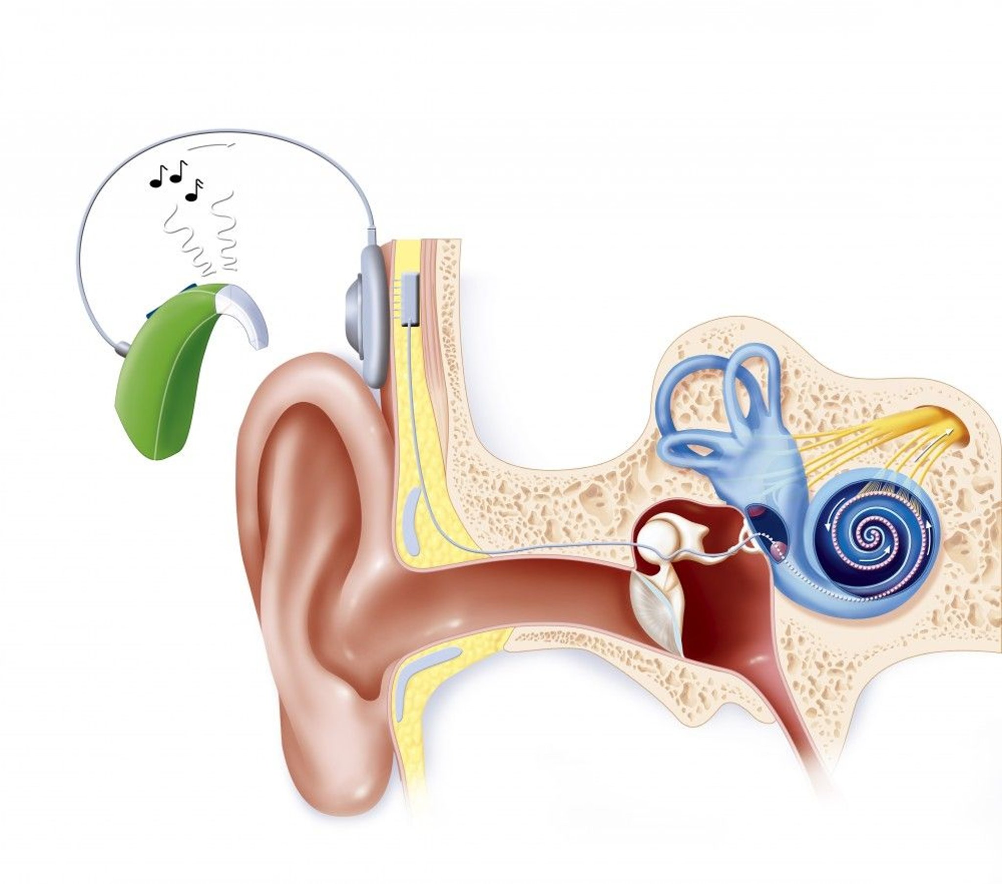
This illustration of a cochlear implant in the ear shows how sound travels to the brain. The transmitter is worn behind the ear (shown here next to the ear), linked to a transmitting antenna placed under the scalp, which transmits sound to a receiver placed under the skin, to the electrode array rolled up in the inner ear duct (in the cochlea). Sound is transmitted to the auditory nerve and to the brain.
JACOPIN/BSIP/SCIENCE PHOTO LIBRARY
This device provides electrical signals directly into the auditory nerve via multiple electrodes implanted in the cochlea. An external microphone and processor convert sound waves to electrical impulses, which are transmitted through the skin electromagnetically from an external induction coil to an internal coil implanted in the skull above and behind the ear. The internal coil connects to electrodes inserted in the scala tympani.
Cochlear implants help with speech-reading by providing information about the intonation of words and the rhythm of speech. Many if not most adults with cochlear implants can discriminate words without visual clues, allowing them to talk on the telephone. Cochlear implants enable deaf people to hear and distinguish environmental sounds and warning signals. They also help deaf people modulate their voice and make their speech more intelligible.
Outcomes with cochlear implants vary, depending on a number of factors, including the:
Length of time between onset of hearing loss and placement of the implant (shorter duration leads to better outcomes)
Cause of the underlying hearing loss
Position of the implant within the cochlea
Brain stem implants
Brain stem implants via electrodes coupled to sound-detection and processing technologies can restore partial auditory function. Implants function similarly to cochlear devices, however, they typically yield lower auditory function. This intervention is indicated for patients in whom cochlear implants are not a therapeutic option, such as those with bilateral acoustic nerve destruction, bilateral temporal bone fractures, neurofibromatosis, or congenital cochlear nerve aplasia.
In general, children with cochlear nerve aplasia who receive the brain stem implant usually have more auditory function restored than patients who receive it following vestibular schwannoma resection (9). Clinical outcomes following the brain stem implant range from, on one end, requiring assistance with lip-reading skills to, on the other end, advanced auditory processing including the ability to understand language without lip-reading (termed "open-set speech" understanding).
Assistive strategies and technologies
Alerting systems that use light let people know when the doorbell is ringing, a smoke detector is sounding, or a baby is crying. Special sound systems transmitting infrared or FM radio signals help people hear in theaters, churches, or other places where competing noise exists. Many television programs carry closed captioning. Telephone communication devices are also available.
Lip-reading or speech-reading is particularly important for people who can hear but have trouble discriminating sounds. Most people get useful speech information from lip-reading even without formal training. Even people with normal hearing can better understand speech in a noisy place if they can see the speaker. To use this information, the listener must be able to see the speaker’s mouth. Health care professionals should be cognizant of this issue and always position themselves appropriately when speaking to the hearing-impaired. Observing the position of a speaker’s lips allows recognition of the consonant being spoken, thereby improving speech comprehension in patients with high-frequency hearing loss. Lip-reading may be learned in aural rehabilitation sessions in which a group of age-matched peers meets regularly for instruction and supervised practice in optimizing communication.
People can exercise control over their listening environment by modifying or avoiding difficult situations. For example, people can visit a restaurant during off-peak hours, when it is quieter. They can ask for a booth, which blocks out some extraneous sounds. In direct conversations, people may ask the speaker to face them. At the beginning of a telephone conversation, they can identify themselves as being hearing-impaired. At a conference, the speaker can be asked to use an assistive listening system, which makes use of either inductive loop, infrared, Bluetooth or FM technology that sends sound through the microphone to a patient’s hearing aid.
People with profound hearing loss often communicate by using sign language. American Sign Language (ASL) is the most common version in the United States. Other forms of linguistic communication that utilize visual inputs include Signed English, Signing Exact English, and Cued Speech. Around the world, it has been estimated that there are over 300 unique sign languages, with different countries, cultures, and villages having their own unique form of sign language.
Treatment references
1. Michels TC, Duffy MT, Rogers DJ. Hearing Loss in Adults: Differential Diagnosis and Treatment. Am Fam Physician. 2019;100(2):98-108.
2. Rigobello R, Shaw J, Ilg D, et al. Clinical Pharmacogenomic MT-RNR1 Screening for Aminoglycoside-Induced Ototoxicity and the Post-Test Counseling Conundrum. Clin Pharmacol Ther. 2023;114(2):262-265. doi:10.1002/cpt.2910
3. Fu X, Wan P, Li P, et al. Mechanism and Prevention of Ototoxicity Induced by Aminoglycosides. Front Cell Neurosci. 2021;15:692762. Published 2021 Jun 15. doi:10.3389/fncel.2021.692762
4. Cribb L, Moreno-Betancur M, Pase MP, et al. Treating Hearing Loss With Hearing Aids for the Prevention of Cognitive Decline and Dementia. Neurology. 2026;106(3):e214572. doi:10.1212/WNL.0000000000214572
5. Yeo BSY, Song HJJMD, Toh EMS, et al. Association of Hearing Aids and Cochlear Implants With Cognitive Decline and Dementia: A Systematic Review and Meta-analysis. JAMA Neurol. 2023;80(2):134-141. doi:10.1001/jamaneurol.2022.4427
6. Johnson JCS, Marshall CR, Weil RS, et al. Hearing and dementia: from ears to brain. Brain. 2021;144(2):391-401. doi:10.1093/brain/awaa429
7. Unwin BK, D'Amora JM, O'Shaughnessy C. What Physicians Need to Know About Over-the-Counter Hearing Aids. Am Fam Physician. 2024;109(3):279-283.
8. Carlson ML. Cochlear Implantation in Adults. N Engl J Med. 2020;382(16):1531-1542. doi:10.1056/NEJMra1904407
9. Garcia A, Haleem A, Poe S, et al. Auditory Brainstem Implant Outcomes in Tumor and Nontumor Patients: A Systematic Review. Otolaryngol Head Neck Surg. 2024;170(6):1648-1658. doi:10.1002/ohn.662
Single-Sided Deafness
Patients with single-sided deafness (SSD) represent a special challenge. In one-on-one situations, hearing and speech understanding is relatively unaffected. However, with noisy backgrounds or complex acoustic environments (eg, classrooms, parties, meetings), patients with SSD are unable to hear and communicate effectively. Further, patients who hear out of only one ear are unable to localize the origin of sounds. The "head shadow" effect is the skull's tendency to block sound coming from the deaf side from reaching the hearing ear. This can result in up to a 30-dB loss of sound energy reaching the hearing ear (as a comparison, a store-bought ear plug results in a 22- to 32-dB drop in hearing, roughly equivalent). For many patients, SSD can be life-altering and lead to significant disability at work and socially.
Treatment for SSD includes contralateral routing of signal (CROS) hearing aids or bone-anchored hearing implants that pick up sound from the deaf side and transfer it to the hearing ear without the loss of sound energy. Although these technologies improve hearing in noisy settings, they do not allow sound localization. Cochlear implants are recommended for use in patients with SSD, particularly if the deaf ear also has severe tinnitus; implants have also been shown to provide sound source localization (1, 2).
Single-sided deafness references
1. Dillon MT, Kocharyan A, Daher GS, et al. American Cochlear Implant Alliance Task Force Guidelines for Clinical Assessment and Management of Adult Cochlear Implantation for Single-Sided Deafness. Ear Hear. 2022;43(6):1605-1619. doi:10.1097/AUD.0000000000001260
2. Lindquist NR, Holder JT, Patro A, et al. Cochlear Implants for Single-Sided Deafness: Quality of Life, Daily Usage, and Duration of Deafness. Laryngoscope. 2023;133(9):2362-2370. doi:10.1002/lary.30452
Treatment of Hearing Loss in Children
In addition to treatment of any cause and the provision of hearing aids, children with hearing loss require support of language development with appropriate therapy. Because children must hear language to learn it spontaneously, most deaf children develop language only with special training, ideally beginning as soon as the hearing loss is identified (an exception would be a deaf child growing up with deaf parents who are fluent sign language users). Deaf infants must be provided with a form of language input. For example, a visually based sign language can provide a foundation for later development of oral language if a cochlear implant is not available. However, for children, there is no substitute for access to the sounds of speech (phonemes) to enable them to integrate acoustic inputs and develop a refined and nuanced understanding of speech and language.
If infants as young as 1 month have profound bilateral hearing loss and cannot benefit from hearing aids, they may be considered for a cochlear implant. Although cochlear implants allow auditory communication in many children with either congenital or acquired deafness, they are generally more effective in children who already have developed language. Children who have postmeningitic deafness eventually develop an ossified inner ear that prevents the placement of an implant; they should receive cochlear implants as soon as possible to allow the implant to be correctly placed and maximize effectiveness. Children whose acoustic nerves have been extensively damaged by tumors may be helped by implantation of brain stem auditory-stimulating electrodes. Children with cochlear implants may have a slightly greater risk of meningitis than children without cochlear implants or adults with cochlear implants.
Children with unilateral deafness should be allowed to use a special system in the classroom, such as a frequency modulated (FM) auditory trainer. With these systems, the teacher speaks into a microphone that sends signals to a hearing aid in the child’s nonaffected ear, improving the child’s impaired baseline ability to hear speech against a noisy background. Cochlear implants have also gained popularity, but the amount of benefit and appropriate timing of implantation is an active area of study.
Lunsotogene parvec-cwha is a gene therapy that may be used for a specific genetic form of hearing loss cause by mutations in the Otoferlin gene (OTOF) (1, 2). OTOF-related hearing loss is responsible for 2 to 8% of inherited, nonsyndromic hearing loss. A dual adeno-associated virus (AAV) vector-based gene therapy delivers a functional copy of the defective gene to inner hair cells to restore their function.
Treatment of hearing loss in children references
1. Valayannopoulos V, Bance M, Carvalho DS, et al. DB-OTO Gene Therapy for Inherited Deafness. N Engl J Med. 2026;394(11):1074-1083. doi:10.1056/NEJMoa2400521
2. Azaiez H, Thorpe RK, Odell AM, Smith RJH. OTOF-Related Hearing Loss. In: Adam MP, Bick S, Mirzaa GM, Pagon RA, Wallace SE, Amemiya A, eds. GeneReviews®. Seattle (WA): University of Washington, Seattle; February 29, 2008.
Prevention of Hearing Loss
Prevention of hearing loss consists mainly of limiting the duration and intensity of noise exposure. People required to expose themselves to loud noise must wear ear protectors (eg, plastic plugs in the ear canals or glycerin-filled muffs over the ears). The Occupational Safety and Health Administration (OSHA) of the U.S. Department of Labor and similar agencies in many other countries have standards regarding the length of time that a person can be exposed to a noise (1). The louder the noise, the shorter should be the permissible safe time of exposure.
Use of ototoxic medications should be avoided when feasible. Increasing the interval between consecutive medication doses does not always clearly decrease the risk of ototoxicity. For example, the risk of ototoxicity was not significantly different with once-daily dosing of aminoglycosides than with more frequent dosing intervals (2).
Prevention references
1. U.S. Department of Labor. Occupational Safety and Health Administration (OSHA). Occupational Noise Exposure. Accessed July 13, 2026.
2. Barza M, Ioannidis JP, Cappelleri JC, Lau J. Single or multiple daily doses of aminoglycosides: a meta-analysis. BMJ. 1996;312(7027):338-345. doi:10.1136/bmj.312.7027.338
Geriatrics Essentials: Hearing Loss
Older adults typically experience a progressive decrease in hearing (presbycusis). In the United States, 22% of people ages 65 to 74 years and 55% of those who are 75 and older have disabling hearing loss (1). Although hearing loss is common with aging, hearing loss in older adults should be evaluated and not simply ascribed to aging; older patients may have a tumor, a neurologic or autoimmune or systemic rheumatic disorder, or an easily correctable conductive hearing loss. Also, research strongly suggests that hearing loss in older adults can facilitate dementia (2), which may be mitigated by properly correcting hearing loss.
Presbycusis
Presbycusis is sensorineural hearing loss that probably results from a combination of age-related deterioration and cell death in various components of the hearing system and the effects of chronic noise exposure.
Hearing loss usually affects the highest frequencies (18 to 20 kHz) early on and gradually affects the lower frequencies; it usually becomes clinically significant when it affects the critical 2- to 4-kHz range at approximately age 55 to 65 years (sometimes sooner). The loss of high-frequency hearing also significantly affects speech comprehension. Although the loudness of speech seems normal, certain consonant sounds (eg, C, D, K, P, S, T) become harder to hear. Consonant sounds are the most important sounds for speech recognition. For example, when “shoe,” “blue,” “true,” “too,” or “new” is spoken, many people with presbycusis can hear the “oo” sound, but most have difficulty recognizing which word has been spoken because they cannot distinguish between consonants. This inability to distinguish consonants causes affected people to often think the speaker is mumbling. A speaker attempting to speak louder usually accentuates vowel sounds (which are low frequency), doing little to improve speech recognition. Speech comprehension is particularly difficult when background noise is present.
Screening
A screening tool is often helpful for older adults because many do not realize they have hearing loss. One tool is the Hearing Handicap Inventory for the Elderly–Screening Version, which asks
Does a hearing problem cause you to feel embarrassed when you meet people?
Does a hearing problem cause you to feel frustrated when talking to a family member?
Do you have difficulty hearing when someone whispers?
Do you feel handicapped by a hearing problem?
Does a hearing problem cause you difficulty when visiting friends, relatives, or neighbors?
Does a hearing problem cause you to attend religious services less often than you would like?
Does a hearing problem cause you to have arguments with family members?
Does a hearing problem cause you difficulty when listening to the television or radio?
Do you feel that any difficulty with your hearing hampers your personal or social life?
Does a hearing problem cause you difficulty when in a restaurant with relatives or friends?
Scoring is “no” = 0 points, “sometimes” = 2 points, and “yes” = 4 points. Scores > 10 suggest significant hearing impairment and necessitate follow-up (3).
Geriatrics essentials references
1. National Institutes of Health: National Institute on Deafness and Other Communication Disorders: Quick Statistics About Hearing, Balance and Dizziness. Updated September 20, 2024. Accessed July 10, 2026.
2. Griffiths TD, Lad M, Kumar S, et al. How Can Hearing Loss Cause Dementia? Neuron. 2020;108(3):401–412. doi:10.1016/j.neuron.2020.08.003
3. Servidoni AB, Conterno LO. Hearing Loss in the Elderly: Is the Hearing Handicap Inventory for the Elderly - Screening Version Effective in Diagnosis When Compared to the Audiometric Test? Int Arch Otorhinolaryngol. 2018;22(1):1–8, 2018. doi:10.1055/s-0037-1601427
Key Points
Cerumen, genetic disorders, infections, aging, and noise exposure are the most common causes of hearing loss.
All patients with hearing loss should have audiologic testing.
Cranial nerve deficits and other neurologic deficits should raise concern and warrant imaging tests.
Treatment modalities include correction of reversible causes, hearing aids, surgical procedures including ossicular reconstruction and cochlear implants, and various assistive technologies.
More Information
The following English-language resources may be useful. Please note that The Manual is not responsible for the content of these resources.
Drug Information for the Topic