


Vaginitis is infectious or noninfectious inflammation of the vaginal mucosa, sometimes with inflammation of the vulva (vulvitis or vulvovaginitis). Symptoms and signs include vaginal discharge, irritation, pruritus, edema, and erythema. Diagnosis is by pelvic examination and evaluation of vaginal fluid. Treatment is directed at the cause and at any severe symptoms.
Vaginitis and vulvitis are common clinical issues encompassing infectious and noninfectious inflammatory conditions; typical clinical manifestations include abnormal vaginal discharge, odor, pruritus, burning, irritation, and/or dyspareunia. Etiologies of vulvovaginitis vary by reproductive stage, reflecting shifts in estrogen exposure, epithelial integrity, and vaginal pH, as well as use of vaginal products or medications.
Pathophysiology of the Vaginal Microbiome
The vaginal microbiome refers to the collection of bacteria that populate the vagina. A healthy vaginal microbiome is a critical component of reproductive tract health and systemic immune balance. Disruptions in the vaginal microbiome can predispose individuals to a host of adverse obstetric and gynecologic outcomes, including but not limited to increased risk for sexually transmitted infections and pelvic inflammatory disease, preterm birth, and postoperative infections (1, 2, 3).
Key components to a healthy vaginal microbiome include all of the following:
Dominance of Lactobacillus species (most commonly L. crispatus, L. iners, L. jensenii, and L. gasseri) among the bacterial population
Maintenance of an acidic pH (generally < 4.5) due to production of lactic acid, hydrogen peroxide, and bacteriocins (peptides produced by bacterial ribosomes that inhibit or kill pathogenic bacteria and viruses)
Stable bacterial community dynamics (absence of shifts in bacterial composition in the setting of variations in environmental conditions, such as menses and intercourse) (4, 5)
The vaginal microbiome is Lactobacillus-dominant in most reproductive-age individuals, though in some people there may be a higher abundance of anaerobic bacteria. Lactobacilli are facultative anaerobes that can grow in the presence or absence of oxygen, though low-oxygen conditions are most favorable for their survival. They are nonmotile gram-positive rods that are typically 0.7 to 1 micrometer in diameter and 1 to 1.5 micrometer in length (6). They produce lactic acid via fermentation, which lowers pH and inhibits the growth of many pathogens.
Vaginal environments that are Lactobacillus-dominant are less easily disrupted by various endogenous or exogenous factors and are associated with lower risk of sexually transmitted infections (including bacterial vaginosis [BV]) and preterm birth. -dominant vaginal environments also exhibit improved mucosal immune homeostasis. A healthy vaginal microbial composition can vary by life stage, hormonal milieu, menses, sexual practices, ethnicity, geography, and antibiotic exposure. Normal microbial states may be heterogeneous but functionally convergent when stable and -rich (7).
Multiple endogenous and exogenous factors can disrupt the vaginal microbiome and thereby predispose to mucosal inflammation, vaginitis, and increased susceptibility to sexually transmitted infections, including HIV. Factors that can reduce abundance and allow expansion of anaerobic polymicrobial communities (bacterial vaginosis [BV]-associated bacterial species, including Gardnerella, Atopobium, Prevotella, Sneathia) include:
Antibiotics
Douching
Spermicides
New sexual partners
Semen exposure
Menses
Hormonal fluctuations (including pregnancy and menopause)
Systemic inflammation (8)
A disrupted vaginal microbiome is dysbiotic, which means it has an imbalance in which beneficial microbes decrease, harmful ones increase, and/or overall diversity decreases. In this state the vaginal tissue demonstrates increased epithelial permeability, proliferation of proinflammatory cytokines, impaired mucosal immunity, and elevated HIV target cell recruitment, thereby increasing biological susceptibility to HIV and other infections. Persistent dysbiosis is also linked to recurrent BV, candidiasis risk modulation, adverse pregnancy outcomes, and persistence or recurrence of inflammation even after treatment.
Correcting vaginal microbiome disruption remains an evolving area of investigation. Current best practice emphasizes treating symptomatic vaginitis, avoiding exposures that worsen dysbiosis, and using evidence-based adjunctive microbial restoration strategies where data exist, while acknowledging that preventive, durable microbiome-directed interventions are an active research frontier. Standard management of BV (eg, metronidazole, clindamycin) or vulvovaginal candidiasis remains first line; however, recurrence is common, suggesting that short-term antimicrobial suppression does not always restore a resilient bacterial community. Evidence suggests that routine douching or use of feminine hygiene products worsens dysbiosis (9). Diet, systemic probiotics, and general wellness interventions have limited proven impact on vaginal microbial community structure (10).
Emerging approaches to actively restore dominance include targeted vaginal probiotics (particularly L. crispatus CTV-05/Lactin-V), vaginal microbiota transplantation, estrogen therapy for patients in hypoestrogenic states, and strategies addressing partner microbial exchange. However, only some of these approaches are clinically available. Most require further validation in large trials (11, 12, 13, 14, 15).
Pathophysiology references
1. White BA, Creedon DJ, Nelson KE, et al. The vaginal microbiome in health and disease. Trends Endocrinol Metab. 2011;22(10):389-393. doi:10.1016/j.tem.2011.06.00
2. Greenbaum S, Greenbaum G, Moran-Gilad J, et al. Ecological dynamics of the vaginal microbiome in relation to health and disease. Am J Obstet Gynecol. 2019;220(4):324-335. doi:10.1016/j.ajog.2018.11.1089
3. Ziogou A, Ziogos E, Giannakodimos I, et al. Bacterial Vaginosis and Post-Operative Pelvic Infections. Healthcare (Basel). 2023;11(9):1218. Published 2023 Apr 25. doi:10.3390/healthcare11091218
4. Hugerth LW, Krog MC, Vomstein K, et al. Defining Vaginal Community Dynamics: daily microbiome transitions, the role of menstruation, bacteriophages, and bacterial genes. Microbiome. 2024;12(1):153. Published 2024 Aug 19. doi:10.1186/s40168-024-01870-5
5. Ravel J, Gajer P, Abdo Z, et al. Vaginal microbiome of reproductive-age women. Proc Natl Acad Sci U S A. 2011;108 Suppl 1(Suppl 1):4680-4687. doi:10.1073/pnas.1002611107
6. Schär-Zammaretti P, Ubbink J. The cell wall of lactic acid bacteria: surface constituents and macromolecular conformations. Biophys J. 2003;85(6):4076-4092. doi:10.1016/S0006-3495(03)74820-6
7. Ma B, Forney LJ, Ravel J. Vaginal microbiome: rethinking health and disease. Annu Rev Microbiol. 2012;66:371-389. doi:10.1146/annurev-micro-092611-150157
8. Morsli M, Gimenez E, Magnan C, et al. The association between lifestyle factors and the composition of the vaginal microbiota: a review. Eur J Clin Microbiol Infect Dis. 2024;43(10):1869-1881. doi:10.1007/s10096-024-04915-7
9. Holdcroft AM, Ireland DJ, Payne MS. The Vaginal Microbiome in Health and Disease-What Role Do Common Intimate Hygiene Practices Play?. Microorganisms. 2023;11(2):298. Published 2023 Jan 23. doi:10.3390/microorganisms11020298
10. Barrientos-Durán A, Fuentes-López A, de Salazar A, et al. Reviewing the Composition of Vaginal Microbiota: Inclusion of Nutrition and Probiotic Factors in the Maintenance of Eubiosis. Nutrients. 2020;12(2):419. Published 2020 Feb 6. doi:10.3390/nu12020419
11. Collins SL, McMillan A, Seney S, et al. Promising Prebiotic Candidate Established by Evaluation of Lactitol, Lactulose, Raffinose, and Oligofructose for Maintenance of a Lactobacillus-Dominated Vaginal Microbiota. Appl Environ Microbiol. 2018;84(5):e02200-17. Published 2018 Feb 14. doi:10.1128/AEM.02200-17
12. McLean NW, Rosenstein IJ. Characterisation and selection of a Lactobacillus species to re-colonise the vagina of women with recurrent bacterial vaginosis. J Med Microbiol. 2000;49(6):543-552. doi:10.1099/0022-1317-49-6-543
13. van de Wijgert JHHM, Verwijs MC, Agaba SK, et al. Intermittent Lactobacilli-containing Vaginal Probiotic or Metronidazole Use to Prevent Bacterial Vaginosis Recurrence: A Pilot Study Incorporating Microscopy and Sequencing. Sci Rep. 2020;10(1):3884. Published 2020 Mar 3. doi:10.1038/s41598-020-60671-6
14. Pagar R, Deshkar S, Mahore J, et al. The microbial revolution: Unveiling the benefits of vaginal probiotics and prebiotics. Microbiol Res. 2024;286:127787. doi:10.1016/j.micres.2024.127787
15. Lev-Sagie A, Goldman-Wohl D, Cohen Y, et al. Vaginal microbiome transplantation in women with intractable bacterial vaginosis. Nat Med. 2019;25(10):1500-1504. doi:10.1038/s41591-019-0600-6
Etiology of Vaginitis
The most common causes of vaginitis vary as reproductive status changes from premenarchal (see Vaginitis in Children) to reproductive age (menarche through menopause) to menopause. Vaginitis can be infectious or noninfectious. Vulvitis and vulvovaginitis have some of the same causes as vaginitis.
In reproductive-age women, species are the predominant constituent of normal vaginal flora. Colonization by these bacteria maintains vaginal pH in an acidic range (3.8 to 4.2), which is the usual range for the vagina. The acidic environment prevents overgrowth of pathogenic bacteria. Also, high serum estrogen levels maintain vaginal epithelial thickness, bolstering local defenses.
In menopause, a marked decrease in estrogen causes thinning of the vaginal epithelium, increasing vulnerability to infection and inflammation.
Infectious vaginitis
Infectious vaginitis is caused by infection with or overgrowth of organisms. The most common types are:
Factors that predispose to overgrowth of bacterial vaginal pathogens include the following:
An alkaline vaginal pH due to menstrual blood, semen, a decrease in lactobacilli, or hypoestrogenism (due to menopause, antiestrogenic medications, or prolonged breastfeeding in some patients)
Poor vulvovaginal hygiene
Vaginal douching
Hypoestrogenic (atrophic) vaginitis
Hypoestrogenism due to menopause, antiestrogenic medications (eg, gonadotropin-releasing hormone agonists or antagonists or aromatase inhibitors), or prolonged breastfeeding in some patients, causes atrophy of the vaginal epithelium with thinning of the tissue and loss of the cornified layer. In addition, decreased serum and tissue estrogen levels results in a more alkaline vaginal pH, which can predispose to overgrowth of vaginal pathogenic bacteria (1). Atrophy alone is not a vaginitis, but it predisposes to inflammation, which may cause symptoms such as pruritus, irritation, and discharge. Genitourinary syndrome of menopause is a set of symptoms (typically including vaginal dryness, burning, itching and irritation) which may include atrophic vaginitis.
Hypersensitivity or irritant vulvovaginitis
Noninfectious vulvitis or vulvovaginitis due to hypersensitivity or irritant reactions is a common cause of vaginitis or vulvovaginitis. This may be caused by reactions to soaps, products applied to the vulva or vagina (eg, lubricants, moisturizers, hygiene products), contraceptive products or devices (eg, spermicide, condoms, diaphragm, cervical cap, vaginal contraceptive ring or sponge), menstrual products (eg, pads, tampons, menstrual cups), incontinence pads, sex toys, bathwater additives, toilet paper, laundry products (eg, detergents, bleach, fabric softeners), fabric dyes, or synthetic fibers.
Prolonged exposure of the vulva to urine or stool can be an irritant and can lead to chronic vulvar inflammation due to chemical irritation or infection. This may occur in patients with chronic urinary or stool incontinence, particularly if they are bedbound. Another etiology is vaginal fistula from the urinary or gastrointestinal tract, which may occur following obstructed labor, hysterectomy, pelvic radiation therapy, or severe pelvic injury.
Desquamative inflammatory vaginitis
Desquamative inflammatory vaginitis (DIV) is a chronic inflammatory vaginitis that is considered a severe inflammatory form of noninfectious vaginitis. Unlike candidiasis or bacterial vaginosis (BV), DIV lacks a dominant infectious pathogen; instead, it reflects mucosal epithelial inflammation and disrupted mucosal defense (2). DIV occurs most commonly in perimenopausal and postmenopausal women but can be seen in women in other reproductive stages (3). It should be considered in menopausal symptomatic women with negative infectious vaginitis testing.
DIV is characterized by copious yellow discharge, burning, dyspareunia, pruritus, and often vaginal erythema with petechiae or friability (4). Vaginal pH is elevated (usually > 5.5). Diagnosis is based on history and pelvic examination and microscopy after exclusion of specific infectious etiologies. On microscopy there is a loss of lactobacilli and predominance of inflammatory cells (particularly neutrophils) with abundant parabasal epithelial cells.
Treatment is with at least 2 weeks of anti-inflammatory and antimicrobial therapy, such as intravaginal clindamycin and/or topical/vaginal glucocorticoids. Relapse is common and some patients require prolonged suppressive regimens (4).
Aerobic vaginitis
Aerobic vaginitis (AV) is a somewhat controversial diagnosis but is felt to be a distinct type of inflammatory vaginitis (5, 6, 7). Though it has overlapping features with BV, it is characterized by depletion of lactobacilli and overgrowth of aerobic or facultative organisms—most commonly Escherichia coli, Enterococcus faecalis, Staphylococcus aureus, and group B Streptococcus (8). Clinically, patients often present with yellow or green inflammatory discharge, burning, soreness, dyspareunia, and pronounced vaginal redness or ulceration, sometimes mimicking lichen planus or erosive vaginitis (9). Vaginal pH is typically elevated, and microscopy demonstrates reduced lactobacilli, increased leukocytes, toxic leukocytes, and immature epithelial cells (parabasal cells). Unlike BV, which is polymicrobial and largely anaerobic, AV represents a proinflammatory dysbiosis with epithelial damage and has been associated with adverse reproductive outcomes including preterm birth, prelabor rupture of membranes, and postpartum complications. Management often requires targeted antimicrobial therapy (eg, topical clindamycin or other directed therapy) sometimes combined with anti-inflammatory therapy (eg, glucocorticoids) and strategies to restore healthy lactobacilli, though standardized treatment algorithms continue to evolve.
Etiology references
1. The NAMS 2020 GSM Position Statement Editorial Panel. The 2020 genitourinary syndrome of menopause position statement of The North American Menopause Society. Menopause. 2020;27(9):976-992. doi:10.1097/GME.0000000000001609
2. Donders GG. Definition and classification of abnormal vaginal flora. Best Pract Res Clin Obstet Gynaecol. 2007;21(3):355-373. doi:10.1016/j.bpobgyn.2007.01.002
3. Reichman O, Sobel J. Desquamative inflammatory vaginitis. Best Pract Res Clin Obstet Gynaecol. 2014;28(7):1042-1050. doi:10.1016/j.bpobgyn.2014.07.003
4. Mason MJ, Winter AJ. How to diagnose and treat aerobic and desquamative inflammatory vaginitis. Sex Transm Infect. 2017;93(1):8-10. doi:10.1136/sextrans-2015-052406
5. Donders GG, Vereecken A, Bosmans E, et al. Definition of a type of abnormal vaginal flora that is distinct from bacterial vaginosis: aerobic vaginitis. BJOG. 2002;109(1):34-43. doi:10.1111/j.1471-0528.2002.00432.x
6. Sonthalia S, Aggarwal P, Das S, et al. Aerobic vaginitis - An underdiagnosed cause of vaginal discharge - Narrative review. Int J STD AIDS. 2020;31(11):1018-1027. doi:10.1177/0956462420913435
7. Donders GGG, Bellen G, Grinceviciene S, et al. Aerobic vaginitis: no longer a stranger. Res Microbiol. 2017;168(9-10):845-858. doi:10.1016/j.resmic.2017.04.004
8. Miller JM, Binnicker MJ, Campbell S, et al. Guide to Utilization of the Microbiology Laboratory for Diagnosis of Infectious Diseases: 2024 Update by the Infectious Diseases Society of America (IDSA) and the American Society for Microbiology (ASM). Clin Infect Dis. Published online March 5, 2024. doi:10.1093/cid/ciae104
9. Han C, Wu W, Fan A, et al. Diagnostic and therapeutic advancements for aerobic vaginitis. Arch Gynecol Obstet. 2015;291(2):251-257. doi:10.1007/s00404-014-3525-9
Symptoms and Signs of Vaginitis
Vaginitis presents with an abnormal vaginal discharge, which must be distinguished from physiologic discharge.
Physiologic vaginal discharge is commonly white or clear, thin or mucoid, odorless, and nonirritating. The overall volume is small, but it can result in vaginal wetness that dampens underwear. In reproductive-age women, physiologic discharge may occur daily or intermittently. Many women have a clear, sticky discharge during ovulation due to ovulatory cervical mucus.
Discharge caused by vaginitis is usually accompanied by additional symptoms, which may include: pruritus, odor, discomfort (sometimes burning), or mild bleeding from inflammation. Pruritus may be severe, even interfering with sleep. Volume of discharge can range from a small amount to copious. Examination findings may include vulvar or vaginal erythema or edema.
In menopausal atrophic vaginitis, dyspareunia is common, discharge is scant, and vaginal tissue may appear smooth (loss of rugae) and pale.
Although symptoms vary among particular types of vaginitis, there is much overlap (see table ).
Common Types of Vaginitis
Disorder | Typical Symptoms | Diagnostic Methods | Microscopic Findings | Differential Diagnosis |
|---|---|---|---|---|
Discharge is white-gray or green, thin, homogeneous, smoothly coats the vaginal walls, and malodorous (fishy-smelling) Sometimes mild vulvovaginal irritation Often asymptomatic | Amsel criteria (at least 3 of 4 of the following):
NAAT DNA hybridization probe | Saline wet mount*: > 20% of vaginal epithelial cells are clue cells (vaginal epithelial cells with adherent coccobacilli); increased coccobacilli; decreased lactobacilli | Trichomonal vaginitis | |
White, thick discharge, sometimes clumpy or curd-like Vaginal and sometimes vulvar pruritus, burning or irritation, or dyspareunia | Typical discharge and symptoms, pH < 4.5, and microscopy NAAT DNA hybridization probe Culture (gold standard) if clinical suspicion is high and microscopic findings are negative or symptoms recur or persist after treatment | 10% Potassium hydroxide wet mount†: Budding yeast, pseudohyphae, or hyphae | Contact irritant or allergic vulvitis Vulvodynia | |
Yellow-green thin, malodorous, profuse discharge Burning, irritation, sometimes dysuria, dyspareunia, or postcoital bleeding | pH usually > 4.5, negative whiff test, and detection of trichomonads by microscopy NAAT (gold standard) DNA hybridization probe Rapid antigen test Culture if microscopy or molecular diagnostic tests are not available | Saline wet mount*: Motile trichomonads (flagellated protozoa), increased PMNs | Bacterial vaginosis Desquamative inflammatory vaginitis | |
Copious discharge Vaginal pain, burning, dyspareunia Most common in perimenopausal and postmenopausal women | pH > 4.5, negative whiff test, and characteristic microscopy findings | Saline wet mount*: Increased PMNs, parabasal cells; decreased lactobacilli | Erosive lichen planus | |
Hypoestrogenic (atrophic) vaginitis | White, yellow, or brown, scant, watery, sometimes malodorous discharge Vaginal dryness, irritation, burning; dyspareunia; postcoital bleeding; sometimes vulvar fissures Occurs in patients in menopause or with other causes of hypoestrogenism (ie, antiestrogenic medications) | pH > 4.5, negative whiff test | Saline wet mount*: Decreased lactobacilli; increased PMNs; parabasal cells (immature vaginal epithelial cells with relatively large nuclei) | Desquamative inflammatory vaginitis Erosive lichen planus |
* Saline wet mount slide is prepared with 0.9% sodium chloride. | ||||
† Potassium hydroxide wet mount slide is prepared with 10% potassium hydroxide solution. | ||||
NAAT = nucleic acid amplification test; PMN = polymorphonuclear leukocyte. Reference: Vaginitis in Nonpregnant Patients: ACOG Practice Bulletin, Number 215. Obstet Gynecol. 2020;135(1):e1-e17. doi:10.1097/AOG.0000000000003604 | ||||


Normal findings, trichomonal vaginitis with frothy discharge, and candidal vaginitis with thick, white discharge are shown (left to right).
By permission of the publisher. From Spitzer M, Mann M. In Atlas of Clinical Gynecology: Gynecologic Pathology. Edited by M Stenchever (series editor) and B Goff. Philadelphia, Current Medicine, 1998. (Courtesy of the International Society for the Study of Vulvar Disease, from the collection of the late Dr. Herman Gardner.)
Vulvitis can cause erythema, pruritus, and sometimes tenderness and discharge from the vulva.
Diagnosis of Vaginitis
History and pelvic examination
Vaginal pH
Saline and potassium hydroxide (KOH) wet mounts if available
Sometimes nucleic acid amplification testing (NAAT) or other molecular diagnostic tests, or culture
Vaginitis is diagnosed using clinical criteria and in-office or laboratory testing.
The most cost-effective way to diagnose bacterial vaginosis or vaginal candidiasis is microscopic examination of vaginal fluid (called a wet mount) in an outpatient clinic. NAATs and DNA hybridization probes are available, but are typically more expensive and results are often not available immediately (1, 2, 3, 4, 5). For candidal infection, culture may be performed if microscopy is inconclusive or symptoms recur or persist after treatment; antimicrobial sensitivity should be tested to detect fluconazole-resistant Candida.
Nucleic acid amplification testing (NAAT) is the preferred method to diagnose trichomonal vaginitis, but microscopy may be used. Culture is used when NAAT and microscopy are not available.
Bacterial culture is not routinely recommended in the evaluation of vaginitis in adults; it typically shows vaginal flora and is likely to be nondiagnostic or may have an incidental finding of growth of a dominant bacterium that may not be contributing to symptoms. Special circumstances may warrant evaluation to detect a specific organism, such as Group A Streptococcus (GAS). Clinical scenarios that suggest GAS vaginitis include prepubescent girls who present with severe vulvovaginitis symptoms or adult women with persistent vulvovaginal symptoms that test negative for routine vulvovaginal infections and/or fail to respond to standard treatments for yeast infections or bacterial vaginosis. Risk factors for GAS vaginitis include household or personal history of dermal or respiratory infection due to Group A Streptococcus, sexual contact with a partner with a GAS infection, and lactational or menopausal vaginal atrophy (6).
Patients diagnosed with bacterial vaginosis or trichomonal vaginitis are at increased risk of other sexually transmitted infections. The patient should be counseled about this risk, partner notification and treatment, and safer sex. They should also be offered testing for other STIs, including Neisseria gonorrhoeae and Chlamydia trachomatis, common causes of sexually transmitted pelvic inflammatory disease (PID) (7).
During the pelvic examination, presence of a foreign body should excluded (eg, a retained tampon). If contact irritant or allergic vulvitis is suspected, the patient should systematically eliminate potential irritants or allergens to identify the cause. Severe dermatitis or allergic reactions require evaluation by an allergist or dermatologist.
Wet mount
Prior to collecting vaginal fluid for microscopic examination, a pelvic examination is performed using a water-lubricated speculum (gel lubricants can be bacteriostatic). A sample of vaginal discharge is collected on a swab and tested with pH paper; pH is measured in 0.2 intervals from 4.0 to 6.0 (normal vaginal pH is 3.8 to 4.2).
If point-of-care microscopy is available, a sample of discharge on a swab is placed on 2 slides. Each slide is prepared with a different fluid (added by a dropper to the vaginal discharge sample):
Saline wet mount: 0.9% sodium chloride
Potassium hydroxide (KOH) wet mount: 10% potassium hydroxide
The saline wet mount is examined microscopically as soon as possible to detect trichomonads, which can become immotile and more difficult to recognize within minutes after slide preparation. The slide is also examined for clue cells (vaginal epithelial cells with adherent coccobacilli that partially obscure their cell margins) and polymorphonuclear leukocytes.
The KOH wet mount is checked for a fishy odor (whiff test), which results from amines produced in trichomonal vaginitis or bacterial vaginosis. Potassium hydroxide is also used to test for Candida; KOH dissolves most cellular material except for yeast hyphae, making identification easier.

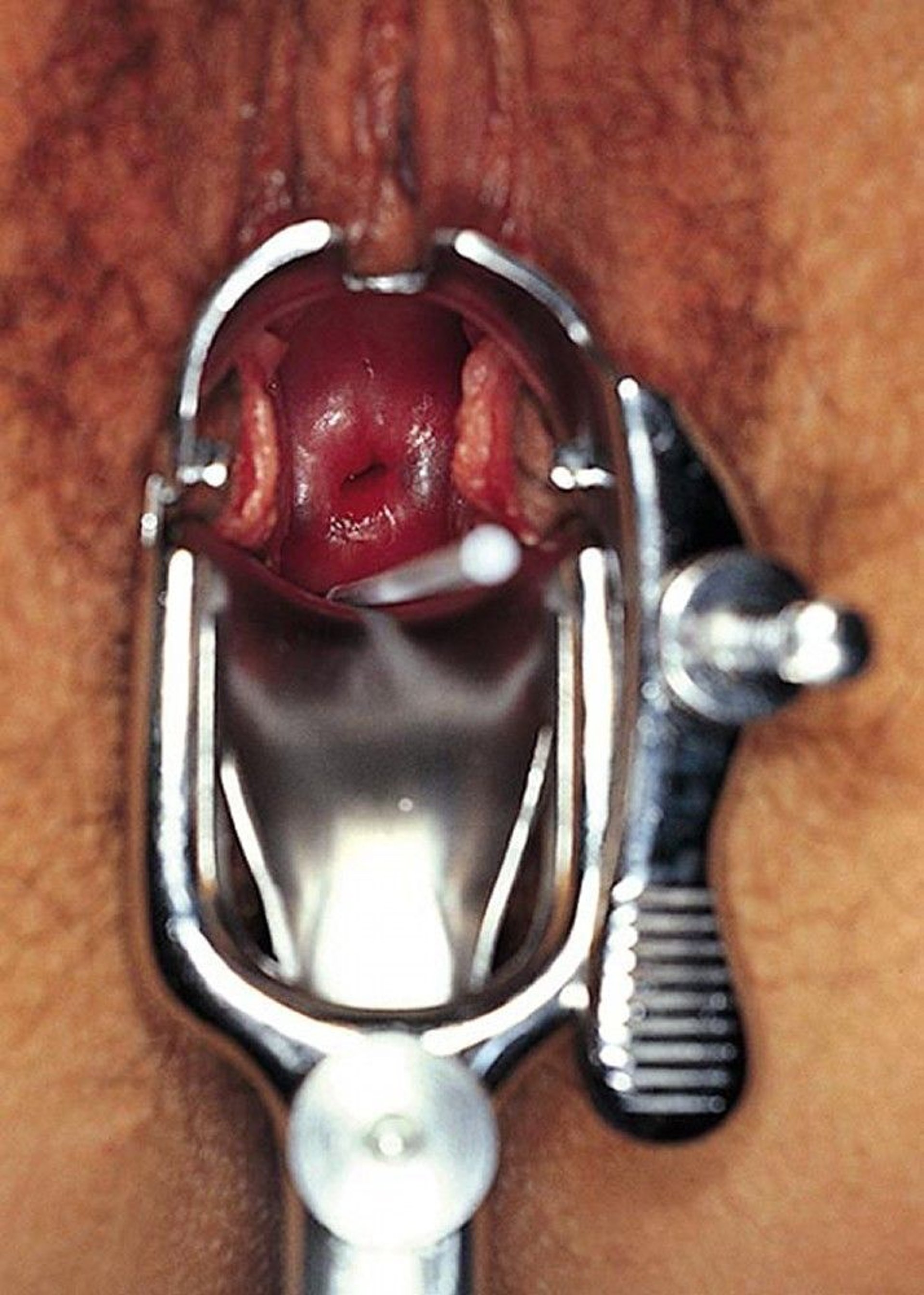
For wet-mount microscopy, a cotton-tipped applicator is used to obtain specimens from secretions that pool in the posterior vagina behind the cervix.
By permission of the publisher. From Judson F. In Atlas of Infectious Diseases: Sexually Transmitted Diseases. Edited by G Mandell (series editor) and MF Rein. Philadelphia, Current Medicine, 1996.


Wet mount examination using saline shows large nucleated vaginal squamous epithelial cells, small bacteria that are barely visible, and intermediate-sized white blood cells. Magnification is 400X.
By permission of the publisher. From Hillier S. In Atlas of Infectious Diseases: Sexually Transmitted Diseases. Edited by G Mandell (series editor) and MF Rein. Philadelphia, Current Medicine, 1996.


Trichomonads on wet mount are about the size of white blood cells but may be slightly smaller and more ovoid, have flagella, and, if examined immediately, be motile.
Image obtained from the Public Health Image Library of the Centers for Disease Control and Prevention.
Differential diagnosis of vaginitis
Other causes of vaginal discharge or irritation should be excluded, including:
Cervicitis: Cervical discharge due to cervicitis can resemble that of vaginitis (eg, herpes simplex virus cervicitis or vulvovaginitis).
Cancer: Discharge that is watery, bloody, or both may result from vulvar, vaginal, or cervical cancer; cancers can be differentiated from vaginitis by examination, Papanicolaou (Pap) tests, and/or biopsy, as appropriate.
Skin disorders: Vaginal pruritus and/or discharge may result from vulvar dermatoses (eg, lichen planus, lichen sclerosus) or other vulvar skin disorders (eg, psoriasis, tinea versicolor), which can usually be differentiated from infectious vaginitis by history and skin findings.
Diagnosis references
1. Cartwright CP, Lembke BD, Ramachandran K, et al. Development and validation of a semiquantitative, multitarget PCR assay for diagnosis of bacterial vaginosis. J Clin Microbiol. 2012;50(7):2321–2329. doi: 10.1128/JCM.00506-12
2. Schwebke JR, Gaydos CA, Nyirjesy P, et al. Diagnostic performance of a molecular test versus clinician assessment of vaginitis. J Clin Microbiol. 2018;56(6):e00252-18. doi: 10.1128/JCM.00252-18
3. Gaydos CA, Beqaj S, Schwebke JR, et al. Clinical validation of a test for the diagnosis of vaginitis. Obstet Gynecol 2017;130(1):181–189. doi: 10.1097/AOG.0000000000002090
4. Schwebke J, Merriweather A, Massingale S, et al. Screening for Trichomonas vaginalis in a large high-risk population: Prevalence among men and women determined by nucleic acid amplification testing. Sex Transm Dis. 2018;45(5):e23-e24. doi: 10.1097/OLQ.0000000000000757
5. Coleman JS, Gaydos CA. Molecular diagnosis of bacterial vaginosis: An update. J Clin Microbiol. 2018;56(9):e00342–e00318. doi: 10.1128/JCM.00342-18
6. Verstraelen H, Verhelst R, Vaneechoutte M, et al. Group A streptococcal vaginitis: an unrecognized cause of vaginal symptoms in adult women. Arch Gynecol Obstet. 2011;284(1):95-98. doi:10.1007/s00404-011-1861-6
7. Workowski KA, Bachmann LH, Chan PA, et al. Sexually Transmitted Infections Treatment Guidelines, 2021. MMWR Recomm Rep. 2021;70(4):1-187. Published 2021 Jul 23. doi:10.15585/mmwr.rr7004a1
Treatment of Vaginitis
Treatment of etiology
Symptom management
Infectious vaginitis (eg, bacterial vaginosis, candidal vaginitis, trichomoniasis) is treated with antimicrobials.
If a foreign body is present, it is removed.
If chronic vulvar inflammation is due to being bedbound or incontinent, vulvar hygiene and skin care measures may help.
Infectious, irritant, or allergic vulvovaginitis may cause significant discomfort until adequately treated. Intermittent use of ice packs or warm sitz baths with or without baking soda may reduce soreness and pruritus.
If symptoms of noninfectious conditions are moderate or severe or do not respond to other measures, vaginal glucocorticoids (eg, topical 1% hydrocortisone ointment) can be applied to the symptomatic areas of the vulva or vagina. Oral antihistamines may be suggested to decrease pruritus and cause drowsiness, helping patients sleep.
Key Points
Vaginitis is infectious or noninfectious inflammation of the vaginal mucosa, sometimes with inflammation of the vulva (vulvitis or vulvovaginitis).
Symptoms include vaginal discharge, irritation, pruritus, and/or erythema.
Diagnose vaginitis based on pelvic examination and evaluation of vaginal fluid with microscopy or NAAT, and sometimes candidal culture.
Treat infectious and other specific causes, treat symptoms, and discuss any preventive measures with the patient.
Drug Information for the Topic







