

Sprains are tears in ligaments; strains are tears in muscles. Tears (ruptures) may also occur in tendons.
In addition to sprains, strains, and tendon injuries, musculoskeletal injuries include:
Joint dislocations and subluxations
Musculoskeletal injuries are common and vary greatly by mechanism, severity, and treatment. The extremities, spine, and pelvis can all be affected.
Musculoskeletal injuries may occur in isolation or as part of multisystem trauma (see Approach to the Trauma Patient). Most musculoskeletal injuries result from blunt trauma, but penetrating trauma can also damage musculoskeletal structures.
(See also Approach to Sports Injuries.)

Pathophysiology of Sprains and Other Soft-Tissue Injuries
Sprains and strains
Tears in ligaments or muscles may be graded as:
1st degree: Minimal (fibers are stretched but intact, or only a few fibers are torn)
2nd degree: Partial (some to almost all fibers are torn)
3rd degree: Complete (all fibers are torn)
Tendon injuries
Tendon tears are categorized as partial or complete.
With complete tears, the motion produced by the detached muscle is usually lost.
Partial tears can result from a single traumatic event (eg, penetrating trauma) or repeated stress, causing tendinopathy. Motion is often intact, but partial tears may progress to complete tears, particularly when significant or repetitive force is applied.
Healing
Many partial tears in ligaments, tendons, or muscles heal spontaneously.
Complete tears often require surgery to restore anatomy and function.
Prognosis and treatment vary greatly depending on the location and severity of the injury.
Complications
Serious complications of sprains, strains, and tendon injuries are unusual but may cause permanent limb dysfunction.
Acute complications (associated injuries) include the following:
Bleeding accompanies all significant soft-tissue injuries.
Vascular injuries rarely occur as a result of sprains. However, a severe sprain of the knee with tears to one or more of the ligaments (eg, anterior cruciate, posterior cruciate, medial collateral, lateral collateral) may actually be a spontaneously reduced dislocation, which may be accompanied by a limb-threatening arterial injury.
Nerve injuries may occur if the nerves adjacent to a tendon or ligament are stretched or damaged by the same mechanism of injury that causes a strain or sprain. There are several classification systems to describe the severity of peripheral nerve injury. In Seddon's classification (1), neurapraxia is a mild injury with focal demyelination or ischemia without structural damage to the axon. Following neurapraxia, there is temporary loss of motor and sensory function that typically resolves within days or weeks. Axonotmesis describes a more severe peripheral nerve injury, which includes both demyelination and axonal injury, with resultant degeneration of the distal nerve segment (Wallerian degeneration); however, the connective tissues surrounding the nerve (epineurium, perineurium, and endoneurium) remain intact. Depending on the extent of the damage, after axonotmesis, the nerve may regenerate over weeks to years. Neurotmesis is the most severe peripheral nerve injury, with complete transection of the nerve. This type of injury may require surgical repair.
Compartment syndrome: Rarely, swelling is severe enough to contribute to compartment syndrome. Tissue pressure increases in a closed fascial space, disrupting the vascular supply and reducing tissue perfusion. Untreated compartment syndrome can lead to ischemia and as the tissue necroses, rhabdomyolysis, hyperkalemia, and infection occur. It can also cause contractures, sensory deficits, and paralysis. Compartment syndrome threatens limb viability (possibly requiring amputation). Contractures may develop after necrotic tissue heals.
Long-term complications include the following:
Instability: Various ligament injuries, particularly 3rd-degree sprains, can lead to joint instability. Instability can be disabling and increases the risk of osteoarthritis.
Stiffness and impaired range of motion: Stiffness is more likely if a joint needs prolonged immobilization. The knee, elbow, and shoulder are particularly prone to posttraumatic stiffness, especially in older adults.
Osteoarthritis: Injuries that result in joint instability predispose to repeated joint stresses that can damage joint cartilage and result in osteoarthritis.
Pathophysiology reference
1. Seddon HJ. Three types of nerve injury. Brain. 66(4):237–288, 1942. doi.org/10.1093/brain/66.4.237
Evaluation of Sprains and Other Soft-Tissue Injuries
Evaluation for serious injuries
History and physical examination
Sometimes radiographs to check for fractures
Diagnosis of sprains, strains, and tendon injuries should include a thorough history and physical examination, which are often sufficient for diagnosis.
In the emergency department, if the mechanism of injury suggests potentially severe or multiple injuries (as in a high-speed motor vehicle crash or fall from a height), patients are first evaluated from head to toe for serious injuries to all organ systems and, if needed, are resuscitated (see Approach to the Trauma Patient).
Patients should be evaluated for fractures and dislocations due to the potential for simultaneous ligament, tendon, and muscle injuries, since the mechanism of injury is similar.
The joint above and below the injured joint should also be examined.
History
History focuses on the:
Mechanism of injury
Past injuries
Timing of pain onset
Extent and duration of pain before, during, and after activity
Clinicians should also ask about use of medications (eg, fluoroquinolones [1], glucocorticoids [2]) that increase the risk of tendon tears.
The mechanism (eg, direction and magnitude of force) may suggest the type of injury. However, many patients do not remember or cannot describe the exact mechanism.
If a patient reports a deformity that has resolved before the patient is medically evaluated, the deformity should be assumed to be a true deformity that spontaneously reduced.
A perceived snap or pop at the time of injury may signal a ligament or tendon injury (or a fracture). Serious ligamentous injuries usually cause immediate pain; pain that begins hours to days after the injury suggests minor injury.
Physical examination
Examination includes:
Vascular and neurologic assessment
Inspection for deformity, swelling, ecchymoses, open wounds, and decreased or abnormal motion
Palpation for tenderness, crepitus, and gross defects in bone or tendon
Examination of the joints above and below the injured area
After fracture and dislocation are excluded (clinically or by imaging), stress testing of the affected joints for pain and instability
If muscle spasm and pain limit physical examination (particularly stress testing), examination is sometimes easier after the patient is given a systemic analgesic or local anesthetic. Or the injury can be immobilized until muscle spasm and pain subside, usually for a few days, and then the patient can be reexamined.
Deformity suggests dislocation, subluxation (partial separation of bones in a joint), or fracture.
Swelling commonly indicates a significant musculoskeletal injury but may require several hours to develop. If no swelling occurs within this time, severe ligament disruption is unlikely.
Tenderness accompanies nearly all injuries, and for many patients, palpation anywhere around the injured area causes discomfort. However, a noticeable increase in tenderness in one localized area (point tenderness) suggests a sprain (or fracture). Localized ligamentous tenderness and pain when the joint is stressed are consistent with sprain. With some complete muscle or tendon tears, a defect may be palpable in the affected structure.
Gross joint instability suggests severe ligamentous disruption (or dislocation, which may have spontaneously reduced).
Stress testing is performed to evaluate the stability of an injured joint; however, if a fracture is suspected or patients have marked pain, swelling, or spasm, stress testing is deferred until radiographs exclude fracture. Bedside stress testing involves passively opening the joint in a direction usually perpendicular to the normal range of motion (stressing). Because muscle spasm during acutely painful injuries may mask joint instability, the surrounding muscles are relaxed as much as possible, and examinations are begun gently, then repeated, with slightly more force each time. Findings are compared with those for the opposite, normal side but can be limited by their subjective nature.
Findings can help differentiate between 2nd- and 3rd-degree sprains:
2nd-degree sprains: Stress is painful, and joint opening is limited.
3rd-degree sprains: Stress is less painful because the ligament is completely torn and is not being stretched, and joint opening is significant.
If muscle spasm is severe despite use of analgesia or anesthetic injection, the joint should be immobilized with a splint and the examination should be repeated a few days later, when the spasm has subsided.
Pearls & Pitfalls
|
Some partial tendon tears escape initial clinical detection because function appears intact. Any of the following suggests partial tendon tears:
Tendon tenderness
Pain when the joint is moved through its range of motion
Dysfunction
Weakness
Palpable defects
Partial tendon tears may progress to complete tears if patients continue to use the injured part. If the mechanism of injury or examination suggests a partial tendon injury or if the examination is inconclusive, a splint should be applied to limit motion and thus the potential for further injury. Subsequent examination, occasionally supplemented with MRI, may further delineate the extent of injury.
On examination, attention should be paid to joints or other areas that are affected in injuries that are commonly missed during evaluation (see table ).
Physical Examination for Some Commonly Missed Soft-Tissue Injuries
Symptom | Characteristic History | Finding | Injury |
|---|---|---|---|
Shoulder pain | Seizure Electric shock | Restriction of passive external rotation with the elbow flexed | Posterior shoulder (glenohumeral) dislocation, possibly bilateral |
History of shoulder dislocation in patients > 40 | Inability to maintain a position at 90° of abduction when slight downward pressure is applied (drop-arm test) | Acute complete rotator cuff tear | |
Various mechanisms (eg, pile-on injury in football, direct blow to joint) | Tenderness over the sternoclavicular joint | Sternoclavicular joint injury | |
Most often, fall on the point of the shoulder | Tenderness over the acromioclavicular area | Acromioclavicular strain or disruption (shoulder separation) | |
Knee pain or swelling | Various mechanisms | Weak or absent active knee extension and normal knee radiographs | Quadriceps tendon rupture Patellar tendon rupture |
For other commonly missed injuries, see tables Examination for Some Commonly Missed Fractures and Examination for Some Commonly Missed Injuries. | |||
If physical examination is normal in a joint that patients identify as painful, the cause may be referred pain. For example, patients with a sternoclavicular joint injury may feel pain in their shoulder. Thus, clinicians should always examine the joint above and below the injury.
Imaging
Not all limb injuries require imaging. Clinical decision tools can assist in determining the need for imaging. For example, not all ankle sprains require radiographs during the initial evaluation because the probability of finding a fracture that would require a change in treatment is acceptably low. For ankle sprains, the Ottawa ankle rules is a clinical decision tool that can help determine which patients are more likely to have a fracture requiring specific treatment. If imaging is needed, radiographs are performed first.
Radiographs, which show primarily bone (and joint effusion secondary to bleeding or occult fracture), may be performed to check for dislocations and fractures; radiographs do not show direct evidence of sprains but may show abnormal anatomic relationships that suggest sprains or other soft-tissue injuries. Radiographs should include at least 2 views taken in different planes (usually anteroposterior and lateral views).
Additional views (eg, oblique) may be performed when:
The evaluation suggests fracture and 2 projections are negative.
Additional views are routine for certain joints (eg, a mortise view for evaluating an ankle, an oblique view for evaluating a foot).
Certain abnormalities are suspected (eg, dislocation).
For lateral views of digits, the digit of interest should be separated from the others.
MRI can be performed to identify soft-tissue injuries, including ligament, tendon, cartilage, and muscle injuries.
MRI or CT may also be performed to check for subtle fractures (eg, CT for subtle tibial plateau fractures in patient's with traumatic knee pain and negative radiographs who are unable to bear weight on the affected leg despite appropriate pain management).
Evaluation references
1. Alves C, Mendes D, Marques FB. Fluoroquinolones and the risk of tendon injury: a systematic review and meta-analysis. Eur J Clin Pharmacol. 2019;75(10):1431-1443. doi:10.1007/s00228-019-02713-1
2. Spoendlin J, Meier C, Jick SS, Meier CR. Oral and inhaled glucocorticoid use and risk of Achilles or biceps tendon rupture: a population-based case-control study. Ann Med. 2015;47(6):492-498. doi:10.3109/07853890.2015.1074272
Treatment of Sprains and Other Soft-Tissue Injuries
Treatment of associated injuries
Reduction as indicated, splinting, and analgesia
RICE (rest, ice, compression, and elevation) or PRICE (including protection) as indicated
Usually immobilization
Sometimes surgery
Initial treatment
Serious associated problems, if present, are treated first. Hemorrhagic shock is treated immediately. Injuries to arteries are surgically repaired unless they affect only small arteries with good collateral circulation. Severed nerves are surgically repaired; for neuropraxia and axonotmesis, initial treatment is usually observation, supportive measures, and sometimes physical therapy.
Suspected open fractures or dislocations require:
Sterile wound dressings
Tetanus prophylaxis
Broad-spectrum antibiotics (eg, cefazolin for Grade I injuries and ceftriaxone for Grade II and Grade III injuries), which should be initiated within one hour of arrival to the emergency department (Broad-spectrum antibiotics (eg, cefazolin for Grade I injuries and ceftriaxone for Grade II and Grade III injuries), which should be initiated within one hour of arrival to the emergency department (1)
Surgery to irrigate and debride them (and thus prevent infection).
Most moderate and severe injuries, particularly grossly unstable ones, are immobilized immediately by splinting (immobilization with a nonrigid or noncircumferential device) to decrease pain and to prevent further injury to soft tissues by unstable injuries.
Pain is treated as soon as possible; nonsteroidal anti-inflammatory drugs are sufficient for many injuries, sometimes opioids are required.
After initial treatment, soft-tissue injuries are treated symptomatically and are immobilized as indicated.
Many 3rd-degree sprains and tendon tears require surgical repair.
PRICE
Patients who have soft-tissue injuries, with or without other musculoskeletal injuries, may benefit from PRICE (protection, rest, ice, compression, elevation), although this practice is not supported by strong evidence.
Protection helps prevent further injury. It may involve limiting the use of an injured part, applying a splint or cast, and/or using crutches.
Rest may prevent further injury and speed healing.
Ice and compression may minimize swelling and pain. Ice is enclosed in a plastic bag or towel and applied intermittently during the first 24 to 48 hours (for 15 to 20 minutes, as often as possible). Injuries can be compressed with a splint, an elastic bandage, or, for certain injuries likely to cause severe swelling, a Jones compression dressing. The Jones dressing is 4 layers; layers 1 (the innermost) and 3 are cotton batting, and layers 2 and 4 are elastic bandages.
Elevating the injured limb above the heart for the first 2 days, ideally in a position that provides an uninterrupted downward path, allows gravity to help drain edema fluid and minimize swelling.
After 48 hours, periodic application of warmth (eg, a heating pad) for 15 to 20 minutes may relieve pain and speed healing.
Immobilization
Immobilization decreases pain and facilitates healing by preventing further injury.
First-degree sprains are immobilized briefly if at all. Early mobilization is best. Mild 2nd-degree sprains are often immobilized with a sling or splint for a few days. Severe 2nd-degree and some 3rd-degree sprains and tendon tears are immobilized for days or weeks, sometimes with a cast. Many 3rd-degree sprains require surgery; usually, immobilization is only adjunctive therapy.
A cast is usually used for injuries that require weeks of immobilization. Rarely, swelling under a cast is severe enough to contribute to compartment syndrome.
A splint (see figure ) can be used to immobilize some stable injuries, including some suspected but unproven fractures, rapidly healing fractures, sprains, and other injuries that require immobilization for several days or less. A splint is noncircumferential, allowing for some swelling, which decreases the risk for compartment syndrome in the acute management of an orthopedic injury. Some injuries that ultimately require casting are immobilized initially with a splint until most of the swelling resolves.


© Elsevier Inc. All Rights Reserved.
This video is for personal informational use. Users are prohibited from copying, reproducing, licensing, subscribing, selling, leasing or distributing this video.

© Elsevier Inc. All Rights Reserved.
This video is for personal informational use. Users are prohibited from copying, reproducing, licensing, subscribing, selling, leasing or distributing this video.


© Elsevier Inc. All Rights Reserved.
This video is for personal informational use. Users are prohibited from copying, reproducing, licensing, subscribing, selling, leasing or distributing this video.

© Elsevier Inc. All Rights Reserved.
This video is for personal informational use. Users are prohibited from copying, reproducing, licensing, subscribing, selling, leasing or distributing this video.


Joint Immobilization as Acute Treatment: Some Commonly Used Techniques

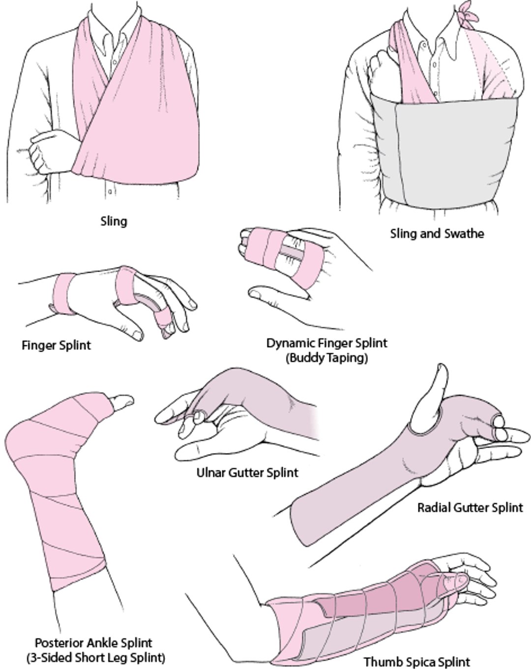
A sling provides some degree of support and limits mobility; it can be useful for injuries that are adversely affected by complete immobilization (eg, for shoulder injuries, which, if completely immobilized, can rapidly lead to adhesive capsulitis [frozen shoulder]).


A swathe (a piece of cloth or a strap) may be used with a sling to prevent the arm from swinging outward, especially at night. The swathe is wrapped around the back and over the injured part.
Prolonged immobilization (> 3 to 4 weeks for young adults) of a joint can cause stiffness, contractures, and muscle atrophy. These complications may develop rapidly and may be permanent, particularly in older adults. Some rapidly healing injuries are best treated with resumption of active motion within the first few days or weeks; such early mobilization may minimize contractures and muscle atrophy, thus accelerating functional recovery. Physical therapists can advise patients as to what they can do during immobilization to maintain as much function as possible (eg, elbow, wrist, and hand range-of-motion exercises if the shoulder is immobilized).
After immobilization, physical therapists can help patients regain or improve range of motion and muscle strength and can provide exercises to strengthen and stabilize the injured joint and thus help prevent recurrence and long-term impairment. Early range-of-motion exercises are important for all patients, especially those > 40 years old (2). More complications and morbidity can occur for immobilized patients as they age, particularly for anyone > 40 years old.
Surgery
Many 3rd-degree sprains and tendon tears require surgical repair.
Arthroscopic surgery is sometimes used. This procedure is used most often to repair ligaments or menisci in the knee.
Treatment references
1. Goldman AH, Tetsworth K. AAOS Clinical Practice Guideline Summary: Prevention of Surgical Site Infection After Major Extremity Trauma. J Am Acad Orthop Surg. 2023;31(1):e1-e8. doi:10.5435/JAAOS-D-22-00792
2. Braun C, McRobert CJ. Conservative management following closed reduction of traumatic anterior dislocation of the shoulder. Cochrane Database Syst Rev. 2019;5(5):CD004962. Published 2019 May 10. doi:10.1002/14651858.CD004962.pub4
Geriatrics Essentials: Sprains and Other Soft-Tissue Injuries
Older adults are predisposed to musculoskeletal injuries in general because of the following:
A tendency to fall frequently (eg, due to age-related loss of proprioception, adverse effects of medications or drugs on proprioception or postural reflexes, orthostatic hypotension)
Impaired protective reflexes during falls
For any musculoskeletal injury in older adults, the goal of treatment is rapid return to activities of daily living.
Immobility (joint immobilization) is more likely to have adverse effects (eg, stiffness, contractures, muscle atrophy) in older adults.
Early mobilization and physical therapy are essential to recovery of function.
Coexisting disorders (eg, arthritis) can interfere with recovery.
Key Points
Compartment syndrome and injuries that disrupt arterial supply threaten limb viability and may ultimately threaten life; these problems are very rare with sprains and strains.
Check for fractures and dislocations, and consider spontaneously reduced dislocations, as well as ligament, tendon, and muscle injuries; sometimes part of this evaluation is deferred until fracture is excluded.
Consider referred pain and examine the joints above and below the injured area, particularly if physical findings are normal in a joint that patients identify as painful (eg, shoulder pain in patients with a sternoclavicular joint injury).
Radiographs are not necessary for many injuries, including ankle sprains.
MRI can be used to diagnose soft-tissue injuries, but are usually not needed in the immediate workup.
Immediately treat serious associated injuries and splint unstable injuries, and treat pain as soon as possible.
Treat most minor injuries with PRICE (protection, rest, ice, compression, elevation).
Provide patients with explicit, written instructions about cast care.
Encourage patients, especially older adults, to perform the recommended exercises to maintain range of motion and muscle strength.
Drug Information for the Topic


