


Myocarditis is inflammation of the myocardium (heart muscle), usually with necrosis, of cardiac myocytes. Myocarditis may be caused by many disorders (eg, infection, cardiotoxins, medications, and systemic disorders such as sarcoidosis) but is often idiopathic. Symptoms can vary and can include fatigue, dyspnea, edema, palpitations, and sudden death. Diagnosis is based on symptoms and clinical findings of abnormal cardiac biomarkers, cardiac imaging, and electrocardiography (ECG), often in the absence of cardiovascular risk factors. Endomyocardial biopsy, when indicated, confirms clinical diagnosis of myocarditis. Treatment depends on the cause, but general measures include medications and devices to treat heart failure and arrhythmias and rarely mechanical circulatory support (eg, intra-aortic balloon pump, left ventricular assist device) or transplantation. Immunosuppression is used in certain types of myocarditis (eg, hypersensitivity myocarditis, giant cell myocarditis, myocarditis caused by sarcoidosis).
Pathophysiology of Myocarditis
Myocarditis is inflammation of the myocardium, usually with necrosis of cardiac myocytes. Biopsy-proven myocarditis typically demonstrates inflammatory infiltrate of the myocardium with lymphocytes, neutrophils, eosinophils, giant cells, granulomas, or a mixture.
The pathophysiology of myocarditis remains a subject of research. Potential mechanisms that lead to myocardial injury include:
Direct cardiomyocyte injury caused by an infectious or other cardiotoxic agent
Myocardial injury caused by an autoimmune or inflammatory reaction to an infectious or other cardiotoxic agent
Myocardial inflammation can be diffuse or focal. Inflammation can extend into the pericardium, causing myopericarditis. The extent of myocardial involvement and extension into adjacent pericardium determines the type of symptoms. Diffuse involvement can lead to heart failure, arrhythmias and sometimes sudden cardiac death. Focal involvement is less likely to cause heart failure but can lead to arrhythmias and sudden cardiac death. Involvement of the pericardium leads to chest pain and other symptoms typical of pericarditis. Some patients remain asymptomatic whether myocardial involvement is focal or diffuse.
Etiology of Myocarditis
Myocarditis may result from infectious or noninfectious causes. Many cases are idiopathic (see table ).
Infectious myocarditis is most often viral in North America and Western Europe (1, 2). Viruses that cause myocarditis may be cardiotropic, vasculotropic, lymphotropic, directly cardiotoxic, or angiotensin converting enzyme-2 tropic. Commonly implicated viruses include parvovirus B19, human herpes virus 6, enteroviruses, adenoviruses, Epstein-Barr virus, influenza, and coronaviruses (3). Of these, parvovirus B19 and human herpes virus 6 are the most commonly found in the subset of patients undergoing endomyocardial biopsy (4).
In selected populations, infectious myocarditis with other pathogens (eg, bacteria, parasites) is also often associated with rheumatic carditis, Lyme carditis, Chagas disease, or HIV infection (1, 5). Direct myocardial injury due to infection with SARS-CoV-2, with symptoms ranging from mild chest discomfort to fulminant myocarditis, is uncommon in patients with COVID-19 (< 1%), but the risk of myocarditis is 5 to16 times higher in those with infection than in those not infected (6).
Noninfectious causes include systemic autoimmune and inflammatory disorders, cardiotoxins, certain medications, and radiation therapy. Myocarditis caused by medications is termed hypersensitivity myocarditis. Myocarditis after mRNA-based COVID-19 vaccination is very rare (< 0.01% in the highest risk populations of males 12 to 29 years old) and far less common than COVID-associated myocarditis (6, 7, 8). It occurs mostly in adolescent and young adult males, usually within a week of vaccination, and is generally mild.
Eosinophilic myocarditis has various causes. It is an inflammatory cardiomyopathy characterized by eosinophilic infiltration; among the causes is hypersensitivity myocarditis (9).
Genetic substrate plays a significant role in some cases of myocarditis. Genetic variants, including heterozygous mutations in some genes associated with inherited arrhythmia or cardiomyopathy, may increase susceptibility to infectious or other environmental factors that can cause myocarditis (1, 3). There are also rare familial chronic inflammatory cardiomyopathies.
Causes of Myocarditis
Cause | Examples |
|---|---|
Viral infections | Enteroviruses, including coxsackie B virus Adenoviruses Coronaviruses, including SARS-CoV-2 |
Bacterial infections | Lyme disease (Borrelia burgdorferi) Group A streptococci |
Parasitic infections | Chagas disease (Trypanosoma cruzi) |
Fungal infections | |
Autoimmune disorders | Granulomatosis with polyangiitis Idiopathic inflammatory myopathies (eg, dermatomyositis) |
Inflammatory disorders | |
Cardiotoxins | Alcohol Cocaine |
Medications | Clozapine Immune checkpoint inhibitors Penicillins Thiazide diuretics Anthracycline chemotherapy (eg, doxorubicin) |
Radiation therapy | — |
Idiopathic | — |
Giant cell myocarditis
Giant cell myocarditis is a rare form of myocarditis with a fulminant course. The etiology is uncertain but may include an autoimmune mechanism. Biopsy shows characteristic multinucleated giant cells. Patients with giant cell myocarditis present in cardiogenic shock and frequently have intractable ventricular arrhythmias or complete heart block. Giant cell myocarditis has a poor prognosis and is important to identify when an otherwise healthy patient presents in fulminant heart failure or with intractable arrhythmias, because immunosuppressive therapy can help improve survival.
Inflammatory myopericardial syndromes
Inflammatory myopericardial syndromes encompass a spectrum of conditions involving inflammation of the pericardium and/or myocardium that is defined by the predominant site and extent of inflammation and includes pericarditis, myopericarditis (predominant pericarditis with extension to the myocardium), perimyocarditis (predominant myocarditis with extension to the pericardium), and myocarditis (5).
The European Society for Cardiology developed this phenotypic classification framework in an effort to::
Characterize structural phenotyping based on the dominant symptomatology (ie, chest pain/heart failure/arrhythmia/fulminant)
Prioritize cardiac MRI as the primary diagnostic tool (with endomyocardial biopsy limited to selected cases)
Emphasize that therapy is etiology-directed (eg, virus-positive inflammatory cardiomyopathy) rather than purely driven by clinical phenotype
Etiology references
1. Basso C. Myocarditis. N Engl J Med. 2022;387(16):1488-1500. doi:10.1056/NEJMra2114478
2. Bozkurt B, Colvin M, Cook J, et al. Current Diagnostic and Treatment Strategies for Specific Dilated Cardiomyopathies: A Scientific Statement From the American Heart Association. Circulation. 2016;134(23):e579-e646. doi:10.1161/CIR.0000000000000455
3. Tschöpe C, Ammirati E, Bozkurt B, et al. Myocarditis and inflammatory cardiomyopathy: current evidence and future directions. Nat Rev Cardiol. 2021;18(3):169-193. doi:10.1038/s41569-020-00435-x
4. Law YM, Lal AK, Chen S, et al. Diagnosis and Management of Myocarditis in Children: A Scientific Statement From the American Heart Association. Circulation. 2021;144(6):e123-e135. doi:10.1161/CIR.0000000000001001
5. Schulz-Menger J, Collini V, Gröschel J, et al. 2025 ESC Guidelines for the management of myocarditis and pericarditis. Eur Heart J. 2025;46(40):3952-4041. doi:10.1093/eurheartj/ehaf192
6. Boehmer TK, Kompaniyets L, Lavery AM, et al. Association Between COVID-19 and Myocarditis Using Hospital-Based Administrative Data - United States, March 2020-January 2021. MMWR Morb Mortal Wkly Rep. 2021;70(35):1228-1232. doi:10.15585/mmwr.mm7035e5
7. Patone M, Mei XW, Handunnetthi L, et al. Risk of Myocarditis After Sequential Doses of COVID-19 Vaccine and SARS-CoV-2 Infection by Age and Sex. Circulation. 2022;146(10):743-754. doi:10.1161/CIRCULATIONAHA.122.059970
8. Zuin M, Rigatelli G, Bilato C, et al. One-Year Risk of Myocarditis After COVID-19 Infection: A Systematic Review and Meta-analysis. Can J Cardiol. 2023;39(6):839-844. doi:10.1016/j.cjca.2022.12.003
9. Zhong Z, Yang Z, Peng Y, Wang L, Yuan X. Diagnosis and treatment of eosinophilic myocarditis. J Transl Autoimmun. 2021;4:100118. doi:10.1016/j.jtauto.2021.100118
Symptoms and Signs of Myocarditis
The clinical presentation of myocarditis is variable. Patients may be minimally symptomatic or have fulminant heart failure and fatal arrhythmias. Symptoms depend on the etiology of the myocarditis as well as the extent and severity of myocardial inflammation.
Patients may report an antecedent viral syndrome, upper respiratory symptoms, or other nonspecific illness, often 1 to 4 weeks before the onset of myocarditis symptoms. Or, they may report a history of exposure to a toxin or medication known to cause myocarditis.
Heart failure symptoms may include fatigue, dyspnea, and edema. Patients may show signs of fluid overload with crackles, elevated jugular venous pulses, and edema. Cardiac examination may be significant for a third (S3) or fourth (S4) heart sound. Systolic murmurs of mitral regurgitation and tricuspid regurgitation may be present in patients with ventricular enlargement. Pulsus alternans, palpable on physical examination or visible on pulse oximeter or arterial pressure waveforms, indicates severe left ventricle systolic dysfunction.
Sudden cardiac death due to a fatal arrhythmia is sometimes the presenting feature. Patients may experience preceding palpitations or syncope.
When patients have concomitant pericardial inflammation, they may present with chest pain typical of pericarditis. Dull or sharp precordial or substernal pain may radiate to the neck, trapezius ridge (especially the left), or shoulders. Pain ranges from mild to severe. Unlike ischemic chest pain, pain due to pericarditis is usually aggravated by thoracic motion, cough, breathing, or swallowing food; it may be relieved by sitting up and leaning forward. A pericardial friction rub may be auscultated in patients with a pericardial effusion.
Certain clinical findings may be indicative of a specific cause of myocarditis. Infectious myocarditis may be preceded by symptoms of fever, myalgias, and other symptoms depending on the exact pathogen. Medication-related or hypersensitivity myocarditis may be accompanied by a rash. Enlarged lymph nodes may be indicative of sarcoidosis as the underlying etiology. Fulminant heart failure and arrhythmias may be indicative of giant cell myocarditis.
Myocarditis can be acute, subacute, or chronic. There are no set time-frames for each phase. The acute phase typically lasts a few days while the subacute phase lasts weeks to months. If myocarditis does not resolve after a few months, it is called chronic myocarditis. In some cases, myocarditis can lead to dilated cardiomyopathy. Fulminant myocarditis (also called acute fulminant myocarditis) is a sudden, severe, and life-threatening subset of myocarditis that requires inotropic and/or mechanical support prior to recovery (or transplant) (1).
Symptoms and signs reference
1. Kociol RD, Cooper LT, Fang JC, et al. Recognition and Initial Management of Fulminant Myocarditis: A Scientific Statement From the American Heart Association. Circulation. 2020;141(6):e69-e92. doi:10.1161/CIR.0000000000000745
Diagnosis of Myocarditis
Cardiac troponin and complete blood count
Cardiac imaging (echocardiography and cardiac MRI)
Electrocardiography (ECG)
Sometimes, endomyocardial biopsy
Tests to identify cause
Myocarditis should be suspected when otherwise healthy patients with no cardiac risk factors present with symptoms of heart failure or arrhythmias. Cardiac enzymes, cardiac imaging, and ECG are not specific for myocarditis but can be diagnostic in the appropriate clinical setting (1).
The stepwise diagnostic approach begins with recognition of the clinical syndrome of myocarditis, with initial testing including a cardiac troponin, a complete blood count, an echocardiogram, and an electrocardiogram (2). Hemodynamic stability and rhythm are evaluated and managed as needed, and the need for hospitalization or transfer to a heart failure center are assessed. Finally, a definitive diagnostic test, either cardiac MRI or endomyocardial biopsy, is performed (see figure ).
Evaluation and Management of Clinically Suspected Myocarditis
CBC = complete blood count; ECG = electrocardiography; LVAD = left ventricular assist device. Data from Writing Committee, Drazner MH, Bozkurt B, et al. 2024 ACC Expert Consensus Decision Pathway on Strategies and Criteria for the Diagnosis and Management of Myocarditis: A Report of the American College of Cardiology Solution Set Oversight Committee. J Am Coll Cardiol. 2025;85(4):391-431. doi:10.1016/j.jacc.2024.10.080 |


Cardiac troponin can be abnormal in patients with acute myocarditis. Cardiac troponin can be elevated due to inflammation and/or necrosis of cardiac myocytes. High-sensitivity troponin assays have a reported sensitivity of 64 to 100% for acute myocarditis (3). Creatine kinase (CK) isoenzymes are not used in the diagnosis of myocarditis.
Cardiac imaging can be abnormal in patients with myocarditis, and cardiac MRI can be diagnostic.
An echocardiogram can be normal in early or mild myocarditis, but segmental wall motion abnormalities (mimicking myocardial ischemia) can be seen. Left ventricular dilation or other morphologic changes and global systolic dysfunction can also be seen as in dilated cardiomyopathy. Global strain may be abnormal. Diastolic function is often abnormal on echocardiography.
Cardiac MRI is important in the diagnosis of myocarditis and in many cases is the definitive diagnostic modality. Cardiac MRI of patients with myocarditis may show a characteristic pattern of late gadolinium enhancement in the subepicardial and mid-myocardial walls (in contrast to ischemia where late gadolinium enhancement is usually subendocardial with extension to mid-myocardial and epicardial walls). Other features of myocarditis on cardiac MRI are the presence of myocardial edema and myocardial hyperemia relative to skeletal muscle. Formal diagnosis based on cardiac MRI requires at least one T1-based finding supporting myocardial edema and at least one T2-based finding supporting non-ischemic myocardial injury, with findings of pericarditis or left ventricle systolic dysfunction providing supporting evidence (4).
ECG can be normal or abnormal in patients with myocarditis. ST segment abnormalities are common and can mimic myocardial ischemia. ST segment elevation is sometimes seen, but more common findings include nonspecific ST-T wave changes. Atrial and ventricular enlargement may also be reflected on ECG. Patients may experience conduction delays and atrial or ventricular arrhythmias, including sinus tachycardia, ventricular tachycardia, and ventricular fibrillation. Heart block may occur in Lyme carditis, giant cell myocarditis, or sarcoidosis. Low QRS voltage may signal severe edema and/or ventricular dysfunction.

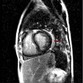
Patchy mid wall (bottom white arrow) and epicardial (top white arrow) delayed enhancement on MRI is consistent with myocarditis. This patient also has a pericardial effusion (red arrow).
Endomyocardial biopsy showing inflammatory infiltrate of the myocardium with necrosis of adjacent myocytes is the gold standard for diagnosis of myocarditis. However, this test has low sensitivity for diagnosis of myocarditis due to sampling error (5). Therefore, a positive biopsy result is diagnostic for myocarditis, but a negative result does not exclude it. In addition, the biopsy carries a risk of complications, including myocardial rupture and death, so is not routinely performed. Endomyocardial biopsy should be performed in cases of fulminant heart failure, ventricular arrhythmias or heart block, or if results would affect management and the potential benefit outweighs the risk (eg, if there is suspicion for giant cell myocarditis where prompt treatment can decrease mortality rates). Endomyocardial biopsy should also be considered in cases of suspected immune checkpoint inhibitor myocarditis with peripheral eosinophilia to evaluate for eosinophilic myocarditis, as well as in cases of dilated cardiomyopathy, high grade heart block, ventricular arrhythmia, and heart failure where the diagnosis is unclear but myocarditis remains a diagnostic possibility (2, 5, 6).
Stage of myocarditis
The 2024 American College of Cardiology guideline outlines a staging system for myocarditis based on clinical presentation as well as cardiac MRI and biopsy findings, that guides disease management, followup, and return to exercise (2) (see table ).
Myocarditis Staging and Management
Stage | Stage A | Stage B | Stage C | Stage D |
|---|---|---|---|---|
Description | At risk | Asymptomatic | Symptomatic | Severe |
Symptoms | No | No | Yes | Yes |
Diagnostic testing (biopsy, CMRI, troponin) | Normal | Biopsy or CMRI meets diagnostic criteria OR abnormal cardiac troponin with additional supportive evidence | Biopsy or CMRI meets diagnostic criteria OR abnormal cardiac troponin with additional supportive evidence | Biopsy or CMRI meets diagnostic criteria OR abnormal cardiac troponin with additional supportive evidence |
Additional testing (if not already done) | None | ECG | ECG; Consider biopsy | ECG; Perform biopsy |
Management | Remove offending agent when appropriate Risk counseling | Remove offending agent when appropriate Treat cause | Consider hospitalization and/or referral to heart failure center Guideline-directed medical heart failure therapy Immunosuppression or other etiology-based treatment | Hospitalize in intensive care unit; refer to heart failure center Hemodynamic stabilization, including mechanical support as needed Arrhythmia treatment Guideline-directed medical heart failure therapy Long-term mechanical support or transplantation if needed |
Follow-up | Monitor for progression | Monitor for symptoms Repeat CMRI and echocardiogram Consider genetic counseling/testing | Serial troponin, CMRI, and echocardiography Consider genetic counseling/testing | Serial troponin, CMRI, and echocardiography Consider genetic counseling/testing |
Activity | N/A | Consider clinical scenario | Restrict strenuous activity for 3–6 months; return to activity per guidelines | Restrict strenuous activity for 3–6 months; return to activity per guidelines |
Writing Committee, Drazner MH, Bozkurt B, et al. 2024 ACC Expert Consensus Decision Pathway on Strategies and Criteria for the Diagnosis and Management of Myocarditis: A Report of the American College of Cardiology Solution Set Oversight Committee. J Am Coll Cardiol. 2025;85(4):391-431. doi:10.1016/j.jacc.2024.10.080 | ||||
CMRI = cardiac magnetic resonance imaging. | ||||
Diagnosis of cause
After myocarditis is diagnosed, tests to determine etiology are performed. In a young, previously healthy adult who presents with symptoms of a viral infection and myocarditis, an extensive evaluation is usually unnecessary. Differentiating viral from idiopathic myocarditis is difficult, expensive, and generally of little practical importance.
A complete blood count (CBC) is helpful to assess for peripheral eosinophilia, which is present in hypersensitivity myocarditis.
Cardiac catheterization or coronary CT scan may be useful for excluding coronary artery disease since myocarditis can mimic myocardial infarction or myocardial ischemia.
In other cases, a biopsy of myocardial tissue may be needed to establish a diagnosis. Acid-fast stains of myocardial tissue are essential if tuberculosis (TB) is considered possible (TB myocarditis can be aggressive and can worsen rapidly with glucocorticoid therapy). Myocardial samples are examined for giant cells, which are characteristic of giant cell myocarditis, and the granulomas that occur in sarcoidosis.
Other tests include acute-phase reactants, routine chemistry tests, cultures, autoimmune tests, and, when appropriate, tests for HIV infection, SARS-CoV-2 infection, histoplasmosis complement fixation or Lyme disease titers (in endemic areas), and antibody tests for coxsackievirus, influenza virus, streptococcus, and other organisms. Nucleic acid antibody testing is performed on biopsy specimens, especially to exclude infection prior to immunosuppressive therapy, and for blood-based diagnosis of other causes, such as acute Chagas disease (6).
Genetic testing should be considered in patients with recurrent myocarditis or pericarditis, persistent troponin elevation, family history of myocarditis or pericarditis, severe ventricular arrhythmia, or certain patterns of late gadolinium enhancement on MRI (6).
Diagnosis references
1. Tschöpe C, Ammirati E, Bozkurt B, et al. Myocarditis and inflammatory cardiomyopathy: current evidence and future directions. Nat Rev Cardiol. 2021;18(3):169-193. doi:10.1038/s41569-020-00435-x
2. Writing Committee, Drazner MH, Bozkurt B, et al. 2024 ACC Expert Consensus Decision Pathway on Strategies and Criteria for the Diagnosis and Management of Myocarditis: A Report of the American College of Cardiology Solution Set Oversight Committee. J Am Coll Cardiol. 2025;85(4):391-431. doi:10.1016/j.jacc.2024.10.080
3. Ammirati E, Moslehi JJ. Diagnosis and Treatment of Acute Myocarditis: A Review. JAMA. 2023;329(13):1098-1113. doi:10.1001/jama.2023.3371
4. Ferreira VM, Schulz-Menger J, Holmvang G, et al. Cardiovascular Magnetic Resonance in Nonischemic Myocardial Inflammation: Expert Recommendations. J Am Coll Cardiol. 2018;72(24):3158-3176. doi:10.1016/j.jacc.2018.09.072
5. Seferović PM, Tsutsui H, McNamara DM, et al. Heart Failure Association of the ESC, Heart Failure Society of America and Japanese Heart Failure Society Position statement on endomyocardial biopsy. Eur J Heart Fail. 2021;23(6):854-871. doi:10.1002/ejhf.2190
6. Schulz-Menger J, Collini V, Gröschel J, et al. 2025 ESC Guidelines for the management of myocarditis and pericarditis. Eur Heart J. 2025;46(40):3952-4041. doi:10.1093/eurheartj/ehaf192
Treatment of Myocarditis
Treatment of heart failure and arrhythmias
Treatment of specific etiologies
The overall approach to the treatment of myocarditis is based on disease severity and the underlying cause (1). Hemodynamic or electrical instability is treated first, with invasive therapies when necessary. Heart failure is managed medically once stable. Cause-specific treatment may include removal of inciting agents, use of immunosuppressive agents, and/or antimicrobial therapy. There should also be ongoing surveillance with biomarkers and imaging to guide clinical decisions. The table summarizes management strategies based on disease stage.
Hemodynamic instability and cardiogenic shock in cases of severe or fulminant myocarditis should be identified and managed with diuresis, afterload reduction, inotropic support, and mechanical circulatory support when indicated. Intra-aortic balloon pump (IABP), left ventricular assist device (LVAD), or transplantation may be necessary. Similarly, atrial and ventricular arrhythmias are treated with antiarrhythmic therapy. Heart block can be treated with temporary pacing but may require insertion of a permanent pacemaker if conduction abnormalities persist.
Once the patient is stabilized, heart failure should managed according to established guideline-directed medical therapy. For patients with a reduced left ventricular ejection fraction, management typically includes a combination of angiotensin-converting enzyme inhibitors (ACE inhibitors), angiotensin receptor/neprilysin inhibitors (ARNIs), beta-blockers, mineralocorticoid receptor antagonists, receptor blockers (ARBs), and sodium-glucose co-transporter 2 (SGLT2) inhibitors.
For non-infectious causes, particularly hypersensitivity myocarditis, removal of the causative agent is a critical part of treatment.
Symptomatic treatment of chest pain may include nonsteroidal anti-inflammatory drugs (NSAIDs) or colchicine, particularly for chest pain with associated pericardial inflammation, although NSAIDS should generally be avoided if left ventricular ejection fraction is significantly reduced (1).
For symptomatic patients meeting diagnostic criteria for myocarditis, strenuous activity including competitive sports is restricted for 3 to 6 months, with the timing of gradual return to play determined by:
Clinical course
Resolution of myocardial inflammation on MRI
Arrhythmia burden on ambulatory ECG (Holter monitor)
Exercise capacity, symptoms, and ectopy during exercise electrocardiography
Treatment of specific causes
Infectious myocarditis
Infectious myocarditis is generally treated with supportive therapy for associated heart failure and arrhythmias. In general, infectious disease consultation should be considered prior to the use of antimicrobial therapy if bacterial myocarditis is suspected (1).
Antiviral therapy has not been shown to be helpful in the treatment of most viral etiologies. However, nirmatrelvir and ritonavir can help treat myocarditis due to SARS-CoV2 infection (2). Glucocorticoids may also help treat myocarditis due to SARS-CoV2 infection (3). Oseltamivir, baloxavir, peramivir, and zanamivir are used to treat myocarditis due to influenza (4, 5, 6).
Lyme carditis is treated with oral doxycycline, amoxicillin, or cefuroxime or IV ceftriaxone (6).
For Chagas disease, benznidazole can be considered in early or mild disease but is of minimal clinical benefit in established chronic Chagas cardiomyopathy (6). Specific arrhythmia therapy, including medications and implantable-cardioverter defibrillators, is required in some patients.
Inflammatory or autoimmune disorders
Immunosuppression, usually beginning with glucocorticoids, is appropriate for patients with eosinophilic or giant cell myocarditis, chronic lymphocytic myocarditis without a viral cause, myocarditis due to autoimmune disorders, granulomatous myocarditis due to sarcoidosis, and immune checkpoint inhibitor (ICI) myocarditis (1, 7). Nucleic acid antibody testing, such as polymerase chain reaction (PCR) performed on biopsy samples, to exclude active infection, is often recommended prior to treatment.
Hypersensitivity myocarditis is treated by immediate discontinuation of the causative medication or cardiotoxin and glucocorticoid therapy.
Patients with giant cell myocarditis have improved survival when treated with immunosuppressants, usually glucocorticoids and calcineurin inhibitors (eg, cyclosporine or tacrolimus) (8).
Myocarditis caused by sarcoidosis can be treated with glucocorticoids or other immunosuppressants (9).
ICI-associated myocarditis is treated with high-dose glucocorticoids and adjunctive immunosuppressive agents are added for severe or steroid-refractory cases (7). Adjunctive immunosuppressive agents include abatacept, alemtuzumab, and antithymocyte globulin.
Immunosuppression using a combination of azathioprine and prednisone has been studied for biopsy-proven virus-negative inflammatory myocarditis (10). Expert opinion supports the notion that immunosuppression should only be a treatment strategy when the phenotype, including virology, supports it (6, 11).
Treating myocarditis with an inflammatory phenotype (ie, fever, elevated C-reactive protein, and/or inflammation seen on cardiac MRI) using interleukin 1 (IL-1) inhibitors such as anakinra remains investigational (12, 13, 14), but their established effectiveness in inflammatory pericarditis offers promise.
Treatment references
1. Writing Committee, Drazner MH, Bozkurt B, et al. 2024 ACC Expert Consensus Decision Pathway on Strategies and Criteria for the Diagnosis and Management of Myocarditis: A Report of the American College of Cardiology Solution Set Oversight Committee. J Am Coll Cardiol. 2025;85(4):391-431. doi:10.1016/j.jacc.2024.10.080
2. Souza KM, Carrasco G, Rojas-Cortés R, et al. Effectiveness of nirmatrelvir-ritonavir for the treatment of patients with mild to moderate COVID-19 and at high risk of hospitalization: Systematic review and meta-analyses of observational studies. PLoS One. 2023;18(10):e0284006. doi:10.1371/journal.pone.0284006
3. WHO Guidelines for Pharmacological Management of Pandemic Influenza A(H1N1) 2009 and Other Influenza Viruses. Geneva: World Health Organization; February 2010.
4. Kamarullah W, Nurcahyani, Mary Josephine C, Bill Multazam R, Ghaezany Nawing A, Dharma S. Corticosteroid Therapy in Management of Myocarditis Associated with COVID-19; a Systematic Review of Current Evidence. Arch Acad Emerg Med. 2021;9(1):e32. doi:10.22037/aaem.v9i1.1153
5. Law YM, Lal AK, Chen S, et al. Diagnosis and Management of Myocarditis in Children: A Scientific Statement From the American Heart Association. Circulation. 2021;144(6):e123-e135. doi:10.1161/CIR.0000000000001001
6. Schulz-Menger J, Collini V, Gröschel J, et al. 2025 ESC Guidelines for the management of myocarditis and pericarditis. Eur Heart J. 2025;46(40):3952-4041. doi:10.1093/eurheartj/ehaf192
7. Ganatra S, Barac A, Armenian S, et al. Diagnosis and Management of Cardiovascular Adverse Effects of Targeted Oncology Therapies: Bruton's Tyrosine Kinase, Immune Checkpoint, and Vascular Endothelial Growth Factor Inhibitors: 2025 ACC Concise Clinical Guidance: A Report of the American College of Cardiology Solution Set Oversight Committee. J Am Coll Cardiol. 2026;87(5):654-682. doi:10.1016/j.jacc.2025.10.018
8. Bang V, Ganatra S, Shah SP, et al. Management of Patients With Giant Cell Myocarditis: JACC Review Topic of the Week. J Am Coll Cardiol. 2021;77(8):1122-1134. doi:10.1016/j.jacc.2020.11.074
9. Baughman RP, Valeyre D, Korsten P, et al. ERS clinical practice guidelines on treatment of sarcoidosis. Eur Respir J. 2021;58(6):2004079. doi:10.1183/13993003.04079-2020
10. Chimenti C, Russo MA, Frustaci A. Immunosuppressive therapy in virus-negative inflammatory cardiomyopathy: 20-year follow-up of the TIMIC trial. Eur Heart J. 2022;43(36):3463-3473. doi:10.1093/eurheartj/ehac348
11. Caforio AL, Pankuweit S, Arbustini E, et al. Current state of knowledge on aetiology, diagnosis, management, and therapy of myocarditis: a position statement of the European Society of Cardiology Working Group on Myocardial and Pericardial Diseases. Eur Heart J. 2013;34(33):2636-2648d. doi:10.1093/eurheartj/eht210
12. Cavalli G, Pappalardo F, Mangieri A, Dinarello CA, Dagna L, Tresoldi M. Treating Life-Threatening Myocarditis by Blocking Interleukin-1. Crit Care Med. 2016;44(8):e751-e754. doi:10.1097/CCM.0000000000001654
13. Maunier L, Charbel R, Lambert V, Tissières P; CLOVIS study group. Anakinra in pediatric acute fulminant myocarditis. Ann Intensive Care. 2022;12(1):80. doi:10.1186/s13613-022-01054-0
14. Morini S, Filomia S, Saponara G, et al. Anakinra in Fulminant Acute Myocarditis: A Case Report and Review of the Literature. J Cardiovasc Pharmacol. 2026;87(1):19-27. doi:10.1097/FJC.0000000000001762
Key Points
Clinical presentation of myocarditis is variable, ranging from subclinical disease to fulminant heart failure, intractable arrhythmias, and sudden cardiac death.
Diagnosis is often based on clinical findings and noninvasive testing, including cardiac MRI; perform endomyocardial biopsy if patients present with severe heart failure or intractable arrhythmias or if findings would change treatment.
Treat patients with heart failure and arrhythmias; immunosuppression or other therapies are added for specific etiologies.
Drug Information for the Topic


