


Epiglottitis is a rapidly progressive bacterial infection of the epiglottis and surrounding tissues that may lead to sudden respiratory obstruction and death. Symptoms include severe sore throat, dysphagia, high fever, drooling, and inspiratory stridor. Diagnosis requires direct visualization of the supraglottic structures; this procedure is not to be performed until full respiratory support is available. Treatment includes airway protection and antibiotics.
Epiglottitis is an acute infection and inflammation of the epiglottis. It used to primarily affect children and was usually caused by Haemophilus influenzae type B. Since the introduction of the H. influenzae type B (HiB) conjugate (Hib) vaccine in 1985 and its widespread use, epiglottitis has been almost eradicated in children. For example, in settings where vaccination is well adopted, the incidence of acute epiglottitis in children has been found to be < 1 case/100,000 annually (1, 2). Epiglottitis is now more frequent in adults, particularly in those with comorbidities (eg, cardiovascular, respiratory, or other infectious diagnoses) (3).
Causal organisms in children and adults include Streptococcus pneumoniae, Staphylococcus aureus, nontypeable H. influenzae, Haemophilus parainfluenzae, beta-hemolytic streptococci, Branhamella catarrhalis, and Klebsiella pneumoniae. H. influenzae type B is still a cause in adults and unvaccinated children. Noninfectious etiologies such as caustic injuries, thermal injuries, and autoimmune disease (eg, Sjögren syndrome) may rarely lead to the development of epiglottitis (4, 5).
Bacteria that have colonized the nasopharynx spread locally to cause supraglottic cellulitis with marked inflammation of the epiglottis, vallecula, aryepiglottic folds, arytenoids, and laryngeal ventricles. With H. influenzae type B, infection may spread hematogenously.
The inflamed supraglottic structures mechanically obstruct the airway, increasing the work of breathing, ultimately causing respiratory failure. Clearance of inflammatory secretions is also impaired.
General references
1. Isakson M, Hugosson S. Acute epiglottitis: epidemiology and Streptococcus pneumoniae serotype distribution in adults. J Laryngol Otol. 2011;125(4):390-393. doi:10.1017/S0022215110002446
2. Guldfred LA, Lyhne D, Becker BC. Acute epiglottitis: epidemiology, clinical presentation, management and outcome. J Laryngol Otol. 2008;122(8):818-823. doi:10.1017/S0022215107000473
3. Shah RK, Stocks C. Epiglottitis in the United States: national trends, variances, prognosis, and management. Laryngoscope. 2010;120(6):1256-1262. doi:10.1002/lary.20921
4. Orhan İ, Aydın S, Karlıdağ T. Infectious and Noninfectious Causes of Epiglottitis in Adults, Review of 24 Patients. Turk Arch Otorhinolaryngol. 2015;53(1):10-14. doi:10.5152/tao.2015.718
5. Hsu CM, Tsai MS, Yang YH, et al. Epiglottitis in Patients With Preexisting Autoimmune Diseases: A Nationwide Case-Control Study in Taiwan. Ear Nose Throat J. 2024;103(1):NP40-NP48. doi:10.1177/01455613211033689
Symptoms and Signs of Epiglottitis
In children with epiglottitis, sore throat, odynophagia, and dysphagia develop abruptly, often with no visible oropharyngeal inflammation. Fatal asphyxia may occur within a few hours of onset. Drooling without a barking cough is a hallmark characteristic and is considered highly specific and sensitive for epiglottitis (1). Additionally, the child typically has signs of toxicity (poor or absent eye contact, cyanosis, irritability, inability to be consoled or distracted) and is febrile and anxious. Dyspnea, tachypnea, and inspiratory stridor may be present, often causing the child to sit upright, lean forward, and hyperextend the neck with the jaw thrust forward and mouth open in an effort to maximize air exchange (tripod position). Relinquishing this position may herald respiratory failure. Suprasternal, supraclavicular, and subcostal inspiratory retractions may be present.
In adults, symptoms are similar to those of children, including sore throat, fever, dysphagia, and drooling, but peak symptoms usually take > 24 hours to develop. Because of the larger diameter of the adult airway, obstruction is less common and less fulminant.
In many children and adults, oropharyngeal inflammation is not visible. However, severe throat pain with a normal-appearing pharynx raises suspicion of epiglottitis. A delay in diagnosis and treatment increases the risk of airway obstruction and death.
Symptoms and signs reference
1. Tibballs J, Watson T. Symptoms and signs differentiating croup and epiglottitis. J Paediatr Child Health. 2011;47(3):77-82. doi:10.1111/j.1440-1754.2010.01892.x
Diagnosis of Epiglottitis
Direct inspection (typically in the operating room)
Radiographs in milder cases with low suspicion
Epiglottitis is primarily a clinical diagnosis that should be suspected in patients with symptoms such as severe sore throat without pharyngitis, and sore throat and inspiratory stridor. Drooling in the absence of a barking cough is an important clinical feature suggesting epiglottitis. Stridor in children may also result from croup (viral laryngotracheal bronchitis—see table ), bacterial tracheitis, and airway foreign body. The tripod position may also occur with peritonsillar abscess or retropharyngeal abscess.
Patients with suspected epiglottitis should be hospitalized.
Differentiating Epiglottitis From Croup
Feature | Epiglottitis | Croup* |
|---|---|---|
Onset | Acute and fulminant in children | More gradual |
Age | Commonly, 2–8 years (particularly if not vaccinated against Haemophilus influenzae type B) and adults | Commonly, 6–36 months |
Barking cough | Uncommon | Characteristic |
Epiglottis | Edematous and cherry red | May be erythematous |
Neck radiograph findings | Enlarged epiglottis (thumb sign) and distention of the hypopharynx | Subglottic narrowing (steeple sign) and a normal-sized epiglottis |
* Also called viral laryngotracheal bronchitis. | ||
Diagnosis requires direct examination, usually with flexible fiberoptic laryngoscopy. (CAUTION:Examination of the pharynx and larynx may precipitate complete respiratory obstruction in children; only specifically trained personnel should directly examine the pharynx and larynx, and the examination should be performed in an operating room, where the most advanced airway intervention is available.) Direct laryngoscopy that reveals a beefy-red (or cherry red), stiff, edematous epiglottis is diagnostic. In some adult patients with epiglottis, flexible fiberoptic laryngoscopy can be performed safely. However, it is not always performed because of the potential for inducing airway collapse. Laryngoscopy in children with epiglottitis should be performed only in a controlled setting (eg, operating room) with immediate airway management capabilities, as it is both diagnostic and potentially life-threatening.
Although plain radiographs may be helpful, they are not highly accurate (1) and must be taken during inspiration, during neck extension, and with no rotation to avoid false-positive results. Lateral neck radiography should not delay definitive management in unstable patients. For example, a child with stridor should not be transported to the x-ray suite due to risk of a life-threatening decompensation and airway collapse either en route or during such positioning.
Cultures from the surface of the epiglottis and blood should then be obtained to identify the causative organism (2).

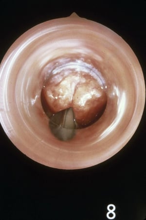
This image shows epiglottitis in an intubated patient. Note the stiff, edematous epiglottis in the upper part of the photo. The vocal cords are visible below and distal to the epiglottis.
This image shows epiglottitis in an intubated patient. Note the stiff, edematous epiglottis in the upper part of the ph
Image provided by Clarence T. Sasaki, MD.

This radiograph shows the enlarged epiglottis (thumb sign—see arrow) characteristic of epiglottitis and distention of the hypopharynx. Note the posteriorly displaced, thickened epiglottis.
This radiograph shows the enlarged epiglottis (thumb sign—see arrow) characteristic of epiglottitis and distention of t
Image provided by Clarence T. Sasaki, MD.

This anterior-posterior radiograph shows characteristic subglottic narrowing of the airway (steeple sign [arrow]) caused by croup.
This anterior-posterior radiograph shows characteristic subglottic narrowing of the airway (steeple sign [arrow]) cause
Image provided by Clarence T. Sasaki, MD.
This image shows epiglottitis in an intubated patient. Note the stiff, edematous epiglottis in the upper part of the photo. The vocal cords are visible below and distal to the epiglottis.
This image shows epiglottitis in an intubated patient. Note the stiff, edematous epiglottis in the upper part of the ph
Image provided by Clarence T. Sasaki, MD.
This radiograph shows the enlarged epiglottis (thumb sign—see arrow) characteristic of epiglottitis and distention of the hypopharynx. Note the posteriorly displaced, thickened epiglottis.
This radiograph shows the enlarged epiglottis (thumb sign—see arrow) characteristic of epiglottitis and distention of t
Image provided by Clarence T. Sasaki, MD.
This anterior-posterior radiograph shows characteristic subglottic narrowing of the airway (steeple sign [arrow]) caused by croup.
This anterior-posterior radiograph shows characteristic subglottic narrowing of the airway (steeple sign [arrow]) cause
Image provided by Clarence T. Sasaki, MD.
Pearls & Pitfalls
|
Diagnosis references
1. Fujiwara T, Miyata T, Tokumasu H, et al. Diagnostic accuracy of radiographs for detecting supraglottitis: A systematic review and meta-analysis. Acute Med Surg. 2016;4 (2):190–197. doi: 10.1002/ams2.256 eCollection 2017 Apr.
2. Alcaide ML, Bisno AL. Pharyngitis and epiglottitis. Infect Dis Clin North Am. 2007 Jun;21(2):449-69, vii. doi: 10.1016/j.idc.2007.03.001. Erratum in: Infect Dis Clin North Am. 2007 Sep;21(3):847-8. PMID: 17561078; PMCID: PMC7126481
Treatment of Epiglottitis
Airway establishment
Antibiotics (eg, ceftriaxone)
Supportive care
The treatment of epiglottitis consists of airway management, empiric intravenous antibiotics, and supportive care.
Airway assessment and management are paramount and take priority over all other interventions. Stridor, respiratory distress, drooling, and epiglottic abscess are important predictors for the need for urgent airway management. In children with stridor, any intervention that could be upsetting and thus could trigger airway obstruction should be avoided until an airway is established. In children with epiglottitis, the airway must be secured immediately. Securing the airway can be quite difficult and should, if possible, be performed by experienced personnel in the operating room. An endotracheal tube is usually required until the patient has been stabilized for 24 to 48 hours (usual total intubation time is < 60 hours for both children and adults). Alternatively, a tracheotomy may be performed. If respiratory arrest occurs before an airway is established, bag-mask ventilation may be a life-saving temporary measure. For emergency care of children with epiglottitis, each institution should have a protocol that involves critical care, otolaryngology, anesthesia, and pediatrics.
Adults whose airway is severely obstructed can be endotracheally intubated during flexible fiberoptic laryngoscopy. Other adults may not require immediate intubation but should be observed for airway compromise in an intensive care unit with an intubation set and cricothyrotomy or tracheostomy tray at the bedside.
Empiric broad-spectrum intravenous antibiotics should be administered promptly upon diagnosis (1). A beta-lactamase–resistant antibiotic, such as a third-generation cephalosporin (eg, ceftriaxone 50 to 75 mg/kg IV once/day [maximum 2 g]), should be used; antibiotic therapy may be narrowed after receiving culture and sensitivity test results.
Supportive care includes the administration of humidified oxygen, which must be administered to all patients with epiglottitis, and proper positioning (typically upright), and avoidance of undue agitation (2).
Epiglottitis caused by H. influenzae type B can be effectively prevented with the H. influenzae type B (HiB) conjugate vaccine.
Treatment references
1. Sur DKC, Plesa ML. Antibiotic Use in Acute Upper Respiratory Tract Infections. Am Fam Physician. 2022;106(6):628-636.
2. Kivekäs I, Rautiainen M. Epiglottitis, Acute Laryngitis, and Croup. Infections of the Ears, Nose, Throat, and Sinuses. 2018;247-255. Published 2018 May 4. doi:10.1007/978-3-319-74835-1_20
Key Points
The incidence of epiglottitis has decreased significantly, particularly in children, because of widespread vaccination against the most common cause, Haemophilus influenzae type B.
Stridor, as well as sore throat with a normal-appearing pharynx, should raise suspicion of epiglottitis.
In children with stridor and suspected epiglottitis, examination of the pharynx or larynx may precipitate complete airway obstruction; only specifically trained personnel should directly examine the pharynx and larynx, and the examination should be performed in an operating room.
If the diagnosis is suspected, flexible fiberoptic laryngoscopy may be performed in the operating room; reserve imaging tests for cases with very low suspicion.
Children typically should have their airway secured by tracheal intubation; if airway obstruction is not severe, adults can often be monitored closely for signs of airway compromise in an intensive care unit.
Administer a beta-lactamase–resistant antibiotic, such as ceftriaxone.
Drug Information for the Topic


