

Trauma to the spine may cause injuries involving the spinal cord, vertebrae, or both. Occasionally, the spinal nerves are affected. For more information see anatomy of the spinal column.
Spinal cord injury may be:
Complete
Incomplete
(See also Approach to the Trauma Patient.)
Etiology of Spinal Trauma
Spinal cord injury
During a typical year, there are approximately 18,000 spinal cord injuries in the United States, or 54 cases per million population (1).
The most common causes of spinal cord injuries are the following:
Motor vehicle crashes
Falls
A United States population database of patients with spinal cord injuries found that motor vehicle crashes accounted for 38% of injuries and falls for 31% (2). The remainder of spinal cord injuries were attributed to violence (13%), sports (9%), and complications of medical or surgical procedures (5%). Approximately 80% of patients were male.
In older patients, falls are the most common cause of spinal cord injury (1, 3). Osteoporotic bones and degenerative joint disease may increase the risk of cord injury at lower impact velocities due to angulations formed by the degenerated joints, osteophytes impinging on the cord, and brittle bone allowing for easy fracture through critical structures.
Spinal cord injuries occur when blunt physical force damages the vertebrae, ligaments, or disks of the spinal column, causing bruising, crushing, or tearing of spinal cord tissue, and when the spinal cord is penetrated (eg, by a gunshot or a knife wound). Such injuries can also cause vascular injury with resultant ischemia or hematoma (typically extradural), leading to further damage. All forms of injury can cause spinal cord edema, further decreasing blood flow and oxygenation. Damage may be mediated by excessive release of neurotransmitters from damaged cells, an inflammatory immune response with release of cytokines, accumulation of free radicals, and apoptosis.
Vertebral injury
Vertebral Column and Spinal Anatomy
Macrovector/stock.adobe.com |


Vertebral injuries may be:
Fractures, which may involve the vertebral body, lamina, and pedicles as well as the spinous, articular, and transverse processes
Dislocations, which typically involve the facets
Subluxations, which may involve ligament rupture without bony injury
In the neck, fractures of the posterior elements and dislocations can damage the vertebral arteries, causing a syndrome resembling a brain stem stroke.
Unstable vertebral injuries are those in which bony and/or ligamentous integrity are disrupted sufficiently that free movement can occur, potentially compressing the spinal cord or its vascular supply and resulting in marked pain and potential worsening of neurologic function. Such vertebral movement may occur even with a shift in patient position (eg, for ambulance transport, during initial evaluation). Stable vertebral fractures are able to resist such movement.

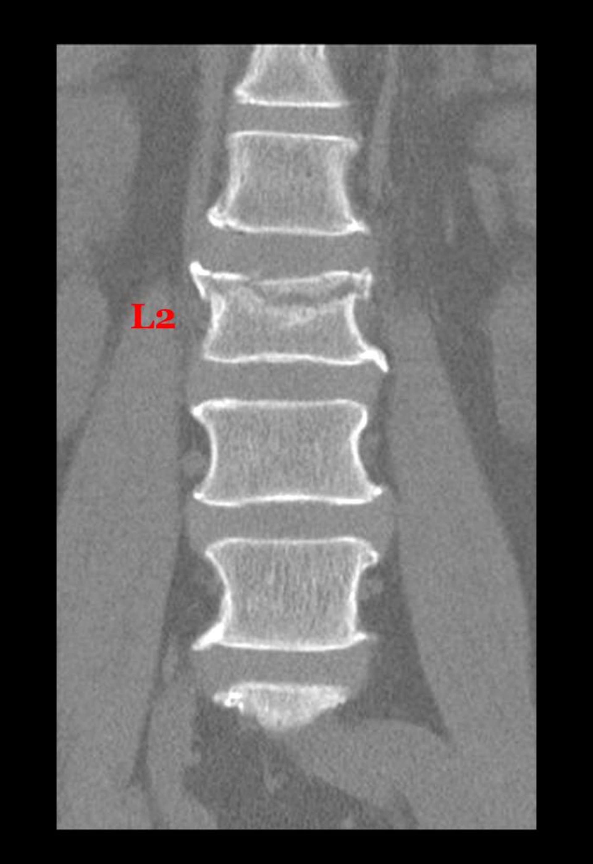
This coronal (frontal) view from a CT shows a fracture through the superior aspect of the body of the 2nd lumbar vertebra.
Living Art Enterprises, LLC /SCIENCE PHOTO LIBRARY
Specific injuries typically vary with mechanism of trauma. Flexion injuries can cause wedge fractures of the vertebral body or spinous process fractures. Greater flexion force may cause bilateral facet dislocation, or if the force occurs at the level of C1 or C2, odontoid fracture, atlanto-occipital or atlantoaxial subluxation, or both fracture and subluxation. Rotational injury can cause unilateral facet dislocation. Extension injury most often causes posterior neural arch fracture. Compression injuries can cause burst fractures of vertebral bodies.
Cauda equina injury
The lower tip of the spinal cord (conus medullaris) is usually at, or above, the level of the L1 vertebra. Spinal nerves below this level comprise the cauda equina. Thus, findings in spinal injuries below this level may mimic those of spinal cord injury, particularly conus medullaris syndrome (see table ).
Etiology references
1. Jain NB, Ayers GD, Peterson EN, et al. Traumatic spinal cord injury in the United States, 1993-2012. JAMA. 2015;313(22):2236-2243. doi:10.1001/jama.2015.6250
2. Chen Y, He Y, DeVivo MJ. Changing Demographics and Injury Profile of New Traumatic Spinal Cord Injuries in the United States, 1972-2014. Arch Phys Med Rehabil. 2016;97(10):1610-1619. doi:10.1016/j.apmr.2016.03.017
3. Bruggink C, van de Ree CLP, van Ditshuizen J, et al. Increased incidence of traumatic spinal injury in patients aged 65 years and older in the Netherlands. Eur Spine J. 2024;33(10):3677-3684. doi:10.1007/s00586-024-08310-w
Symptoms and Signs of Spinal Trauma
The cardinal sign of spinal cord injury is a discrete injury level in which neurologic function above the injury is intact, and function below the injury is absent or markedly diminished. Muscle strength is assessed using the standard 0 to 5 scale. Specific manifestations depend on the exact level (see table ) and whether cord injury is complete or incomplete. Priapism may occur in the acute phase of spinal cord injury.
In addition to motor and sensory function, upper motor neuron signs are an important finding in cord injury. These signs include increased deep tendon reflexes and muscle tone, a plantar extensor response or Babinski sign (upgoing great toe), clonus (most commonly found at the ankle by rapidly flexing the foot upward), and a Hoffman reflex (a positive response is flexion of the terminal phalanx of the thumb after flicking the nail of the middle finger).
Vertebral injury, as with other fractures and dislocations, is typically painful, but patients who are distracted by other painful injuries (eg, long bone fractures) or whose level of consciousness is altered by intoxicants or head injury may not complain of pain.
Effects of Spinal Cord Injury by Location
Vertebral Level of Injury* | Possible Effects† |
|---|---|
At or above C5 | Respiratory paralysis Quadriplegia |
Between C5 and C6 | Paralysis of legs, wrists, and hands Weakened shoulder abduction and elbow flexion Loss of brachioradialis deep tendon reflex |
Between C6 and C7 | Paralysis of legs, wrists, and hands, but shoulder movement and elbow flexion usually possible Loss of biceps jerk reflex |
Between C7 and C8 | Paralysis of legs and hands Loss of triceps jerk reflex |
At C8 to T1 | With transverse lesions, Horner syndrome (ptosis, miotic pupils, facial anhidrosis) Paralysis of legs |
Between T1 and L1 | Paralysis of muscles of the trunk and lower extremities Loss of sensation below the shoulders Loss of bowel and bladder control |
Cauda equina (usually approximately L2 to S5) | Hyporeflexic or areflexic paresis of the lower extremities Usually pain and hyperesthesia in the distribution of the affected nerve roots Usually loss of bowel and bladder control |
At S3 to S5 or conus medullaris at L1 | Complete loss of bowel and bladder control Decreased perineal sensation (saddle anesthesia) Variable degrees of leg weakness depending on location of lesion and associated injury |
* Abbreviations refer to vertebral level. Note that the vertebral level of injury may not correspond directly to the affected spinal cord segment. Because the sensory and motor nerve roots take off from spinal cord 1 to 2 vertebral levels above the foramen and disc level, the noted neurologic level of injury is often higher than the radiographic level of compression and injury. | |
† Priapism, reduced rectal tone, and changes in caudal reflexes may occur with injury at any level. | |
Complete cord injury
Complete spinal cord injury leads to:
Immediate, complete, flaccid paralysis (including loss of anal sphincter tone)
Loss of all sensation and reflex activity
Autonomic dysfunction below the level of the injury
High cervical injury (at or above C5) affects the muscles controlling respiration, causing respiratory insufficiency; ventilator dependence may occur, especially in patients with injuries at or above C3. Autonomic dysfunction due to cervical cord injury can result in bradycardia and hypotension; this condition is termed neurogenic shock. Unlike in other forms of shock, the skin remains warm and dry. Arrhythmias and blood pressure instability may develop.
Flaccid paralysis gradually changes over hours to weeks to spastic paralysis with increased deep tendon reflexes due to loss of descending inhibition. Later, if the lumbosacral cord is intact, flexor muscle spasms appear and autonomic reflexes return.
Incomplete cord injury
In incomplete spinal cord injury, motor and sensory loss occurs, and deep tendon reflexes may be hyperactive, but sensation is preserved at least in segments S4 to S5 (1). Motor and sensory loss may be permanent or temporary, depending on the etiology; function may be lost briefly due to concussion or more lastingly due to a contusion or laceration. Sometimes, however, rapid swelling of the cord results in total neurologic dysfunction that resembles complete cord injury; this condition is termed spinal shock (not to be confused with neurogenic shock). Symptoms resolve over one to several days, but residual disability often remains.
Spinal Cord Anatomy
Macrovector/stock.adobe.com |


Manifestations depend on which portion of the cord is involved; several discrete syndromes are recognized (see table ).
Brown-Séquard syndrome results from unilateral hemisection of the cord. Patients have ipsilateral spastic paralysis and loss of position sense below the lesion, and contralateral loss of pain and temperature sensation.
Anterior cord syndrome results from direct injury to the anterior spinal cord or to the anterior spinal artery. Patients lose motor and pain sensation bilaterally below the lesion. Posterior cord function (vibration, proprioception) is intact.
Central cord syndrome usually occurs in patients with a narrowed cervical spinal canal (congenital or degenerative) after a hyperextension injury. Motor function in the arms is impaired to a greater extent than that in the legs. If the posterior columns are affected, posture, vibration, and light touch are lost. If the spinothalamic tracts are affected, pain, temperature, and, often, light or deep touch are lost. Hemorrhage in the spinal cord resulting from trauma (hematomyelia) is usually confined to the cervical central gray matter, resulting in signs of lower motor neuron damage (muscle weakness and wasting, fasciculations, and diminished tendon reflexes in the arms), which is usually permanent. Motor weakness is often proximal and accompanied by selective impairment of pain and temperature sensation.
Cauda equina lesions
Motor loss or sensory loss, or both, usually partial, occurs in the distal legs. Sensory symptoms are generally bilateral but usually asymmetric, affecting one side more than the other. Sensation is usually diminished in the perineal region (saddle anesthesia). Bowel and bladder dysfunction, either incontinence or retention, may occur. Men may have erectile dysfunction, and women diminished sexual response. Anal sphincter tone is lax, and bulbocavernosus and anal wink reflexes are abnormal. These findings may be similar to those of conus medullaris syndrome.
Complications of spinal cord injury
Sequelae depend on the severity and level of the injury. Breathing may be impaired if the injury is at or above the C5 segment, although patients with injuries as low as T4 may also be at increased risk. Reduced mobility increases the risk of blood clots, urinary tract infections, contractures, atelectasis, pneumonia, and pressure ulcers. Disabling spasticity may develop. Cardiovascular instability is common soon after cervical cord injury and is related to neurogenic shock and autonomic dysreflexia that occur in response to triggering events such as pain or pressure on the body. Chronic neurogenic pain may manifest as burning or stinging.
Symptoms and signs reference
1. ASIA and ISCoS International Standards Committee. The 2019 revision of the International Standards for Neurological Classification of Spinal Cord Injury (ISNCSCI)-What's new?. Spinal Cord. 2019;57(10):815-817. doi:10.1038/s41393-019-0350-9
Diagnosis of Spinal Trauma
Consider spinal cord injury in patients with high-risk injuries (even in the absence of symptoms)
CT, MRI, or upright radiographs
Sometimes CT myelography for patients who are not candidates for MRI
Spinal injuries resulting from trauma are not always obvious. Injury to the spine and spinal cord must be considered in patients with:
Injuries that involve the head
Pelvic fractures
Penetrating injuries in the area of the spine
Injuries sustained in motor vehicle crashes
Severe blunt injuries
Injuries sustained by falling from heights or diving into water
In older patients, spinal column injury must also be considered after minor falls.
Injury to the spine and spinal cord should also be considered in patients with altered sensorium, localized spinal tenderness, painful distracting injuries, or compatible neurologic deficits.
Diagnosis of spine and spinal cord injuries includes assessment of nerve function, including reflex, motor, and sensation, and imaging.
Manifestations of injury may be characterized using the ASIA (American Spinal Injury Association) Impairment Scale or a similar instrument (see table ).
Spinal Injury Impairment Scale*
Level | Impairment |
|---|---|
A | Complete: There is no motor or sensory function, including in the sacral segments S4–S5. |
B | Incomplete: Sensory but not motor function is preserved below the affected neurologic (spinal cord) level, including in the sacral segments S4–S5. |
C | Incomplete: Motor function is preserved below the neurologic level, and more than half of key muscles below the neurologic level have a muscle strength grade of < 3. |
D | Incomplete: Motor function is preserved below the neurologic level, and at least half of key muscles below the neurologic level have a muscle grade of ≥ 3. |
E | Normal: Motor and sensory function are normal. |
* According to the American Spinal Injury Association (ASIA) and the International Spinal Cord Society (ISCoS). Kirshblum S, Snider B, Rupp R, et al; International Standards Committee of ASIA and ISCoS. Updates of the International Standards for Neurologic Classification of Spinal Cord Injury: 2015 and 2019. Phys Med Rehabil Clin N Am. 2020;31(3):319-330. doi:10.1016/j.pmr.2020.03.005 ASIA and ISCoS International Standards Committee. The 2019 revision of the International Standards for Neurological Classification of Spinal Cord Injury (ISNCSCI)-What's new?. Spinal Cord. 2019;57(10):815-817. doi:10.1038/s41393-019-0350-9 | |
Nerve studies
Motor function is tested in all extremities. Sensation testing should involve light touch (posterior column function), pinprick (anterior spinothalamic tract), and position sense (posterior column function). Identification of the sensory level is best done by testing from distal to proximal and by testing thoracic roots on the back to avoid being misled by the cervical "cape"-like distribution (involving sensory changes at the upper back, base of the neck, and proximal upper arms) characteristic of central cord syndrome. Priapism indicates spinal cord damage. Rectal tone may be decreased, and deep tendon reflexes may be exuberant or absent.
Imaging
Traditionally, radiographs are taken of any possibly injured areas. CT is performed of areas that appear abnormal on radiographs and areas at risk of injury based on clinical findings. However, CT is being used increasingly as the primary imaging study for spinal trauma because it has better diagnostic accuracy and, at many trauma centers, can be obtained rapidly as a part of a standardized trauma protocol (1).
MRI helps identify the type and location of cord injury; it is the most accurate study for imaging the spinal cord itself and other soft tissues but may not be immediately available. In patients with implanted devices (eg, pacemaker) who are not candidates for MRI, CT myelography is sometimes performed to identify structures impinging on the spinal cord.

This CT shows a fracture (arrow) through the 7th cervical vertebra just posterior to the vertebral body.
DR P. MARAZZI/SCIENCE PHOTO LIBRARY

This lateral view of the cervical spine shows a fracture of the inferior, anterior aspect of the 6th vertebral body (arrow).
ZEPHYR/SCIENCE PHOTO LIBRARY
If a cervical spine fracture, subluxation, or ligamentous injury is identified, a vascular study is usually warranted (typically CT angiography) to exclude concomitant carotid or vertebral artery injuries (2, 3).
Diagnosis references
1. Expert Panel on Neurological Imaging and Musculoskeletal Imaging, Beckmann NM, West OC, et al. ACR Appropriateness Criteria® Suspected Spine Trauma. J Am Coll Radiol. 2019;16(5S):S264-S285. doi:10.1016/j.jacr.2019.02.002
2. Burlew CC, Biffl WL, Moore EE, et al. Blunt cerebrovascular injuries: Redefining screen criteria in the era of non-invasive diagnosis. J Trauma Acute Care Surg. 72(2):p 539, 2012. | doi: 10.1097/TA.0b013e31824b6133
3. Tobert DG, Le HV, Blucher JA, et al. The Clinical Implications of Adding CT Angiography in the Evaluation of Cervical Spine Fractures: A Propensity-Matched Analysis. J Bone Joint Surg Am. 2018;100(17):1490-1495. doi:10.2106/JBJS.18.00107
Treatment of Spinal Trauma
Immobilization
Maintenance of oxygenation and spinal cord perfusion
Supportive care
Surgical stabilization when appropriate
Long-term symptomatic care and rehabilitation
Immediate care
The initial management of spinal cord injuries includes immediate care to stabilize the patient and prevent further injury to the spine and/or spinal cord (1). In the absence of appropriate immobilization, flexion or extension of the spine can contuse or transect the cord and precipitate paraplegia, quadriplegia, or even death due to spinal injury.
Patients who may have a spinal injury should have the spine immobilized immediately; the neck is held straight manually (inline stabilization) during endotracheal intubation. As soon as possible, the spine is fully immobilized on a firm, flat, padded backboard or similar surface to stabilize the position without excessive pressure. A rigid collar should be used to immobilize the cervical spine. Patients with thoracic or lumbar spine injuries can be carried prone or supine. Those with cervical cord damage that could induce respiratory difficulties should be carried supine, with attention to maintaining a patent airway and avoiding chest constriction. Transfer to a trauma center is desirable.
Medical care should be directed at avoiding hypotension and hypoxia, both of which can further stress the injured cord. Many experts advocate maintaining mildly elevated blood pressure with mean arterial pressure (MAP) ≥ 85 to 90 mm Hg for 5 to 7 days to improve spinal cord perfusion and to reduce hypotensive episodes that may adversely affect recovery (1, 2, 3, 4). MAP targets can be achieved using volume expansion with crystalloids and/or colloids, vasopressors, or a combination. Injuries above T6–T7, because they compromise sympathetic output to the thoracic cardiopulmonary nerves, are treated with vasopressors that have chronotropic and inotropic effects such as norepinephrine and dopamine. Lesions below T7 may respond sufficiently to pure vasoconstrictors such as phenylephrine (). MAP targets can be achieved using volume expansion with crystalloids and/or colloids, vasopressors, or a combination. Injuries above T6–T7, because they compromise sympathetic output to the thoracic cardiopulmonary nerves, are treated with vasopressors that have chronotropic and inotropic effects such as norepinephrine and dopamine. Lesions below T7 may respond sufficiently to pure vasoconstrictors such as phenylephrine (5). Oxygen saturation should be maintained ≥ 90% to prevent cord ischemia. In cervical injuries above C5, which compromise phrenic nerve input, intubation and respiratory support are usually needed.
High dose corticosteroids are not routinely recommended (1, 6). The use of corticosteroids in the setting of acute spinal injuries has been controversial, largely due to limited and conflicting data. Data from multiple randomized trials have not demonstrated a clinical benefit, and have also reported increased adverse events including wound infection, pulmonary embolism, sepsis, and death (1, 7). However, some experts suggest a 24-hour infusion of high-dose methylprednisolone for selected patients who present within 8 hours of their injury. This approach is based on evidence from randomized trials that found a moderate benefit for patients who arrive at the hospital soon after injury without any increase in complications (). However, some experts suggest a 24-hour infusion of high-dose methylprednisolone for selected patients who present within 8 hours of their injury. This approach is based on evidence from randomized trials that found a moderate benefit for patients who arrive at the hospital soon after injury without any increase in complications (8). However, this benefit has not been consistently replicated.
Injuries are treated with rest, analgesics, and muscle relaxants with or without surgery until swelling and local pain have subsided. Additional general treatment for trauma patients is provided as necessary.
Unstable injuries are immobilized until bone and soft tissues have healed in proper alignment; surgery with fusion and internal fixation is sometimes needed. Patients with incomplete cord injuries can have significant neurologic improvement after decompression. In contrast, in complete injury, return of useful neurologic function below the level of the injury is unlikely. Thus, surgery aims to stabilize the spine to allow early mobilization.
Relief of compression caused by bone fragments, epidural hematoma, or acute malalignment may be needed. Many surgeons recommend bedside manual reduction even before MRI (or operation) for translation-rotational or distraction injury of the cervical spine causing active cord compression. However, in general, patients with clear neurologic deficits from spinal trauma should undergo MRI evaluation to define the soft tissue injury and exclude any active compressive pathology prior to any surgical intervention. Early surgery allows for earlier mobilization and rehabilitation. The optimal timing of decompression surgery for incomplete cord injuries is within 24 hours of injury (9). For complete injuries, surgery is also recommended early within the first 24 hours due to similar evidence of clinical benefit compared to incomplete cord injuries. The only randomized controlled trial to investigate the timing of surgery for spinal cord injury analyzed incomplete and complete cord injuries together (10, 11). At 6 months patients who underwent early decompression surgery within 24 hours of injury demonstrated improved neurologic outcomes compared with later surgery.
Other invasive treatments still under investigation include lumbar drain placement for cerebrospinal fluid (CSF) diversion for spinal cord injury and surgical duroplasty during decompression surgery. Both techniques aim to decrease increased intrathecal CSF pressure (and the resulting secondary injury) caused by spinal cord contusion and edema.
Nursing care includes preventing urinary and pulmonary infections and pressure ulcers—eg, by turning the immobile patient every 2 hours (on a prone-positioning bed when necessary). Deep venous thrombosis prophylaxis is required. A removable inferior vena cava filter could be considered in immobile patients who have contraindications to anticoagulation (prophylactic or therapeutic).
Long-term care after spinal cord injury
Medications can effectively control spasticity in some patients. Oral baclofen and tizanidine are typically used for spasticity occurring after spinal cord injury. Intrathecal baclofen may be considered in patients in whom oral medications are ineffective.Medications can effectively control spasticity in some patients. Oral baclofen and tizanidine are typically used for spasticity occurring after spinal cord injury. Intrathecal baclofen may be considered in patients in whom oral medications are ineffective.
Rehabilitation is needed to help patients recover as fully as possible. Rehabilitation, best provided through a team approach, combines physical therapy, skill-building activities, and counseling to meet social and emotional needs. The rehabilitation team is best directed by a physician with training and expertise in rehabilitation (physiatrist); it usually includes nurses, social workers, nutritionists, psychologists, physical and occupational therapists, recreational therapists, and vocational counselors.
Physical therapy focuses on exercises for muscle strengthening, passive stretch exercises to prevent contractures, and appropriate use of assistive devices such as braces, a walker, or a wheelchair that may be needed to improve mobility. Strategies for controlling spasticity, autonomic dysreflexia, and neurogenic pain are taught.
Occupational therapy focuses on redeveloping fine motor skills. Bladder and bowel management programs teach toileting techniques, which may require intermittent catheterization. A bowel regimen, involving timed stimulation with laxatives, is often needed.
Vocational rehabilitation involves assessing both fine and gross motor skills, as well as cognitive capabilities, to determine the likelihood for meaningful employment. The vocational specialist then helps identify possible work sites and determines need for assistive equipment and workplace modifications. Recreational therapists use a similar approach in identifying and facilitating participation in hobbies, athletics, and other activities.
Emotional care aims to combat the depersonalization and the almost unavoidable depression that occur after losing control of the body. Emotional care is fundamental to the success of all other components of rehabilitation and must be accompanied by efforts to educate the patient and encourage active involvement of family and friends.
Investigational treatments
Treatments to promote nerve regeneration and minimize scar tissue formation in the injured cord are under study. Such treatments include implantation of a polymer scaffold at the level of cord injury as well as injections of autologous, incubated macrophages; human-derived embryonic stem cell oligodendrocytes; neural stem cells; and trophic factors. Stem cell research is being performed; many preliminary studies have shown promising results (12, 13, 14).
Implantation of an epidural stimulator is another treatment modality under investigation to improve voluntary movement after spinal cord injury (15). During epidural stimulation, electrical pulses are delivered to the surface of the spinal cord below the injury.
Treatment references
1. Walters BC, Hadley MN, Hurlbert RJ, et al. Guidelines for the management of acute cervical spine and spinal cord injuries: 2013 Updates. Neurosurgery. 60(CN_suppl_1):82–91. doi: 10.1227/01.neu.0000430319.32247.7f
2. Hawryluk G, Whetstone W, Saigal R, et al. Mean arterial blood pressure correlates with neurological recovery after human spinal cord injury: Analysis of high frequency physiologic data. J Neurotrauma. 32(24):1958–1967, 2015. doi: 10.1089/neu.2014.3778
3. Vale FL, Burns J, Jackson AB, et al. Combined medical and surgical treatment after acute spinal cord injury: Results of a prospective pilot study to assess the merits of aggressive medical resuscitation and blood pressure management. J Neurosurg. 87(2):239–246, 1997. doi: 10.3171/jns.1997.87.2.0239
4. Kwon BK, Tetreault LA, Martin AR, et al. A Clinical Practice Guideline for the Management of Patients With Acute Spinal Cord Injury: Recommendations on Hemodynamic Management. Global Spine J. 2024;14(3_suppl):187S-211S. doi:10.1177/21925682231202348
5. Muzevich KM, Voils SA. Role of vasopressor administration in patients with acute neurologic injury. Neurocrit Care. 2009;11(1):112-119. doi:10.1007/s12028-009-9214-z
6. Tetreault LA, Kwon BK, Evaniew N, et al. A Clinical Practice Guideline on the Timing of Surgical Decompression and Hemodynamic Management of Acute Spinal Cord Injury and the Prevention, Diagnosis, and Management of Intraoperative Spinal Cord Injury: Introduction, Rationale, and Scope. Global Spine J. 2024;14(3_suppl):10S-24S. doi:10.1177/21925682231183969
7. Liu Z, Yang Y, He L, et al. High-dose methylprednisolone for acute traumatic spinal cord injury: A meta-analysis. . High-dose methylprednisolone for acute traumatic spinal cord injury: A meta-analysis.Neurology. 2019;93(9):e841-e850. doi:10.1212/WNL.0000000000007998
8. Fehlings MG, Wilson JR, Tetreault LA, et al. A clinical practice guideline for the management of patients with acute spinal cord injury: Recommendations on the use of methylprednisolone sodium succinate. . A clinical practice guideline for the management of patients with acute spinal cord injury: Recommendations on the use of methylprednisolone sodium succinate.Global Spine J.7(3 Suppl):203S-211S, 2017. doi: 10.1177/2192568217703085
9. Badhiwala JH, Wilson JR, Witiw CD, et al. The influence of timing of surgical decompression for acute spinal cord injury: a pooled analysis of individual patient data. Lancet Neurol. 2021;20(2):117-126. doi:10.1016/S1474-4422(20)30406-3
10. Fehlings MG, Vaccaro A, Wilson JR, et al. Early versus delayed decompression for traumatic cervical spinal cord injury: Results of the Surgical Timing in Acute Spinal Cord Injury Study (STASCIS). PLoS One. 7(2):e32037, 2012. doi:10.1371/journal.pone.0032037
11. Fehlings MG, Tetreault LA, Hachem L, et al. An Update of a Clinical Practice Guideline for the Management of Patients With Acute Spinal Cord Injury: Recommendations on the Role and Timing of Decompressive Surgery. Global Spine J. 2024;14(3_suppl):174S-186S. doi:10.1177/21925682231181883
12. Khan FI, Ahmed Z. Experimental Treatments for Spinal Cord Injury: A Systematic Review and Meta-Analysis. Cells. 2022;11(21):3409. Published 2022 Oct 28. doi:10.3390/cells11213409
13. Hosseini SM, Borys B, Karimi-Abdolrezaee S. Neural stem cell therapies for spinal cord injury repair: an update on recent preclinical and clinical advances. Brain. 2024;147(3):766-793. doi:10.1093/brain/awad392
14. Saremi J, Mahmoodi N, Rasouli M, et al. Advanced approaches to regenerate spinal cord injury: The development of cell and tissue engineering therapy and combinational treatments. Biomed Pharmacother. 2022;146:112529. doi:10.1016/j.biopha.2021.112529
15. Chalif JI, Chavarro VS, Mensah E, et al. Epidural Spinal Cord Stimulation for Spinal Cord Injury in Humans: A Systematic Review. J Clin Med. 2024;13(4):1090. Published 2024 Feb 14. doi:10.3390/jcm13041090
Prognosis for Spinal Trauma
Transection of the spinal cord results in irreparable injury and permanent loss of neurologic function below the injury site. Nerve root avulsion also causes permanent loss of function; less severe traumatic injuries from compression or stretching of the nerve tissue can result in recovery of function, depending on the degree of injury to the axons, endoneurium, and epineurium. (See Seddon's classification [1] and Sunderland's classification [2].) Return of some movement or sensation during the first week after injury heralds a favorable recovery. Dysfunction remaining after 6 to 9 months is likely to be permanent; however, the American Spinal Injury Association (ASIA) grade may improve by one grade for up to one year after injury (3, 4). Typically, the most rapid rate of motor recovery occurs in the first 3 months after injury (3). Some research demonstrates return of some function in previous complete spinal cord injuries with spinal cord stimulation.
Pneumonia is a frequent cause of death in people with a high cervical cord injury, especially in those who are ventilator dependent (5).
Prognosis references
1. Seddon HJ. Three types of nerve injury. Brain. 66(4):237–288, 1942. doi.org/10.1093/brain/66.4.237
2. Sunderland S. A classification of peripheral nerve injuries producing loss of function. Brain. 74(4):491–516, 1951. https://doi.org/10.1093/brain/74.4.491
3. Kirshblum S, Snider B, Eren F, Guest J. Characterizing Natural Recovery after Traumatic Spinal Cord Injury. J Neurotrauma. 2021;38(9):1267-1284. doi:10.1089/neu.2020.7473
4. Wu L, Marino RJ, Herbison GJ, Ditunno JF Jr. Recovery of zero-grade muscles in the zone of partial preservation in motor complete quadriplegia. Arch Phys Med Rehabil. 1992;73(1):40-43.
5. Reyes MRL, Elmo MJ, Menachem B, et al. A Primary Care Provider's Guide to Managing Respiratory Health in Subacute and Chronic Spinal Cord Injury. Top Spinal Cord Inj Rehabil. 2020;26(2):116-122. doi:10.46292/sci2602-116
Key Points
In addition to patients with obvious spinal trauma, suspect spinal cord injuries in patients at increased risk for spinal injury, including older patients who may have had a fall and patients with an altered sensorium, neurologic deficits suggesting cord injury, or localized spinal tenderness.
To ensure recognition of incomplete spinal cord injuries, test motor function and sensory function (including light touch, pinprick, and position sensation) and check for disproportionate weakness in the upper extremities.
Immediately immobilize the spine in patients at risk.
Arrange for immediate CT or, if available, MRI.
Arrange for surgery within 24 hours of injury if patients have incomplete cord injuries.
Treat irreversible spinal cord injury with multimodal rehabilitation and medications that control spasticity.
Drug Information for the Topic


