


Bronchoscopy is the introduction of an endoscope into the airways.
Flexible fiberoptic bronchoscopy (rather than rigid bronchoscopy) is used for virtually all diagnostic, and most therapeutic, indications.

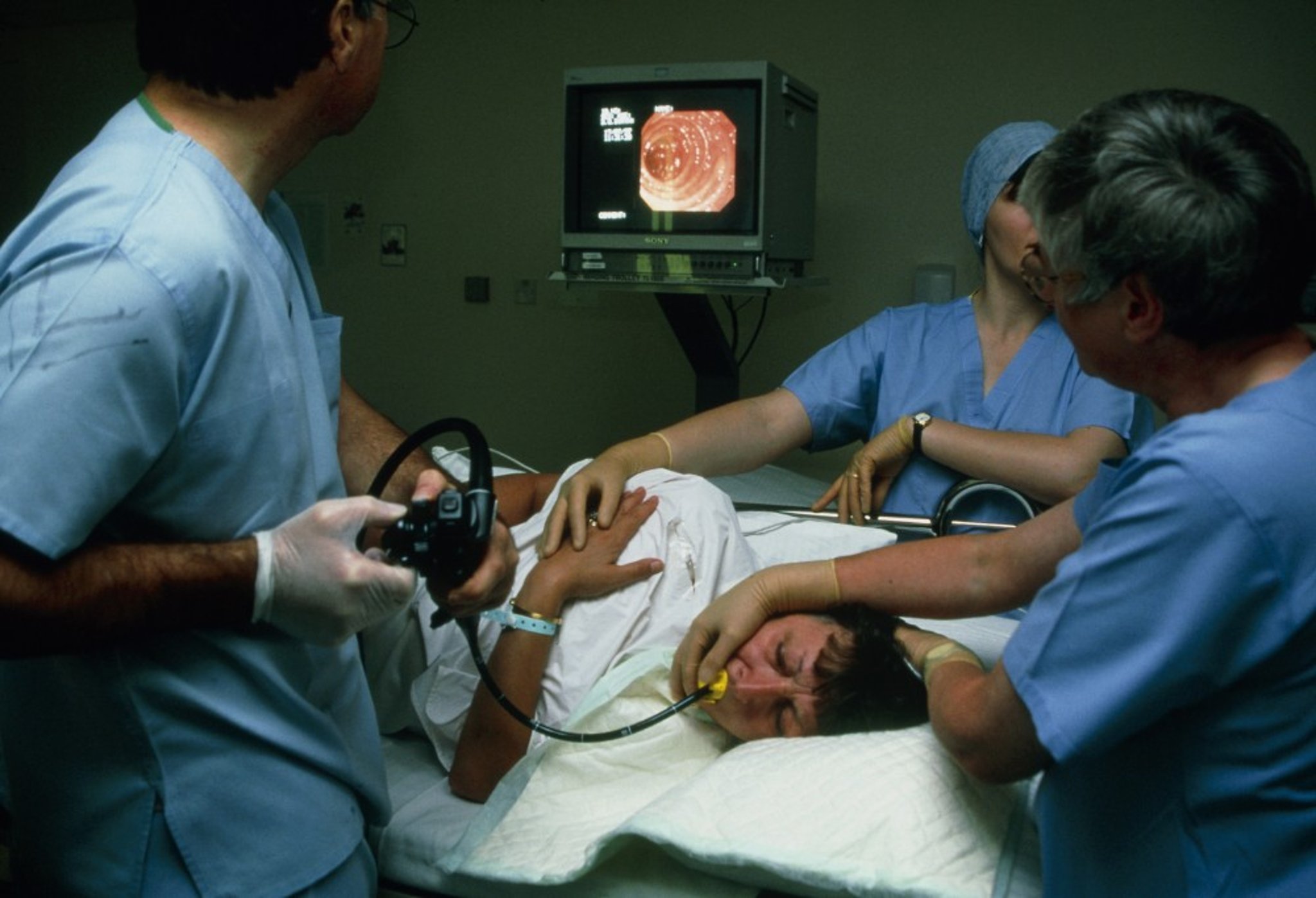
ANTONIA REEVE/SCIENCE PHOTO LIBRARY
Indications for Bronchoscopy
Flexible bronchoscopes facilitate airway visualization and documentation of findings (see table ).
Diagnostically, flexible fiberoptic bronchoscopy allows for:
Direct airway visualization down to, and including, subsegmental bronchi
Sampling of respiratory secretions and cells via bronchial washings, brushings, and lavage of peripheral airways and alveoli
Biopsy of endobronchial, parenchymal, and mediastinal structures
Therapeutic uses include:
Suctioning of retained secretions
Placing an endobronchial tube, stent, or valve
Removing foreign objects
Using balloon dilation to relieve airway stenoses
Guide endotracheal intubation
Indications for Flexible Fiberoptic Bronchoscopy
Procedure | Indication |
|---|---|
Diagnostic | Abnormal chest radiograph: To diagnose the etiology of pneumonia* in a patient who is immunocompromised; in a patient who is immunocompetent and has recurrent or nonresolving disease; or in a patient with a paratracheal/mediastinal/hilar mass, parenchymal mass, or nodule, especially in a proximal lung section Atelectasis (persistent)* Cough (persistent, unexplained)* Diffuse lung process (transbronchial lung biopsy) Evaluation for rejection in a recipient of a lung transplant Evaluation of airway in a patient with burns Evaluation for bronchial disruption in a patient with chest trauma Positive sputum cytology in a patient with a normal chest radiograph* Suspected tracheoesophageal fistula Wheeze (localized/fixed) |
Therapeutic† | Aspiration of retained secretions*, ‡ Bronchopulmonary lavage (pulmonary alveolar proteinosis) Laser resection of tumor§ Lung volume reduction Management of bronchopleural fistula Photodynamic therapy§ Placement of an airway stent§ Placement of endotracheal tube in a difficult situation (cervical injury, abnormal anatomy) Placement of an endobronchial valve Removal of foreign body§ |
* Flexible fiberoptic bronchoscopy is indicated only after failure of less invasive investigations and treatments. | |
† Criner GJ, Eberhardt R, Fernandez-Bussy S, et al. Interventional Bronchoscopy. Am J Respir Crit Care Med. 2020;202(1):29-50. doi:10.1164/rccm.201907-1292SO | |
‡ Flexible fiberoptic bronchoscopy is not a substitute for chest physiotherapy, bronchodilator nebulization, and nasotracheal suctioning; it should be reserved for hypoxemia (in a patient receiving mechanical ventilation) and/or lobar atelectasis secondary to impacted secretions refractory to conventional therapy. | |
§ Rigid bronchoscopy provides more control for instrumentation than flexible bronchoscopy. | |
Rigid bronchoscopy is used only when a wider aperture and channels are required for better visualization and instrumentation, such as when:
Investigating vigorous pulmonary hemorrhage (in which the rigid bronchoscope can better identify the bleeding source and, with its larger suction channel, can better suction the blood and prevent asphyxiation)
Viewing and removing aspirated foreign bodies in young children (in some cases) (1)
Viewing obstructive endobronchial lesions for possible laser debulking or stent placement
Treating an airway obstruction via dilation
Indications reference
1. Safia A, Abd Elhadi U, Bader R, et al. Flexible versus Rigid Bronchoscopy for Tracheobronchial Foreign Body Removal in Children: A Comparative Systematic Review and Meta-Analysis. J Clin Med. 2024;13(18):5652. doi:10.3390/jcm13185652
Contraindications to Bronchoscopy
Absolute contraindications to bronchoscopy include:
Acute respiratory failure with hypercapnia (unless the patient is intubated and ventilated)
High-grade tracheal obstruction
Inability to adequately oxygenate the patient during the procedure
Untreatable life-threatening arrhythmias
Relative contraindications to bronchoscopy include:
Recent myocardial infarction
Inability or unwillingness to hold still for the procedure
Uncorrectable coagulopathy
Transbronchial biopsy should be performed with caution in patients with increased risk of bleeding. Inspection of the airways is safe in these patients, however.
Procedure for Bronchoscopy
Bronchoscopy should be performed only by a pulmonologist or trained surgeon in a monitored setting, typically a bronchoscopy suite, operating room, or intensive care unit.
Except in true emergencies, patients should receive nothing by mouth for a period of time before bronchoscopy (with the specific duration varying by center) and have IV access, intermittent blood pressure monitoring, and continuous pulse oximetry and cardiac monitoring. Supplemental oxygen should be used.
Patients usually receive conscious sedation with short-acting benzodiazepines, opioids, or both before the procedure to decrease anxiety, discomfort, and cough. In some centers, general anesthesia (eg, deep sedation with propofol and airway control via endotracheal intubation or use of a laryngeal mask airway) is commonly used before bronchoscopy. Dexmedetomidine has more recently been studied as a sedative agent for bronchoscopy.
The pharynx and vocal folds are anesthetized with nebulized or aerosolized lidocaine (1 or 2%, to a maximum of 250 to 300 mg for a 70-kg patient). The bronchoscope is lubricated and passed either through a nostril, the mouth with use of an oral airway or bite block, or an artificial airway such as an endotracheal tube. After inspecting the nasopharynx and larynx, the clinician passes the bronchoscope through the vocal folds during inspiration, into the trachea, and then further distally into the bronchi.
Several ancillary procedures can be performed as needed, with or without fluoroscopic guidance:
Bronchial washing: Saline is injected through the bronchoscope and subsequently aspirated from the airways.
Bronchial brushing: A brush is advanced through the bronchoscope and used to abrade suspect lesions to obtain cells.
Bronchoalveolar lavage: 50 to 200 mL of sterile saline is infused into the distal bronchoalveolar tree and subsequently suctioned out, retrieving cells, protein, and microorganisms located at the alveolar level. Local areas of pulmonary edema created by lavage may cause transient hypoxemia.
Endobronchial and transbronchial biopsy: Endobronchial biopsy obtains a tissue sample from a lesion that can be seen in the airway lumen. Transbronchial biopsy uses forceps that are advanced through the bronchoscope and airway to obtain samples from one or more sites in the lung parenchyma. Transbronchial biopsy can be performed without fluoroscopic guidance, although the use of fluoroscopy may be associated both with higher diagnostic yields and lower incidence of significant pneumothorax (1, 2).
Transbronchial needle aspiration: A retractable needle is inserted through the bronchoscope and can be used to sample enlarged mediastinal lymph nodes or masses. Endobronchial ultrasound (EBUS) can be used to help guide the needle biopsy.
Patients who are not critically ill are typically given supplemental oxygen and observed for 2 to 4 hours after the procedure. Return of a gag reflex and maintenance of oxygen saturation when not receiving supplemental oxygen are the primary indices of recovery.
Procedure references
1. Gould MK, Donington J, Lynch WR, et al. Evaluation of individuals with pulmonary nodules: when is it lung cancer? Diagnosis and management of lung cancer, 3rd ed: American College of Chest Physicians evidence-based clinical practice guidelines. Chest. 2013;143(5 Suppl):e93S-e120S. doi:10.1378/chest.12-2351
2. Smyth CM, Stead RJ. Survey of flexible fibreoptic bronchoscopy in the United Kingdom. Eur Respir J. 2002;19(3):458-463. doi:10.1183/09031936.02.00103702
Complications of Bronchoscopy
Serious complications are uncommon; minor bleeding from a biopsy site and fever occur in less than 15% of patients (1, 2, 3).
Patients may have an increase in cough after bronchoalveolar lavage.
Rarely, topical anesthesia causes laryngospasm, bronchospasm, seizures, or cardiac arrhythmias or arrest.
Bronchoscopy itself may cause:
Arrhythmias (most commonly premature atrial contractions, ventricular premature beats, or bradycardia)
Hypoxemia in patients with compromised gas exchange
Minor laryngeal edema or injury with hoarseness
Transmission of infection from suboptimally sterilized equipment (very rare)
Mortality is 1 in 7500 to 25,000 patients (1, 4). Older adults and patients with serious comorbidities (eg, severe chronic obstructive pulmonary disease [COPD], coronary artery disease, pneumonia with hypoxemia, advanced cancers) are at greatest risk.
Transbronchial biopsy can cause pneumothorax (approximately 1% to 6%) or significant hemorrhage (approximately 1% to 4%), but doing the procedure can often avoid the need for thoracotomy (5, 6). In general, complications rates are higher in patients with comorbid disease and those undergoing interventional procedures via bronchoscopy (7).
Complications references
1. Asano F, Aoe M, Ohsaki Y, et al. Deaths and complications associated with respiratory endoscopy: a survey by the Japan Society for Respiratory Endoscopy in 2010. Respirology. 2012;17(3):478-485. doi:10.1111/j.1440-1843.2011.02123.x
2. Carr IM, Koegelenberg CF, von Groote-Bidlingmaier F, et al. Blood loss during flexible bronchoscopy: a prospective observational study. Respiration. 2012;84(4):312-318. doi:10.1159/000339507
3. Hackner K, Riegler W, Handzhiev S, et al. Fever after bronchoscopy: serum procalcitonin enables early diagnosis of post-interventional bacterial infection. BMC Pulm Med 2017;17(1):156. doi:10.1186/s12890-017-0508-1
4. Jin F, Mu D, Chu D, Fu E, Xie Y, Liu T. Severe complications of bronchoscopy. Respiration 2008;76(4):429-433. doi:10.1159/000151656
5. Gupta N, Finlay GA, Kotloff RM, et al. Lymphangioleiomyomatosis Diagnosis and Management: High-Resolution Chest Computed Tomography, Transbronchial Lung Biopsy, and Pleural Disease Management. An Official American Thoracic Society/Japanese Respiratory Society Clinical Practice Guideline. Am J Respir Crit Care Med. 2017;196(10):1337-1348. doi:10.1164/rccm.201709-1965ST
6. Tukey MH, Wiener RS. Population-based estimates of transbronchial lung biopsy utilization and complications. Respir Med. 2012;106(11):1559-1565. doi:10.1016/j.rmed.2012.08.008
7. Stahl DL, Richard KM, Papadimos TJ. Complications of bronchoscopy: A concise synopsis. Int J Crit Illn Inj Sci. 2015;5(3):189-195. doi:10.4103/2229-5151.164995
Drug Information for the Topic


