

Mediastinoscopy is the direct visual examination of the area inside the middle of the chest between the lungs (the mediastinum) through a viewing tube (mediastinoscope). Mediastinotomy is surgical opening of the mediastinum.
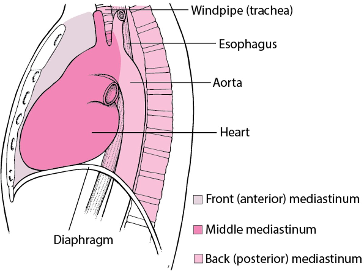
The mediastinum contains the heart, windpipe (trachea), esophagus, thymus, and lymph nodes. Most often, mediastinoscopy is used to diagnose the cause of enlarged lymph nodes deep in the chest or to evaluate how far lung cancer has spread before chest surgery (thoracotomy) is done.
Locating the Mediastinum

Mediastinoscopy is done in an operating room with the person under general anesthesia. A small incision is made in the notch just above the breastbone (sternum). The instrument then is passed down into the chest in front of the windpipe, allowing the doctor to observe the contents of the mediastinum next to the windpipe and to obtain specimens for diagnostic tests if necessary.
Mediastinotomy gives direct access to structures that are inaccessible by mediastinoscopy.
Although mediastinoscopy and mediastinotomy are usually very safe, occasionally complications develop, including reactions to anesthetic agents, infection, and injury to the lung causing air to leak into the pleural space (pneumothorax). Rarer but more serious complications include bleeding caused by damage to one of the large blood vessels around the heart and injury to a nerve in the chest (which can cause hoarseness).
(See also Medical History and Physical Examination for Lung Disorders and Overview of Pleural and Mediastinal Disorders.)


