


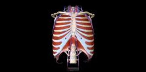
Chest (also called the thorax) injuries most often affect the ribs, breastbone, collarbone, muscles and other soft tissues, heart, lungs, blood vessels, and upper part of the abdomen. Sometimes the esophagus or shoulder blade is also injured.
Chest injuries cause about 25% of deaths due to severe injury. Many of the injuries that can cause death during the first minutes or hours after injury can be treated or stabilized in the emergency department without the need for major surgery.
Causes of Chest Injuries
The chest can be injured by blunt force (such as in motor vehicle crashes, falls, or sports injuries) or by an object that penetrates it (such as a bullet or knife).
Chest injuries are often serious or immediately life threatening because they interfere with breathing or circulation. Some injuries damage the ribs and chest muscles (called the chest wall) severely enough to make it difficult for the lungs to inflate normally.
Damage to the lungs themselves interferes with gas exchange, the main function of the lungs in which oxygen is acquired and carbon dioxide is expelled.
Chest injuries can cause circulatory problems if they result in a lot of bleeding. Bleeding is often inside the chest wall, which also interferes with breathing. Also, injury to the heart can affect circulation by interfering with the heart's ability to pump blood to the body.
Chest injuries that are common or can be severe include the following:
Cardiac tamponade (pressure on the heart caused by accumulation of blood around it)
Hemothorax (blood between the lung and chest wall)
Pulmonary contusion (bruising of the lung)
Injury to the aorta (a tear in the large artery that carries blood from the heart to the body)
Simple pneumothorax (air between the lung and chest wall, sometimes called a collapsed lung)
Did You Know...
|
Symptoms of Chest Injuries
The injured area is usually tender or painful. Pain is worse when people inhale. The chest may be bruised. Sometimes people are short of breath. If the injury is severe, they may feel very short of breath, drowsy, or confused, and the skin may be cold, sweaty, or blue. Such symptoms may develop when the lungs malfunction severely (respiratory failure) or people are in shock. People in shock typically have dangerously low blood pressure and feel as if their heart is racing.
Other symptoms depend on the specific chest injury. For example, sometimes air accumulates under the skin in people with pneumothorax. Affected skin feels crackly and makes a crackling sound when touched. The veins in the neck are sometimes enlarged if blood or fluid accumulates in the sac around the heart and interferes with the heart's ability to pump blood (called cardiac tamponade) or if tension pneumothorax develops.
Diagnosis of Chest Injuries
A doctor's evaluation
Imaging
Usually, a chest injury is obvious. However, the severity of chest injuries cannot be determined without a doctor's evaluation.
First, doctors use a stethoscope to determine whether all parts of the lungs are receiving air, and they carefully examine the neck and chest for injuries. When someone is having difficulty breathing, a doctor places a sensor on the person's finger (pulse oximeter) to measure the amount of oxygen in the blood. Sometimes doctors measure the amount of oxygen and carbon dioxide in the blood with a blood test (arterial blood gas measurement).
A chest x-ray is almost always done. Chest x-ray shows most cases of pneumothorax, hemothorax, and collarbone fractures, as well as some rib fractures. However, a rapid ultrasound procedure is usually needed to look at the heart and lungs. This procedure, called E-FAST (Extended Focused Assessment With Sonography in Trauma), is used in trauma centers and emergency departments. Computed tomography (CT), ultrasound, and/or aortography may be done if doctors suspect injury to the aorta.
Sometimes blood tests and electrocardiography (ECG) are also done.
Treatment of Chest Injuries
Support of breathing and circulation
Treatment of the specific injury
Injuries that are immediately life threatening are treated as quickly as possible. The specific treatment depends on the injury.
For all injuries, doctors take measures to support breathing and circulation if necessary. People may be given oxygen (for example, by nasal prongs, by face mask, or through a breathing tube) and intravenous fluids or sometimes blood transfusions. People with severe chest injuries are admitted to the hospital.
People may be given pain relievers (analgesics) to lessen pain.
For some injuries, a tube must be inserted into the chest (thoracostomy, or chest tube insertion) to drain blood (in hemothorax) or air (in pneumothorax) from the chest. This procedure helps collapsed lungs reinflate. Insertion can usually be done using only local anesthesia.


