


Optic Neuritis
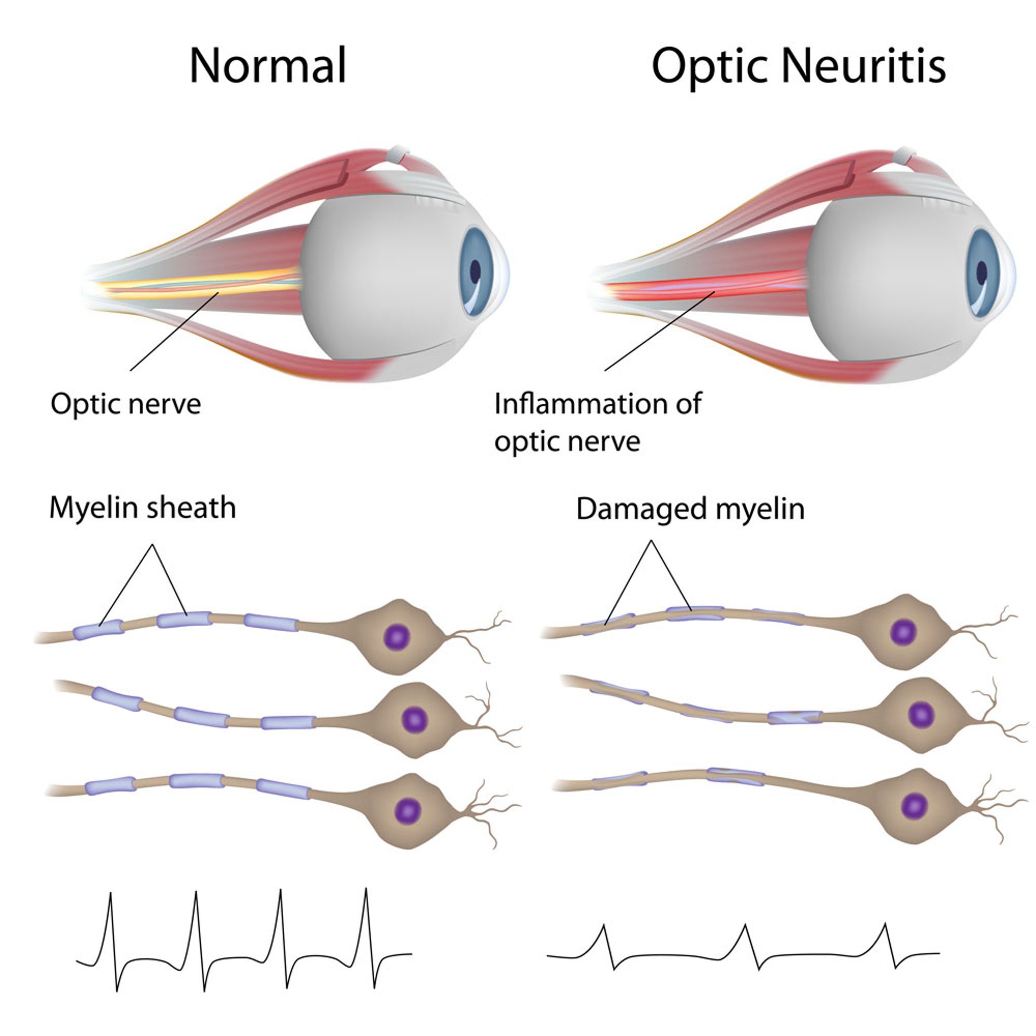
In this illustration, the images in the left column show a healthy optic nerve and healthy myelin sheaths.
The images in the right column show an inflamed optic nerve and damaged myelin.
The bottom line graphics show visual evoked potentials (VEP). A VEP measures how quickly and strongly visual signals travel from the eye to the brain. In optic neuritis, these signals are usually slower, and in some cases weaker.
Alila Medical Media/stock.adobe.com
In these topics