


Limb pain may affect all or part of an extremity (for joint pain, see Pain in and Around a Single Joint and Pain in Multiple Joints). Pain may be constant or intermittent, and unrelated to motion or precipitated by it. Accompanying symptoms and signs often suggest a source.
Etiology of Limb Pain
The most common causes of pain in a limb are also readily apparent by history.
Musculoskeletal injuries and overuse
This discussion covers extra-articular limb pain unrelated to injury or strain. Pain that is in only one joint or in multiple joints is discussed elsewhere.
There are many causes (see table ) but the most common are the following:
Deep venous thrombosis (DVT)
Radiculopathy
Uncommon but serious causes that require immediate diagnosis and treatment include
Deep soft-tissue infection
Acute coronary ischemia (manifesting with only referred arm pain)
Some Causes of Nontraumatic Limb Pain
Cause | Suggestive Findings | Diagnostic Approach |
|---|---|---|
Musculoskeletal and soft tissue | ||
Focal erythema (or darker color on dark skin), warmth, tenderness, swelling Sometimes fever | Clinical evaluation Sometimes blood and tissue cultures (eg, when patients are immunocompromised) | |
Deep soft-tissue infection (eg, myonecrosis, necrotizing subcutaneous infection) | Deep, constant pain, typically out of proportion to other findings Erythema (or darker color on dark skin), warmth, tenderness, tense swelling, fever Sometimes crepitation, foul discharge, bullae or necrotic areas, signs of systemic toxicity (eg, delirium, tachycardia, pallor, shock) | Blood and tissue cultures Radiography Sometimes MRI |
Deep, constant, often nocturnal pain Bone tenderness, fever Often risk factors (eg, immunocompromise, parenteral illicit drug use, known contiguous or remote source for infection) | Radiography, MRI, and/or CT Sometimes bone culture | |
Bone tumor (primary or metastatic) | Deep, constant, often nocturnal pain Bone tenderness Often a known cancer | Radiography, MRI, and/or CT |
Vascular | ||
Swelling, often warmth and/or redness, sometimes venous distension Often risk factors (eg, hypercoagulable state, recent surgery or immobility, cancer) | Ultrasound Sometimes D-dimer testing | |
Mild discomfort with swelling, erythema, and warmth of distal lower extremity Sometimes shallow ulcerations | Clinical evaluation | |
Acute ischemia (typically due to arterial embolism, dissection, or thrombosis but sometimes due to massive iliofemoral venous thrombosis that completely obstructs blood flow in the limb) | Sudden, severe pain Signs of distal limb ischemia (eg, coolness, pallor, pulse deficits, delayed capillary refill) Sometimes chronic ischemic skin changes (eg, atrophy, hair loss, pale color, ulceration) After several hours, neurologic deficits and muscle tenderness Sometimes known peripheral vascular disease | Immediate arteriography |
Intermittent leg pain triggered predictably by exertion and relieved by rest (intermittent claudication), sometimes rest pain which may worsen with leg elevation Low ankle-brachial blood pressure index, chronic ischemic skin changes | Ultrasound Sometimes arteriography | |
Neurologic | ||
Plexopathy (brachial or lumbar) | Usually weakness, often decreased reflexes Sometimes numbness in a nerve plexus distribution | Usually electrodiagnostic testing (electromyography and nerve conduction velocity) Sometimes MRI |
Pain and paresthesias beginning in neck or shoulder and extending to medial aspect of arm and hand | Clinical evaluation Sometimes, electrodiagnostic testing and/or MRI | |
Radiculopathy (eg, caused by herniated intervertebral disk or bone spurs) | Pain and sometimes sensory deficits following a dermatomal distribution and often worsening with movement Often neck or back pain Usually weakness and diminished deep tendon reflexes in a nerve root distribution | Usually MRI Sometimes electromyography and nerve conduction velocity |
Painful polyneuropathy (eg, alcoholic neuropathy) | Chronic, burning pain, typically in both hands or both feet Sometimes sensory abnormalities such as hypoesthesia, hyperesthesia, and/or allodynia (pain with non-noxious stimuli) | Clinical evaluation Sometimes electromyography and nerve conduction velocity |
Burning pain, hyperesthesia, allodynia, vasomotor abnormalities Typically a prior injury (may be remote) | Clinical evaluation | |
Other | ||
Acute coronary ischemia (causing referred arm pain) | Absence of explanatory physical findings at the site of pain; other suggestive findings (eg, history suggesting coronary artery disease, sweating and/or dyspnea occurring simultaneously with arm pain) | ECG and serum troponin Sometimes stress testing or coronary angiography |
Myofascial pain syndrome | Chronic pain and tenderness along a taut band of muscle, worsening with movement and with pressure on a trigger point (focal area separate from site of pain) | Clinical evaluation |
Evaluation of Limb Pain
It is important to exclude acute arterial occlusion.
History
History of present illness should address the duration, intensity, location, quality, and temporal pattern of pain. Recent injury, excessive and/or unusual use, and factors that worsen pain (eg, limb movement, walking) and relieve pain (eg, rest, certain positions) should be noted. Any associated neurologic symptoms (eg, numbness, paresthesias) should be identified.
Review of systems should seek symptoms of possible causes, including back or neck pain (radiculopathy), fever (infections such as osteomyelitis, cellulitis, or deep soft-tissue infection), dyspnea (DVT with pulmonary embolism, myocardial infarction), and chest pain or sweating (myocardial ischemia).
Past medical history should identify known risk factors, including cancer (metastatic bone tumors); immunocompromising disorders or medications (infections); hypercoagulable states (DVT); diabetes; peripheral vascular disease, hypercholesterolemia, and/or hypertension (acute or chronic ischemia); osteoarthritis or rheumatoid arthritis (radiculopathy); and prior injury (complex regional pain syndrome). Family and social history should address family history of early vascular disease and cigarette smoking (limb or myocardial ischemia) and illicit use of parenteral drugs (infections).
Physical examination
Vital signs are reviewed for fever (suggesting infection) and tachycardia and/or tachypnea (compatible with DVT with pulmonary embolism, myocardial infarction, and infection with sepsis).
The painful limb is inspected for color, edema, and any skin or hair changes, and palpated for pulses, temperature, tenderness, and crepitation (a subtle crackling sensation indicating soft-tissue gas). Strength, sensation and deep tendon reflexes are compared between affected and unaffected sides. Systolic blood pressure (BP) is measured in the ankle of the affected extremity and compared with systolic BP of an arm; the ratio of the two is the ankle-brachial index.
Red flags
Sudden, severe pain
Signs of acute limb ischemia (eg, coolness, pallor, pulse deficits, delayed capillary refill)
Dyspnea, chest pain, and/or sweating
Signs of systemic toxicity (eg, delirium, tachycardia, shock, pallor)
Crepitation, tenseness, foul discharge, bullae, necrosis
Risk factors for deep venous thrombosis
Neurologic deficits
Interpretation of findings

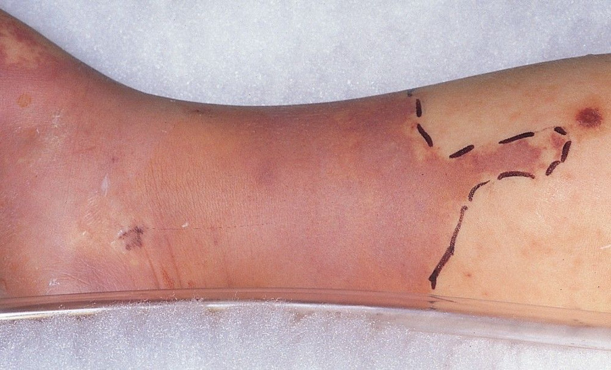
This photo shows focal erythema and swelling, usually accompanied by warmth and tenderness, characteristic of focal cellulitis. Erythema may be purplish and can mimic venous congestion. Note the clinician has marked the border of the cellulitis with a pen to facilitate recognition of spread or resolution.
© Springer Science+Business Media
It can be helpful to categorize pain by acuity of symptom onset and then further narrow the differential diagnosis based on presence or absence of findings of
Ischemia
Inflammation
Neurologic abnormalities
Sudden, severe pain suggests acute ischemia or acute radiculopathy (eg, due to sudden disc herniation). Acute ischemia causes generalized limb pain and manifests with weak or absent pulse, delayed capillary refill (≥ 2 seconds or, with unilateral symptoms, longer than the opposite side), coolness, and pallor; ankle-brachial index is typically < 0.3. Such vascular signs are absent with radiculopathy, in which pain instead follows a dermatomal distribution and is often accompanied by back or neck pain and diminished deep tendon reflexes. However, in both cases, weakness may be present. Acute ischemia due to massive venous thrombosis (phlegmasia cerulea dolens) usually causes edema, which is not present in ischemia due to arterial occlusion.
In subacute pain (ie, of 1 to a few days' duration), erythema and tenderness, often accompanied by swelling, and/or warmth, suggest an inflammatory cause. If these findings are focal or circumscribed, cellulitis is likely. Generalized, circumferential swelling is more suggestive of DVT or, much less commonly, deep tissue infection. Patients with a deep tissue infection typically appear quite ill and may have blisters, necrosis, or crepitation. Findings in DVT vary widely; swelling and warmth may be minimal or absent. Neurologic findings of weakness, paresthesias, and/or sensory abnormalities suggest radiculopathy or plexopathy. If neurologic findings follow a dermatomal pattern, radiculopathy is more likely.
Chronic pain can be difficult to diagnose. If neurologic findings are present, causes include radiculopathy (dermatomal distribution), plexopathy (plexus distribution), neuropathy (stocking-glove distribution), and complex regional pain syndrome (variable distribution). Complex regional pain syndrome should be suspected if vasomotor changes (eg, pallor, mottling, coolness) are present, particularly in patients with previous injury to the affected extremity. Myofascial pain syndrome causes no neurovascular abnormalities and classically manifests with a palpably tense band of muscle in the area of pain, and pain may be reproduced by pressure on a trigger point near but not overlying the area of pain. In patients with essentially no clinical findings, cancer and osteomyelitis should be considered, particularly in those with risk factors.

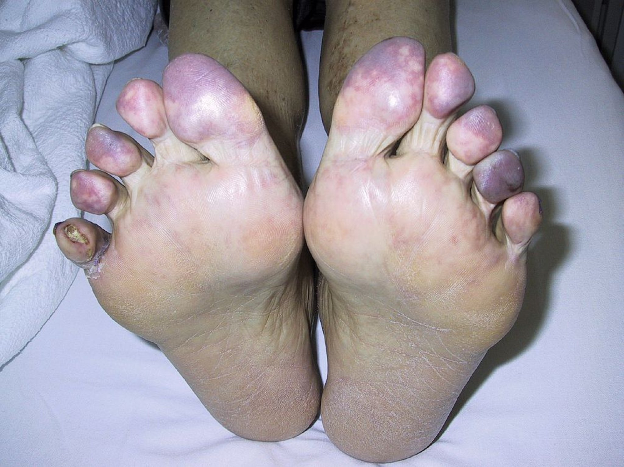
This patient developed painful blue toes and ulceration 6 weeks after coronary angiography and stenting. Aortofemoral angiography revealed diffuse atherosclerosis of the aorta and femoral and popliteal arteries, suggesting that cholesterol emboli caused ischemia of the toes.
© Springer Science+Business Media
Intermittent pain occurring consistently with a given degree of exertion (eg, whenever walking > 3 blocks) and relieved with a few minutes of rest suggests peripheral arterial disease. Such patients typically have an ankle-brachial blood pressure (BP) index of ≤ 0.9; an index ≤ 0.4 indicates severe disease. However, arterial stiffness can produce falsely negative ankle-brachial index values. Because the toe arteries are less susceptible to stiffening, the toe-brachial BP index can be measured instead in patients with suspected peripheral arterial disease and in whom the ankle arteries are likely not compressible (eg, patients with advanced diabetes or aging). Patients with exertional symptoms and normal or borderline ankle-brachial BP index (> 0.9 but < 1.40) should have repeat ankle-brachial BP index measurement after exercise on a treadmill. Patients with peripheral arterial disease may have chronic skin changes (eg, atrophy, hair loss, pale color, ulceration).
Testing
Cellulitis, myofascial pain, painful polyneuropathy, and complex regional pain syndrome can often be diagnosed clinically. Testing (see table ) is usually necessary for other suspected causes of pain.
Treatment of Limb Pain
Primary treatment is directed at the cause. Analgesics can help relieve pain.
Key Points
Acute limb ischemia should be considered in patients with sudden, severe pain.
Presence or absence of findings of ischemia, inflammation, and neurologic abnormalities plus the acuity of onset help narrow the differential diagnosis.


