Acute coronary syndromes usually result from acute obstruction of a coronary artery. Consequences depend on degree and location of obstruction and range from unstable angina to non–ST-segment elevation myocardial infarction (NSTEMI), to ST-segment elevation myocardial infarction (STEMI), sometimes leading to sudden cardiac death. Symptoms are similar in each of these syndromes and include chest discomfort with or without dyspnea, nausea, and diaphoresis. Diagnosis is by electrocardiography (ECG) and biomarkers. Immediate treatment is with antiplatelets, anticoagulants, nitrates and, for STEMI and intermediate- and high-risk NSTEMI, revascularization via percutaneous intervention, fibrinolytic medications (for STEMI only), or occasionally, coronary artery bypass graft surgery. Subsequent care includes lipid management, beta-blockers, cardiac rehabilitation, risk factor management, and dual antiplatelet therapy.
Classification of Acute Coronary Syndromes
Acute coronary syndromes (ACS) include (1):
Unstable angina
Non–ST-segment elevation myocardial infarction (NSTEMI)
ST-segment elevation myocardial infarction (STEMI)
These syndromes all involve acute coronary ischemia and are distinguished based on symptoms, ECG findings, and cardiac troponin levels. It is helpful to distinguish the syndromes because prognosis and treatment vary. Unstable angina and NSTEMI are also collectively referred to as non-ST elevation ACS (NSTE-ACS).
Unstable angina (historically referred to as acute coronary insufficiency, preinfarction angina, or intermediate syndrome) is defined as transient myocardial ischemia with reduced coronary blood flow, in the absence myocardial necrosis (circulating cardiac troponin < 99th percentile).
Symptoms of unstable angina include:
Rest angina that is prolonged (usually > 20 minutes)
New-onset angina of at least class 3 severity in the Canadian Cardiovascular Society (CCS) classification (see table )
Increasing angina, that is, previously diagnosed angina that has become distinctly more frequent, more severe, longer in duration, or lower in threshold (eg, increased by ≥ 1 CCS class or to at least CCS class 3)
However, these symptoms occur across the range of acute coronary syndromes, which are differentiated based on ECG changes and cardiac troponin levels.
ECG changes such as ST-segment depression, ST-segment elevation, or T-wave inversion may occur during unstable angina, but they are transient. Troponin may be detectable with high-sensitivity troponin tests (hs-cTn), but, by definition, is < 99th percentile of the upper reference limit (URL) (1, 2). Unstable angina is, as the name suggests, clinically unstable and often a prelude to myocardial infarction or arrhythmias or, less commonly, to sudden death.
Non–ST-segment elevation MI (NSTEMI, subendocardial MI) is myocardial necrosis (evidenced by circulating cardiac troponin levels ≥ 99th percentile) without acute ST-segment elevation that meets criteria for STEMI. ECG changes such as ST-segment depression, T-wave inversion, or both may be present. NSTEMI is characterized by ischemia that is ongoing rather than transient as in unstable angina, and myocardial necrosis that is typically, though not universally, subendocardial.
ST-segment elevation MI (STEMI, transmural MI) is myocardial necrosis with ECG changes showing ST-segment elevation that is not quickly reversed by nitroglycerin. The presence of a new left bundle branch block is not considered a STEMI equivalent in isolation (1, 3). Cardiac troponin is elevated, and myocardial necrosis is transmural.
Both types of MI may or may not produce Q waves on the ECG.
General references
1. Rao SV, O'Donoghue ML, Ruel M, et al. 2025 ACC/AHA/ACEP/NAEMSP/SCAI Guideline for the Management of Patients With Acute Coronary Syndromes: A Report of the American College of Cardiology/American Heart Association Joint Committee on Clinical Practice Guidelines. Circulation. 2025;151(13):e771-e862. doi:10.1161/CIR.0000000000001309
2. Januzzi JL Jr, Mahler SA, Christenson RH, et al. Recommendations for Institutions Transitioning to High-Sensitivity Troponin Testing: JACC Scientific Expert Panel. J Am Coll Cardiol. 2019;73(9):1059-1077. doi:10.1016/j.jacc.2018.12.046
3. Writing Committee, Kontos MC, de Lemos JA, et al. 2022 ACC Expert Consensus Decision Pathway on the Evaluation and Disposition of Acute Chest Pain in the Emergency Department: A Report of the American College of Cardiology Solution Set Oversight Committee. J Am Coll Cardiol. 2022;80(20):1925-1960. doi:10.1016/j.jacc.2022.08.750
Etiology of Acute Coronary Syndromes
The most common cause of acute coronary syndromes is (1):
Disruption of an unstable atherosclerotic plaque with thrombosis in the atherosclerotic coronary artery
Atheromatous plaque sometimes becomes unstable or inflamed, causing it to erode or rupture, exposing thrombogenic material, which activates platelets and the coagulation cascade and produces an acute thrombus. Platelet activation involves a conformational change in membrane glycoprotein (GP) IIb/IIIa receptors, allowing cross-linking (and thus aggregation) of platelets. The resultant thrombus abruptly interferes with blood flow to parts of the myocardium.
Even atheromas causing minimal obstruction can rupture and result in thrombosis; in one large prospective imaging trial, the majority of patients having myocardial infarction or cardiovascular death did not have evidence of significant coronary stenosis at baseline (2, 3). Thus, although the severity of stenosis helps predict symptoms, it does not always predict acute thrombotic events.
Spontaneous thrombolysis occurs in up to one-quarter of patients (4); 24 hours later, thrombotic obstruction is found in only approximately one-third (5). However, in virtually all cases, obstruction lasts long enough to cause varying degrees of tissue necrosis.
Myocardial infarction in the absence of obstructive coronary artery disease (MINOCA)
Myocardial infarction with nonobstructive coronary arteries (MINOCA) is found in approximately 5 to 6% of patients with acute MI who undergo coronary angiography (6). Patients with MINOCA tend to be younger, female, and without dyslipidemia, and they have myocardial necrosis without significant coronary atherosclerosis.
Causes of MINOCA include:
Coronary artery embolism
Coronary spasm
Spontaneous coronary artery dissection
Coronary arterial embolism (as distinct from the typical atherothrombosis by having a non-coronary source and the possibility of other non-thrombotic material such as air) can occur in mitral stenosis, aortic stenosis, infective endocarditis, nonbacterial thrombotic endocarditis (marantic endocarditis), or atrial fibrillation.
Cocaine use and other causes of coronary spasm can sometimes result in myocardial infarction. Spasm-induced MI may occur in normal or atherosclerotic coronary arteries.
Spontaneous coronary artery dissection is a non-traumatic tear in the coronary intima with creation of a false lumen. Blood flowing through the false lumen expands it, which restricts blood flow through the true lumen, sometimes causing coronary ischemia or infarction. Dissection may occur in atherosclerotic or non-atherosclerotic coronary arteries. Non-atherosclerotic dissection is more likely in pregnant or postpartum patients and/or patients with fibromuscular dysplasia or other connective tissue disorders.
Plaque disruption in atherosclerotic coronary arteries without significant narrowing can also cause MINOCA.
Etiology references
1. Rao SV, O'Donoghue ML, Ruel M, et al. 2025 ACC/AHA/ACEP/NAEMSP/SCAI Guideline for the Management of Patients With Acute Coronary Syndromes: A Report of the American College of Cardiology/American Heart Association Joint Committee on Clinical Practice Guidelines. Circulation. 2025;151(13):e771-e862. doi:10.1161/CIR.0000000000001309
2. Arbab-Zadeh A, Fuster V. From Detecting the Vulnerable Plaque to Managing the Vulnerable Patient: JACC State-of-the-Art Review. J Am Coll Cardiol. 2019;74(12):1582-1593. doi:10.1016/j.jacc.2019.07.062
3. Hoffmann U, Ferencik M, Udelson JE, et al. Prognostic Value of Noninvasive Cardiovascular Testing in Patients With Stable Chest Pain: Insights From the PROMISE Trial (Prospective Multicenter Imaging Study for Evaluation of Chest Pain). Circulation. 2017;135(24):2320-2332. doi:10.1161/CIRCULATIONAHA.116.024360
4. Kanji R, Gue YX, Memtsas V, Spencer NH, Gorog DA: Biomarkers of Thrombotic Status Predict Spontaneous Reperfusion in Patients With ST-Segment Elevation Myocardial Infarction. J Am Coll Cardiol. 2023;81(19):1918-1932. doi:10.1016/j.jacc.2023.03.388
5. DeWood MA, Spores J, Notske R, et al. Prevalence of total coronary occlusion during the early hours of transmural myocardial infarction. N Engl J Med. 1980;303(16):897-902. doi:10.1056/NEJM198010163031601
6. Tamis-Holland JE, Jneid H, Reynolds HR, et al. Contemporary diagnosis and management of patients with myocardial infarction in the absence of obstructive coronary artery disease: A scientific statement from the American Heart Association. Circulation. 2019;139(18):e891-e908. doi: 10.1161/CIR.0000000000000670
Pathophysiology of Acute Coronary Syndromes
Initial consequences vary with size, location, and duration of obstruction and range from transient ischemia to infarction. Measurement of troponin with a high sensitivity test indicates that some cell necrosis probably occurs even with mild ischemia; thus, ischemic events occur on a continuum, and classification into subgroups, although useful, is somewhat arbitrary. Sequelae of the acute event depend primarily on the mass and type of cardiac tissue infarcted.
Myocardial dysfunction
Ischemic (but not infarcted) tissue has impaired contractility and relaxation, resulting in hypokinetic or akinetic segments; these segments may expand or bulge during systole (called paradoxical motion). The size of the affected area determines the clinical effect, which ranges from minimal or mild heart failure to cardiogenic shock; usually, large parts of myocardium must be ischemic to cause significant myocardial dysfunction. Some degree of heart failure occurs in up to approximately one-third of patients hospitalized with acute myocardial infarction (1, 2). It is termed ischemic cardiomyopathy if low cardiac output and heart failure persist. Ischemia involving the papillary muscle may lead to mitral valve regurgitation. Dysfunctional wall motion can allow mural thrombus formation.
Myocardial infarction (MI)
Myocardial infarction is myocardial necrosis resulting from abrupt reduction in coronary blood flow to part of the myocardium. Infarcted tissue is permanently dysfunctional; however, there is a zone of potentially reversible ischemia adjacent to infarcted tissue. MI affects predominantly the left ventricle (LV), but damage may extend into the right ventricle (RV) or the atria.
Infarction may be:
Transmural: Transmural infarcts involve the whole thickness of myocardium from epicardium to endocardium and are usually characterized by abnormal Q waves on ECG.
Nontransmural (subendocardial): Nontransmural infarcts do not extend through the ventricular wall and cause only ST-segment and T-wave (ST-T) abnormalities on ECG.
Because the transmural depth of necrosis cannot be precisely determined clinically, infarcts are classified as STEMI or NSTEMI by the presence or absence of ST-segment elevation, acutely, on the ECG.
Necrosis of a significant portion of the interventricular septum or ventricular wall may rupture, with dire consequences. A ventricular aneurysm or pseudoaneurysm may form.
Electrical dysfunction
Electrical dysfunction can be significant in any form of acute coronary syndrome. Ischemic and necrotic cells are incapable of normal electrical activity, resulting in various ECG changes (predominantly ST-T abnormalities), arrhythmias, and conduction disturbances. ST-T abnormalities of ischemia include ST-segment depression (often downsloping from the J point), T-wave inversion, ST-segment elevation (often referred to as injury current), and peaked T waves in the hyperacute phase of infarction. Conduction disturbances can reflect damage to the sinus node, the atrioventricular (AV) node, or specialized conduction tissues. Most changes are transient; some are permanent.
Types of ST-Segment Depression
alfa md/stock.adobe.com |


Pathophysiology references
1. Faridi KF, Bhalla N, Atreja N, et al. New Heart Failure After Myocardial Infarction (From the National Cardiovascular Data Registries [NCDR] Linked With All-Payer Claims). Am J Cardiol. 2021;151:70-77. doi:10.1016/j.amjcard.2021.04.019
2. Udell JA, Bahit MC, Campbell P, et al. Prevention of heart failure after acute myocardial infarction. Lancet. 2025;406(10508):1154-1170. doi:10.1016/S0140-6736(25)01394-7
Symptoms and Signs of Acute Coronary Syndromes
Symptoms of acute coronary syndromes depend somewhat on the extent and location of obstruction and are quite variable. Painful stimuli from thoracic organs, including the heart, can cause discomfort described as pressure, tearing, gas with the urge to eructate, indigestion, burning, aching, stabbing, and sometimes sharp needle-like pain. Many patients describe their symptoms as discomfort rather than pain. Except when infarction is massive, recognizing the amount of ischemia by symptoms alone is difficult.
Symptoms of ACS are similar to those of angina and are discussed in more detail in sections on unstable angina and acute myocardial infarction. Of note, some patients with coronary artery disease (particularly patients with diabetes) have silent myocardial ischemia, which is typically detected during stress testing.
Complications
After the acute event, many complications can occur. They may involve:
Electrical dysfunction (eg, conduction defects, arrhythmias, sudden cardiac death)
Myocardial dysfunction (eg, heart failure, interventricular septum or free wall rupture, ventricular aneurysm, pseudoaneurysm, mural thrombus formation, cardiogenic shock)
Valvular dysfunction (typically mitral regurgitation)
Electrical dysfunction can be significant in any form of ACS, but usually, large parts of myocardium must be ischemic to cause significant myocardial dysfunction. Other complications of ACS include recurrent ischemia, left ventricular thrombus, and pericarditis. Pericarditis can occur during or immediately after MI or more than one week later (post-MI syndrome, or Dressler syndrome).
Diagnosis of Acute Coronary Syndromes
Early and serial ECGs
Serial cardiac troponin
Immediate coronary angiography for patients with STEMI or NSTE-ACS with complications
Delayed angiography for patients with high- or intermediate-risk NSTE-ACS)
Angiography or noninvasive risk stratification for patients with low-risk NSTE-ACS
Acute coronary syndromes should be considered in males, usually those > age 30 years, and females usually > age 40 years (younger in patients with diabetes), whose main symptom is chest pain or discomfort. Pain must be differentiated from the pain of disorders such as pneumonia, pulmonary embolism, pericarditis, rib fracture, costochondral separation, esophageal spasm, acute aortic dissection, renal calculus, splenic infarction, or various abdominal disorders. In patients with previously diagnosed hiatus hernia, peptic ulcer, or a gallbladder disorder, the clinician must be wary of attributing new symptoms to these disorders. (For approach to diagnosis, see also Chest Pain.)
The initial diagnostic approach is the same when any ACS is suspected: initial and serial ECGs and serial cardiac biomarker measurements, which distinguish among unstable angina, NSTEMI, and STEMI. Every emergency department should have a triage system to immediately identify patients with chest pain for rapid assessment and ECG. Pulse oximetry and chest radiograph (particularly to look for mediastinal widening, which suggests aortic dissection) are also performed.
ECG
ECG is the most important test and should be performed as soon as possible (within 10 minutes of presentation to medical care, whether in or out of the hospital) (1). It is the center of the decision pathway because emergent cardiac catheterization is indicated for patients with acute STEMI but not normally for those with NSTEMI. In addition, fibrinolytics benefit patients with STEMI but may increase risk for those with NSTEMI.
STEMI
For STEMI, initial ECG is usually diagnostic, showing ST-segment elevation ≥ 1 mm in 2 or more contiguous leads subtending the damaged area (see figure ) (1). In leads V2-V3 the elevation must be 1.5 to 2.5 mm depending on age and gender.
Leads in addition to the standard 12 may be used to better detect right ventricular (RV) (V4R) or posterior (V7-V9) infarction.
Acute Lateral Left Ventricular Infarction (Tracing Obtained Within a Few Hours of Illness Onset)
There is striking hyperacute ST-segment elevation in leads I, aVL, V4, and V6 and reciprocal depression in other leads. |

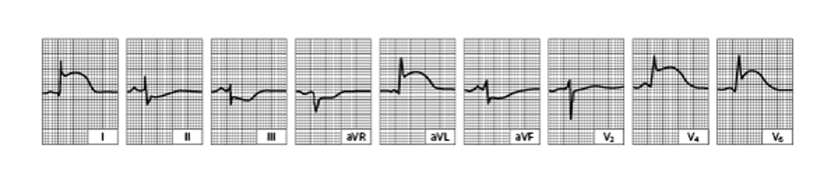
The ECG must be read carefully because ST-segment elevation may be subtle, particularly in the inferior leads (II, III, aVF); sometimes the reader’s attention is mistakenly focused on leads with ST-segment depression. If symptoms are characteristic, ST-segment elevation on ECG has a reported specificity of 94 to 98% and a sensitivity of 55 to 56% for diagnosing MI (2, 3). Serial tracings (obtained every 8 hours for 1 day, then daily) showing a gradual evolution toward a stable, more normal pattern or development of abnormal Q waves over a few days (see figure ) tend to confirm the diagnosis. However, pathologic Q waves are not necessary for the diagnosis of STEMI or transmural infarction.
Inferior (Diaphragmatic) Left Ventricular Infarction (After the First 24 Hours)
Significant Q waves develop with decreasing ST-segment elevation in leads II, III, and aVF. |


NSTEMI
Because nontransmural infarcts are usually in the subendocardial or midmyocardial layers, they do not produce diagnostic ST-segment elevation (or usually Q waves) on the ECG. Instead, they commonly produce only varying degrees of ST-T abnormalities that are less striking, variable, or nonspecific and sometimes difficult to interpret (NSTEMI).
Typical ECG findings in NSTEMI include:
Horizontal or downsloping ST depression
T wave inversion
Transient ST elevation


Panels A, B, and D show ST depression; panel C shows T wave inversion.
凌鲲 宋/stock.adobe.com
If such abnormalities resolve (or worsen) on repeat ECGs, ischemia is very likely. However, when repeat ECGs are unchanged, acute MI is unlikely and, if still suspected clinically, requires other evidence to make the diagnosis. A normal ECG taken when a patient is free of pain does not exclude unstable angina; a normal ECG taken when a patient is experiencing pain, although it does not exclude angina, suggests that the pain is not ischemic.
Cardiac troponin
Cardiac troponins (cTn, referring to subunits T and I, which are specific to cardiac muscle) are the preferred biomarkers used in the diagnosis of acute coronary syndromes. An abnormal cTn level, using an appropriate cutoff, is the primary distinguishing factor between unstable angina and myocardial infarction. Highly sensitive cTn assays are preferred (1).
Troponin is released into the bloodstream after myocardial cell necrosis and, to a lesser extent, during stress and ischemia.
Standard cardiac troponin (cTn) assays, which have been in use for many years, are sensitive and specific, but they are unlikely to detect cardiac troponins except in patients who have an acute cardiac disorder such as a myocardial infarction. Thus, a "positive" cTn test (ie, above the limit of detection) is very specific.
Highly sensitive assays of cardiac troponin (hs-cTn) can detect small amounts of troponin in many healthy people. Thus, troponin levels detected with hs-cTn tests need to be referenced to the normal range and are defined as "elevated" only when higher than 99% of the reference population (1). Furthermore, although an elevated troponin level indicates myocardial cell injury, it does not indicate the cause of the damage (although any troponin elevation increases the risk of adverse outcomes in many disorders) (4). In addition to acute coronary syndromes, many other cardiac and non-cardiac disorders can elevate cardiac troponin levels (see table ); not all elevated levels detected with hs-cTn assays represent myocardial infarction, and not all myocardial necrosis results from an acute coronary syndrome event, even when the etiology is ischemic. However, by detecting lower levels of troponin, hs-cTn assays enable earlier identification of MI than other assays, and they have replaced other cardiac biomarker tests in guidelines and in practical use in many centers.
For troponin T, an abnormal (> 99th percentile cutoff) highly sensitive assay has a sensitivity of 95% and a specificity of 80% for detecting myocardial infarction, compared with a sensitivity of 83% and specificity of 93% for the standard assay (4). For troponin I, the highly sensitive assay has a sensitivity of 82% and a specificity of 92%, compared with a sensitivity of 79% and specificity of 95% for the standard assay.
Patients suspected of having an acute coronary syndrome should have an hs-cTn assay performed on presentation and again 1 to 2 hours later. Troponin should be measured at 0 and 3 to 6 hours if a standard cTn assay is used. Because troponin elevation lags behind the onset of myocardial injury, angiography and treatment in patients with STEMI, or unstable patients with a NSTE-ACS, should not be delayed because of an initially normal troponin level.
An hs-cTn level must be interpreted based on the patient's pre-test probability of disease, which is estimated clinically based on:
Risk factors for ACS
Symptoms
ECG findings
A high pre-test probability plus an elevated troponin level detected with an hs-cTn assay is highly suggestive of ACS, whereas a low pre-test probability plus a normal hs-cTn assay result is unlikely to represent ACS (5). Diagnosis is more challenging when test results are discordant with pre-test probability, in which case serial hs-cTn assays often help. A patient with low pre-test probability and an initially slightly elevated troponin level detected with a hs-cTn assay that remains stable on repeat testing probably has non-ACS cardiac disease (eg, heart failure, stable coronary artery disease). However, if the repeat level rises significantly (ie, > 20 to 50%), the likelihood of ACS becomes much higher. If a patient with high pre-test probability has a normal troponin level detected with a hs-cTn assay and that rises > 50% on repeat testing, ACS is likely; but continued normal levels (often including at 6 hours and beyond when suspicion is high) suggest need to pursue an alternate diagnosis.
Causes of Elevated Troponin Levels
Type | Conditions |
|---|---|
Myocardial ischemia | |
ACS | Classic AMI |
Non-ACS (coronary) | Increased demand (stable coronary artery disease lesion) Coronary artery spasm, embolism, or dissection Procedure-related (PCI, CABG) Cocaine or methamphetamine |
Non-ACS (noncoronary) | Hypoxia Global ischemia Hypoperfusion Cardiothoracic surgery |
Sudden cardiac death | — |
Direct myocardial damage or stress (primarily nonischemic) | |
Cardiac disorders | Cardiomyopathy (eg, hypertrophic, viral) Tachyarrhythmias Injury (ablation procedures, cardiac contusion, cardioversion, electrical shock) Cancer Infiltrative disorders (eg, amyloidosis) |
Systemic disorders | Toxicity (eg, anthracyclines) Trauma (eg, severe burns) Extreme exertion Subarachnoid hemorrhage |
Analytical | |
Assay-based | Poor performance Calibration errors |
Sample-based | Heterophile antibody Interference by substances |
ACS = acute coronary syndrome; AMI = acute MI; CABG = coronary artery bypass grafting; MI = myocardial infarction; NSTEMI = non–ST-segment elevation MI; PCI = percutaneous coronary intervention; STEMI = ST-segment elevation MI. | |
Coronary angiography
Coronary angiography most often combines diagnosis with percutaneous coronary intervention (PCI—ie, angioplasty, stent placement). When possible, emergency coronary angiography and PCI are performed as soon as possible after the onset of acute myocardial infarction (primary PCI). In many studies, a shorter interval to PCI ("door to balloon" or "onset to balloon" time) is associated with significantly lower morbidity and mortality and improved long-term outcomes (6, 7, 8, 9).
Invasive angiography is obtained urgently for patients with STEMI and for those with cardiogenic shock, new or worsening heart failure, chest pain refractory to treatment, or hemodynamic instability (including due to arrhythmia). Patients with high- or intermediate-risk NSTEMI typically undergo angiography within the first 24 to 72 hours of hospitalization to detect lesions that may require treatment; those with low-risk NSTE-ACS, including unstable angina, may undergo angiography or noninvasive testing such as stress testing or coronary CT angiography (see Risk Stratification in NSTE-ACS) (1).
Other tests
Routine laboratory tests are nondiagnostic but, if obtained, show nonspecific abnormalities compatible with tissue necrosis (eg, increased erythrocyte sedimentation rate, moderately elevated white blood cell count with a shift to the left). A fasting lipid profile should be obtained as soon as possible, and preferably within the first 24 hours (before low-density lipoprotein [LDL] levels begin to decrease), for all patients hospitalized with ACS.
Myocardial imaging is not needed to make the diagnosis if cardiac biomarkers or ECG are positive. However, in patients with myocardial infarction, bedside echocardiography is invaluable for detecting mechanical complications. Before or shortly after discharge, patients with symptoms suggesting an ACS but with nondiagnostic ECGs and normal cardiac biomarker levels, in whom angiography is not performed should undergo a stress imaging test (radionuclide or echocardiographic imaging with pharmacologic or exercise stress) or coronary CT angiography (1). Imaging abnormalities in such patients indicate increased risk of complications in the next 3 to 6 months and suggest need for angiography (10), which should be performed before discharge or soon thereafter, with revascularization as necessary.
Right heart catheterization using a balloon-tipped pulmonary artery catheter can be used to measure right heart, pulmonary artery, and pulmonary artery occlusion pressures and cardiac output. This procedure is not routinely recommended and should be performed only if patients have significant complications (eg, severe heart failure, hypoxia, hypotension) and by clinicians experienced with catheter placement and management protocols.
Risk stratification and management strategy for NSTE-ACS
For patients with NSTE-ACS, risk scores (Thrombolysis in Myocardial Infarction [TIMI] or the Global Registry of Acute Coronary Events [GRACE]) (11, 12) can be used in combination with other clinical features to determine the timing of angiography versus a noninvasive initial approach. As with any decision pathway, individual patient factors must be taken into account.


For NSTE-ACS patients with cardiogenic shock, new or worsening heart failure, chest pain refractory to treatment, or hemodynamic instability (including due to arrhythmia), coronary angiography with the intent to revascularize is indicated urgently (within 2 hours) (see table ).
For NSTE-ACS patients with a GRACE score > 140, steeply rising troponin values, or ongoing dynamic ST-segment changes, coronary angiography with the intent to revascularize is recommended within 24 hours.
For NSTE-ACS patients with a GRACE score 109 to 140, no ongoing ischemic symptoms, and stable (or down trending) troponin levels, coronary angiography with the intent to revascularize is recommended before discharge or within 72 hours.
For NSTE-ACS patients with a GRACE score < 109 and TIMI score < 2, with no ongoing ischemic changes, a troponin level < 99th percentile, and no dynamic ST changes, coronary angiography may be performed, or a noninvasive approach with a stress test or coronary CT angiogram may be used.
Management Strategy for NSTE-ACS
Risk Classification | Clinical Features | Management Strategy |
|---|---|---|
Low risk | Any of:
| Routine invasive: Coronary angiography before hospital discharge OR Selective invasive: Initial noninvasive risk stratification stress test or coronary CT angiogram, followed by coronary angiography if indicated |
Intermediate risk | Any of:
| Routine invasive: Coronary angiography before hospital discharge (< 72 hours) |
High risk | Any of:
| Routine invasive: Coronary angiography < 24 hours |
Very high risk (unstable patient) | Any of:
| Immediate invasive: Coronary angiography with intent to revascularize < 2 hours |
GRACE = Global Registry of Acute Coronary Events; NSTE-ACS = non-ST elevation acute coronary syndrome; TIMI = Thrombolysis in Myocardial Infarction; Tn = cardiac troponin; VF = ventricular fibrillation; VT = ventricular tachycardia | ||
Rao SV, O'Donoghue ML, Ruel M, et al. 2025 ACC/AHA/ACEP/NAEMSP/SCAI Guideline for the Management of Patients With Acute Coronary Syndromes: A Report of the American College of Cardiology/American Heart Association Joint Committee on Clinical Practice Guidelines. Circulation. 2025;151(13):e771-e862. doi:10.1161/CIR.0000000000001309 | ||
Diagnosis references
1. Rao SV, O'Donoghue ML, Ruel M, et al. 2025 ACC/AHA/ACEP/NAEMSP/SCAI Guideline for the Management of Patients With Acute Coronary Syndromes: A Report of the American College of Cardiology/American Heart Association Joint Committee on Clinical Practice Guidelines. Circulation. 2025;151(13):e771-e862. doi:10.1161/CIR.0000000000001309
2. Menown IB, Mackenzie G, Adgey AA. Optimizing the initial 12-lead electrocardiographic diagnosis of acute myocardial infarction. Eur Heart J. 2000;21(4):275-283. doi:10.1053/euhj.1999.1748
3. Wang JJ, Pahlm O, Warren JW, Sapp JL, Horáček BM. Criteria for ECG detection of acute myocardial ischemia: Sensitivity versus specificity. J Electrocardiol. 2018;51(6S):S12-S17. doi:10.1016/j.jelectrocard.2018.08.018
4. Eggers KM, Lindahl B. Application of Cardiac Troponin in Cardiovascular Diseases Other Than Acute Coronary Syndrome. Clin Chem. 2017;63(1):223-235. doi:10.1373/clinchem.2016.261495
5. Badertscher P, Boeddinghaus J, Nestelberger T, et al. Effect of Acute Coronary Syndrome Probability on Diagnostic and Prognostic Performance of High-Sensitivity Cardiac Troponin. Clin Chem 2018;64(3):515-525. doi:10.1373/clinchem.2017.279513
6. Foo CY, Bonsu KO, Nallamothu BK, et al. Coronary intervention door-to-balloon time and outcomes in ST-elevation myocardial infarction: a meta-analysis. Heart. 2018;104(16):1362-1369. doi:10.1136/heartjnl-2017-312517
7. Mills EHA, Møller AL, Engstrøm T, et al. Time From Distress Call to Percutaneous Coronary Intervention and Outcomes in Myocardial Infarction. JACC Adv. 2024;3(7):101005. doi:10.1016/j.jacadv.2024.101005
8. Nallamothu BK, Normand SL, Wang Y, et al. Relation between door-to-balloon times and mortality after primary percutaneous coronary intervention over time: a retrospective study. Lancet. 2015;385(9973):1114-1122. doi:10.1016/S0140-6736(14)61932-2
9. Park J, Choi KH, Lee JM, et al. Prognostic Implications of Door-to-Balloon Time and Onset-to-Door Time on Mortality in Patients With ST -Segment-Elevation Myocardial Infarction Treated With Primary Percutaneous Coronary Intervention. J Am Heart Assoc. 2019;8(9):e012188. doi:10.1161/JAHA.119.012188
10. Møller JE, Hillis GS, Oh JK, Reeder GS, Gersh BJ, Pellikka PA. Wall motion score index and ejection fraction for risk stratification after acute myocardial infarction. Am Heart J 2006;151(2):419-425. doi:10.1016/j.ahj.2005.03.042
11. Fox KA, Dabbous OH, Goldberg RJ, et al. Prediction of risk of death and myocardial infarction in the six months after presentation with acute coronary syndrome: prospective multinational observational study (GRACE). BMJ. 2006;333(7578):1091. doi:10.1136/bmj.38985.646481.55
12. Fox KA, Fitzgerald G, Puymirat E, et al. Should patients with acute coronary disease be stratified for management according to their risk? Derivation, external validation and outcomes using the updated GRACE risk score. BMJ Open. 2014;4(2):e004425. doi:10.1136/bmjopen-2013-004425
Treatment of Acute Coronary Syndromes
Prehospital care: Oxygen, aspirin, and nitrates and triage to an appropriate medical center
In-hospital pharmacologic therapy: Additional antiplatelet agents, analgesics, anticoagulants, and in some cases other medications
Often, reperfusion with percutaneous coronary intervention (sometimes with fibrinolytics, or coronary artery bypass grafting)
Supportive care, including management of shock and hemodynamic or electrical instability
Post-discharge cardiac rehabilitation and chronic management of coronary artery disease
Treatment, including pharmacologic therapy, is designed to relieve distress, interrupt thrombosis, reverse ischemia, limit infarct size, reduce cardiac workload, and prevent and treat complications. An acute coronary syndrome is a medical emergency; outcome is greatly influenced by rapid diagnosis and treatment.
Treatment occurs simultaneously with diagnosis.
Contributing disorders (eg, anemia, heart failure) are aggressively treated.
Because the chest pain of myocardial infarction usually subsides within 12 to 24 hours, any chest pain that remains or recurs later is investigated. It may indicate such complications as recurrent ischemia, pericarditis, pulmonary embolism, pneumonia, gastritis, or ulcer.
Prehospital care
Oxygen
Aspirin
Nitrates
Triage to appropriate medical center
A reliable IV route must be established, oxygen given (typically 2 L by nasal cannula) if patients are hypoxemic (oxygen saturation < 90%), and continuous ECG monitoring started. Prehospital interventions by emergency medical personnel—including ECG, chewed aspirin (160 to 325 mg), pain management with nitrates (see Medications for Acute Coronary Syndromes), triage to the appropriate hospital where timely primary percutaneous coronary intervention (PCI) is available, or early thrombolysis when primary PCI is unavailable—can reduce risk of mortality and complications (1, 2).
Although opioids have long been used to treat pain in patients with acute coronary syndromes, data suggest that morphine and fentanyl attenuate activity of some P2Y12 receptor inhibitors and may contribute to worse patient outcomes (3, 4, 5).
Early electrocardiography, data from other diagnostic testing, and response to treatment can help determine the need for and timing of revascularization. Patients with ACS, particularly STEMI, are preferentially brought by emergency medical services to primary PCI-capable centers.
Hospital admission
Risk-stratify patient and choose a reperfusion strategy (PCI, fibrinolytics, or coronary artery bypass grafting [CABG] for patients with STEMI and PCI or CABG for patients with unstable angina or NSTE-ACS)
Pharmacologic therapy with antiplatelets, anticoagulants, and other medications based on reperfusion strategy
On arrival to the emergency department, the patient's diagnosis is confirmed (in some centers patients with STEMI may be transferred directly to the cardiac catheterization laboratory for initial assessment). Pharmacologic therapy and choice of revascularization depend on the type of acute coronary syndrome as well as the clinical picture (see figure , Pharmacologic Treatment for Unstable Angina and Pharmacologic Treatment of Myocardial Infarction, and in more detail at Medications for Acute Coronary Syndrome, and choice of reperfusion strategy is further discussed in Revascularization for Acute Coronary Syndromes).
Approach to Acute Coronary Syndromes
a Use morphine or fentanyl judiciously if nitroglycerin is contraindicated or if the patient has symptoms despite nitroglycerin therapy; they may attenuate the effect of P2Y12 inhibitors. |
b See Antiplatelet Agents for more detail. |
c Based on risk scores, troponin, symptoms, and ECG changes. |
d Unstable patients include those with cardiogenic shock, new or worsening heart failure symptoms, refractory ischemic symptoms, and hemodynamic or electrical instability. |
e 90 minutes if patient is in the United States and calls 911 or presents to a PCI-capable hospital; 120 minutes with hospital-to-hospital transfer. |
f CABG may be preferred to PCI for patients with the following: High complexity coronary disease that involves the left main coronary artery, diabetes and multivessel disease involving the left anterior descending artery, multivessel or diffuse disease, or severe left ventricular dysfunction with multivessel or complex left main disease. |
ACE = angiotensin-converting enzyme inhibitor; ARB = angiotensin II receptor blocker; CABG = coronary artery bypass grafting; CCTA = coronary CT angiography; NSTE–ACS = non–ST-segment elevation–acute coronary syndrome; MINOCA = myocardial infarction with non-obstructive coronary arteries; PCI = percutaneous intervention; SL = sublingual; STEMI = ST-segment elevation myocardial infarction. |
Data from Rao SV, O'Donoghue ML, Ruel M, et al. 2025 ACC/AHA/ACEP/NAEMSP/SCAI Guideline for the Management of Patients With Acute Coronary Syndromes: A Report of the American College of Cardiology/American Heart Association Joint Committee on Clinical Practice Guidelines. Circulation. 2025;151(13):e771-e862. doi:10.1161/CIR.0000000000001309. |


When the diagnosis is unclear, bedside cardiac biomarker measurements can help identify low-risk patients with a suspected ACS (eg, those with initially negative cardiac biomarkers and nondiagnostic ECGs), who can be managed in 24-hour observation units or chest pain centers. Higher-risk patients should be admitted to a monitored inpatient unit or critical care unit (CCU). Several validated tools can help stratify risk. Thrombolysis in MI (TIMI) risk scores may be the most widely used (6, 7). For more detail, see Risk stratification and management strategy for NSTE-ACS.
Patients with suspected NSTEMI and intermediate or high risk should be admitted to an inpatient care unit or CCU. Those with STEMI or who are unstable should be admitted to a CCU.
Only heart rate and rhythm recorded by single-lead ECG are consistently useful for routine, continuous monitoring. However, some clinicians recommend routine multilead monitoring with continuous ST-segment recording to identify transient, recurrent ST-segment elevations or depressions. Such findings, even in patients without symptoms, suggest ischemia and identify patients at higher risk who may require more aggressive evaluation and treatment.
Qualified nurses can interpret the ECG for arrhythmia and initiate protocols for its treatment. All staff members should know how to do cardiopulmonary resuscitation (CPR).
Supportive care
If patients present in cardiogenic shock, revascularization of the primary involved vessel, by PCI or CABG, is performed (8). Mechanical circulatory support with a microvascular intra-axial flow device to directly unload the left ventricle can be used in patients with refractory or severe shock. Other hemodynamic and rhythm-related issues are addressed.
Patients with successful, uncomplicated primary PCI for acute MI may be ambulated quickly and be safely discharged in 2 to 4 days.
If reperfusion is not successful or complications are present, patients require longer monitoring for hemodynamic and electric instability while also initiating ambulation. Prolonged bed rest results in rapid physical deconditioning, with development of orthostatic hypotension, decreased work capacity, increased heart rate during exertion, and increased risk of deep venous thrombosis. Prolonged bed rest also intensifies feelings of depression and helplessness.
Anxiety, mood changes, and denial are common. A mild tranquilizer (usually a benzodiazepine) is often given, but many experts believe such medications are rarely needed. Reactive depression is common by the third day of illness and is almost universal at some time during recovery.
After the acute phase of illness, the most important tasks are often management of depression, rehabilitation, and institution of long-term preventive programs. Overemphasis on bed rest, inactivity, and the seriousness of the disorder reinforces anxiety and depressive tendencies, so patients are encouraged to sit up, get out of bed, and engage in appropriate activities as soon as possible. The effects of the disorder, prognosis, and individualized rehabilitation program should be explained to the patient.
Maintaining normal bowel function with stool softeners (eg, docusate) to prevent straining is important. Urinary retention is common among older patients, especially after several days of bed rest or if atropine was given. A catheter may be required but can usually be removed when the patient can stand or sit to void.
For patients who smoke, smoking cessation should be addressed during the hospitalization. All caregivers should devote considerable effort to making smoking cessation permanent.
Although acutely ill patients have little appetite, tasty food in modest amounts is good for morale. Patients are usually offered a soft diet of 1500 to 1800 kcal/day with sodium reduction to 2 to 3 g. Sodium reduction is not required after the first 2 or 3 days if there is no evidence of heart failure. Patients are given a diet low in cholesterol and saturated fats, which is used to teach healthy eating.
For patients with diabetes and STEMI, intensive glucose control is not recommended; guidelines call for an insulin-based regimen to achieve and maintain glucose levels < 180 to 200 mg/dL (9.9 to 11.1 mmol/L) while avoiding hypoglycemia (9, 10, 11).
Treatment references
1. Nakayama N, Yamamoto T, Kikuchi M, et al: Prehospital Administration of Aspirin and Nitroglycerin for Patients With Suspected Acute Coronary Syndrome— A Systematic Review. Circ Rep 4(10):449–457, 2022. Published 2022 Jul 28. doi:10.1253/circrep.CR-22-0060
2. Savage ML, Hay K, Vollbon W, et al: Prehospital Activation of the Cardiac Catheterization Laboratory in ST-Segment-Elevation Myocardial Infarction for Primary Percutaneous Coronary Intervention. J Am Heart Assoc 12(14):e029346, 2023. doi:10.1161/JAHA.122.029346
3. Ibrahim K, Shah R, Goli RR, et al. Fentanyl Delays the Platelet Inhibition Effects of Oral Ticagrelor: Full Report of the PACIFY Randomized Clinical Trial. Thromb Haemost. 2018;118(8):1409-1418. doi:10.1055/s-0038-1666862
4. Kubica J, Adamski P, Ostrowska M, et al. Morphine delays and attenuates ticagrelor exposure and action in patients with myocardial infarction: the randomized, double-blind, placebo-controlled IMPRESSION trial. Eur Heart J. 2016;37(3):245-252. doi:10.1093/eurheartj/ehv547
5. Meine TJ, Roe MT, Chen AY, et al. Association of intravenous morphine use and outcomes in acute coronary syndromes: results from the CRUSADE Quality Improvement Initiative. Am Heart J. 2005;149(6):1043-1049. doi:10.1016/j.ahj.2005.02.010
6. Backus BE, Six AJ, Kelder JH, Gibler WB, Moll FL, Doevendans PA. Risk scores for patients with chest pain: evaluation in the emergency department. Curr Cardiol Rev. 2011;7(1):2-8. doi:10.2174/157340311795677662
7. Morrow DA, Antman EM, Charlesworth A, et al. TIMI risk score for ST-elevation myocardial infarction: A convenient, bedside, clinical score for risk assessment at presentation: An intravenous nPA for treatment of infarcting myocardium early II trial substudy. Circulation. 2000;102(17):2031-2037. doi:10.1161/01.cir.102.17.2031
8. Rao SV, O'Donoghue ML, Ruel M, et al. 2025 ACC/AHA/ACEP/NAEMSP/SCAI Guideline for the Management of Patients With Acute Coronary Syndromes: A Report of the American College of Cardiology/American Heart Association Joint Committee on Clinical Practice Guidelines. Circulation. 2025;151(13):e771-e862. doi:10.1161/CIR.0000000000001309
9. American Diabetes Association Professional Practice Committee for Diabetes. 16. Diabetes Care in the Hospital: Standards of Care in Diabetes-2026. Diabetes Care. 2026;49(Supplement_1):S339-S355. doi:10.2337/dc26-S016
10. Byrne RA, Rossello X, Coughlan JJ, et al. 2023 ESC Guidelines for the management of acute coronary syndromes. Eur Heart J. 2023;44(38):3720-3826. doi:10.1093/eurheartj/ehad191
11. U. S. Department of Veterans Affairs. VA/DOD Clinical Practice Guidelines. Management of Type 2 Diabetes Mellitus (2023). Version 6.0. Accessed March 24, 2026.
Post-ACS Treatment and Rehabilitation
Medications: Continuation of antiplatelets, beta-blockers, ACE inhibitors, and statins
Sometimes functional evaluation
Cardiac rehabilitation
Changes in lifestyle: Regular exercise, diet modification, weight loss, smoking cessation
Medications
Several medications clearly reduce mortality risk post-MI and are used unless contraindicated or not tolerated:
Antiplatelet therapy
Beta-blockers, in patients with reduced ejection fraction
Angiotensin-converting enzyme (ACE) inhibitors or angiotensin II receptor blockers (ARBs)
Statins
Other medications that can reduce cardiovascular events include:
PCSK9 inhibitors, ezetimibe, or bempedoic acid, which can be added to statins when further reduction in lipid levels is needed
Antiplatelet therapy is recommended to reduce mortality and reinfarction rates in patients after myocardial infarction (1). Dual antiplatelet therapy with aspirin and a P2Y12 receptor blocker (eg, ticagrelor, clopidogrel) for up to 1 year is recommended (1, 2, 3). A proton pump inhibitor should be given with dual antiplatelet therapy. If bleeding risk is a concern after PCI, patients can be transitioned to monotherapy with aspirin or a P2Y12 inhibitor after 1 month. Patients requiring long-term anticoagulation (eg, patients with atrial fibrillation) who are on "triple" therapy (dual antiplatelet plus an anticoagulant) after PCI can be transitioned to clopidogrel antiplatelet monotherapy along with their oral anticoagulant after 1 to 4 weeks. Having an option other than aspirin for long-term antiplatelet monotherapy represents a change from historical practice.
Beta-blockers are considered standard therapy in patients with reduced ejection fraction (LV ejection fraction ≤ 40%) but not in patients with preserved ejection fraction (1, 4, 5, 6). Most available beta-blockers (eg, acebutolol, atenolol, metoprolol, propranolol, timolol) reduce post-MI mortality rate by approximately 25% (7), but major trials have demonstrated that this benefit does not apply to those with preserved ejection fraction.
ACE inhibitors or ARBs are considered standard therapy for post-MI patients with LV ejection fraction < 40%, diabetes, hypertension, or an anterior STEMI. These medications may provide long-term cardioprotection by improving endothelial function; this benefit is still seen but may be less pronounced in the absence of heart failure, hypertension, diabetes, or chronic kidney disease (8).
Statins are also standard therapy and are routinely prescribed for MI patients with coronary artery disease, regardless of lipid levels (1, 9). Reducing cholesterol levels after MI reduces rates of recurrent ischemic events and mortality in patients with elevated or normal cholesterol levels. The statin should be continued indefinitely, unless significant adverse effects occur, and dose should be increased to the maximally tolerated dose. A non-statin lipid-lowering medication (such as ezetimibe, a PSCK9 inhibitor, or bempedoic acid) should be added for patients whose low-density lipoprotein (LDL) level remains ≥ 55 to 70 mg/dL (≥ 1.81 mmol/L), depending on the individual's target level, after 4 to 8 weeks on maximally tolerated statin therapy, or for those who are not able to tolerate a statin at all.
Mineralocorticoid receptor antagonists (spironolactone or eplerenone) are indicated for patients with left ventricular dysfunction and either heart failure symptoms or diabetes following ACS, to reduce morbidity and mortality (1, 10). In patients with heart failure, data also support sodium–glucose co-transporter 2 (SGLT2) inhibition after MI, regardless of diabetes status, to reduce the risk of worsening heart failure, cardiovascular mortality, or both (11).
Functional evaluation
Patients who did not have coronary angiography during admission, have no high-risk features (eg, heart failure, recurrent angina, ventricular tachycardia or ventricular fibrillation after 24 hours, mechanical complications such as new murmurs, shock), and have an ejection fraction > 40% usually should have stress testing of some sort before or shortly after discharge (see table ).
Functional Evaluation After Acute Coronary Syndrome
Exercise Capacity | If ECG Is Interpretable | If ECG Is Not Interpretable |
|---|---|---|
Able to exercise | Submaximal or symptom-limited stress ECG before or after discharge | Exercise echocardiography or nuclear scanning |
Unable to exercise | Pharmacologic stress testing (echocardiography or nuclear scanning) | Pharmacologic stress testing (echocardiography or nuclear scanning) |
Activity
Physical activity is gradually increased during the first 3 to 6 weeks after discharge. Resumption of sexual activity, often of importance to the patient and partner, and other moderate physical activities may be encouraged. If good cardiac function is maintained 6 weeks after acute myocardial infarction, most patients can return to all their normal activities. A regular exercise program consistent with lifestyle, age, and cardiac status reduces risk of ischemic events and enhances general well-being. Supervised cardiac rehabilitation programs based in clinics or hospitals or home-based programs are recommended for all patients after acute coronary syndromes (1); they decrease cardiovascular mortality after revascularization (12).
Risk factor modification
The acute illness and treatment of ACS should serve as a catalyst for discussion of modifiable cardiovascular risk factors. Evaluating the patient’s physical and emotional statuses and discussing them with the patient, advising about lifestyle (eg, smoking, diet, work and play habits, exercise), and aggressively managing risk factors may improve prognosis.
Post-ACS treatment and rehabilitation references
1. Rao SV, O'Donoghue ML, Ruel M, et al. 2025 ACC/AHA/ACEP/NAEMSP/SCAI Guideline for the Management of Patients With Acute Coronary Syndromes: A Report of the American College of Cardiology/American Heart Association Joint Committee on Clinical Practice Guidelines. Circulation. 2025;151(13):e771-e862. doi:10.1161/CIR.0000000000001309
2. Visseren FLJ, Mach F, Smulders YM, et al. 2021 ESC Guidelines on cardiovascular disease prevention in clinical practice: Developed by the Task Force for cardiovascular disease prevention in clinical practice with representatives of the European Society of Cardiology and 12 medical societies With the special contribution of the European Association of Preventive Cardiology (EAPC). Eur Heart J. 2021;42(34):3227-3337. doi: 10.1093/eurheartj/ehab484
3. Wallentin L, Becker RC, Budaj A, et al. Ticagrelor versus clopidogrel in patients with acute coronary syndromes. N Engl J Med. 2009;361(11):1045-1057. doi: 10.1056/NEJMoa0904327
4. Ibanez B, Latini R, Rossello X, et al. Beta-Blockers after Myocardial Infarction without Reduced Ejection Fraction. N Engl J Med. 2025;393(19):1889-1900. doi:10.1056/NEJMoa2504735
5. Munkhaugen J, Kristensen AMD, Halvorsen S, et al. Beta-Blockers after Myocardial Infarction in Patients without Heart Failure. N Engl J Med. 2025;393(19):1901-1911. doi:10.1056/NEJMoa2505985
6. Yndigegn T, Lindahl B, Mars K, et al. Beta-Blockers after Myocardial Infarction and Preserved Ejection Fraction. N Engl J Med. 2024;390(15):1372-1381. doi:10.1056/NEJMoa2401479
7. Pedersen SB, Nielsen JC, Bøtker HE, Udupi A, Goldberger JJ. Long-Term Follow-Up After Acute Myocardial Infarction According to Beta-Blocker Dose. Am J Med. 2023;136(5):458-465.e3. doi:10.1016/j.amjmed.2023.02.006
8. Heart Outcomes Prevention Evaluation Study Investigators, Yusuf S, Sleight P, et al. Effects of an angiotensin-converting-enzyme inhibitor, ramipril, on cardiovascular events in high-risk patients. N Engl J Med. 2000;342(3):145-153. doi:10.1056/NEJM200001203420301
9. Writing Committee Members, Blumenthal RS, Morris PB, et al. 2026 ACC/AHA/AACVPR/ABC/ACPM/ADA/AGS/APhA/ASPC/NLA/PCNA Guideline on the Management of Dyslipidemia: A Report of the American College of Cardiology/American Heart Association Joint Committee on Clinical Practice Guidelines. Circulation. Published online March 13, 2026. doi:10.1161/CIR.0000000000001423
10. Pitt B, Remme W, Zannad F, et al. Eplerenone, a selective aldosterone blocker, in patients with left ventricular dysfunction after myocardial infarction [published correction appears in N Engl J Med. 2003;348(22):2271]. N Engl J Med. 2003;348(14):1309-1321. doi:10.1056/NEJMoa030207
11. Byrne RA, Rossello X, Coughlan JJ, et al. 2023 ESC Guidelines for the management of acute coronary syndromes. Eur Heart J. 2023;44(38):3720-3826. doi:10.1093/eurheartj/ehad191
12. Dibben G, Faulkner J, Oldridge N, et al. Exercise-based cardiac rehabilitation for coronary heart disease. Cochrane Database Syst Rev. 2011;11(11):CD001800. doi:10.1002/14651858.CD001800.pub4
Key Points
Non-ST-segment elevation acute coronary syndromes (NSTE-ACS, including unstable angina, non–ST-segment elevation myocardial infarction [NSTEMI]), and ST-segment elevation myocardial infarction (STEMI) represent a continuum of myocardial ischemia and necrosis; the distinctions help differentiate prognosis and guide treatment.
Diagnosis is based on serial ECG and cardiac biomarker levels, particularly high-sensitivity cardiac troponin (hs-cTn) assays.
Immediate medical treatment depends on the specific syndrome and patient characteristics but typically involves antiplatelet agents, anticoagulants, and nitrates as needed (eg, for chest pain, hypertension, pulmonary edema).
For STEMI, do emergency PCI when door to balloon-inflation time is < 90 minutes (120 minutes accounting for hospital to hospital transfers); do fibrinolysis if such timely PCI is not available.
For intermediate to high-risk NSTE-ACS, do angiography within 24 to 72 hours of hospitalization to identify coronary lesions requiring PCI or coronary artery bypass grafting (CABG); fibrinolysis is not indicated.
For low-risk NSTE-ACS (including unstable angina), use a selective invasive strategy with stress testing, coronary CT angiography, or angiography prior to discharge.
Following recovery, initiate or continue dual antiplatelet therapy, beta-blockers, angiotensin-converting enzyme inhibitors or angiotensin II receptor blockers, and statins in most cases, unless contraindicated.
Drug Information for the Topic