


Mitral regurgitation (MR) is incomplete closure of the mitral valve causing backflow from the left ventricle (LV) into the left atrium during systole. MR can be primary (common causes are mitral valve prolapse and rheumatic fever) or secondary to LV dilation or infarction. Complications include progressive heart failure, arrhythmias, and endocarditis. Symptoms and signs include palpitations, dyspnea, and a holosystolic apical murmur. Diagnosis is by physical examination and echocardiography. Prognosis depends on LV function and etiology, severity, and duration of MR. Patients with mild, asymptomatic MR may be monitored, but progressive or symptomatic MR requires mitral valve repair or replacement.
(See also Overview of Cardiac Valvular Disorders.)
Etiology of Mitral Regurgitation
Mitral regurgitation, also called mitral insufficiency, may be:
Acute or chronic
Primary (organic) or secondary (functional)
Causes of acute mitral regurgitation include the following:
Ischemic papillary muscle dysfunction or rupture
Infective endocarditis with rupture of the chordae tendineae
Acute rheumatic fever
Myxomatous rupture of the chordae tendineae
Acute dilation of the left ventricle due to myocarditis or myocardial ischemia
Mechanical failure of a prosthetic mitral valve
Common causes of chronic mitral regurgitation are intrinsic valve pathology including mitral valve prolapse (primary MR) or distortion of a normal valve by dilatation and impairment of the left ventricle and/or the mitral annulus (secondary MR).
Primary (organic) MR pathology is most often mitral valve prolapse or rheumatic heart disease. Less common causes are connective tissue disorders, congenital cleft mitral valve, and radiation-induced heart disease.
Secondary (functional) MR occurs when disease of the left ventricle or left atrium (LA) impairs valve function. Ventricular impairment and dilation displace the papillary muscles outward, which tether the otherwise normal leaflets and cause non-coaptation. The causes are myocardial infarction (ischemic chronic secondary MR) or intrinsic myocardial disease (nonischemic chronic secondary MR). One mechanism of secondary mitral regurgitation is atrial functional mitral regurgitation, caused by annular dilatation due to chronic atrial fibrillation with an enlarged left atrium. In the presence of cardiomyopathy, any degree of secondary MR worsens prognosis.
In infants and children, causes of MR include LV dysfunction/dilation as in endocardial fibroelastosis or acute myocarditis, a cleft mitral valve with or without an endocardial cushion defect, papillary muscle dysfunction secondary to anomalous left coronary artery arising from the pulmonary artery (ALCAPA), and myxomatous degeneration of the mitral valve.
MR may coexist with mitral stenosis when thickened valvular leaflets do not close normally (see table ).
Typical Echocardiographic Findings in Different Types of Mitral Regurgitation
Type of Mitral Regurgitation | Mitral Regurgitation Jet Direction | Mitral Leaflet Motion | Left Ventricular Function |
|---|---|---|---|
Primary (intrinsic valve pathology) | Eccentric | Increased (myxomatous leaflets) Decreased (rheumatic) | Initially normal |
Secondary (non-ischemic or global ischemic changes) | Central | Decreased Symmetrically tented | Globally reduced |
Secondary (regional ischemic changes focal) | Eccentric | Decreased Asymmetrically tented | Regionally reduced (focal scar due to coronary occlusion) |
Secondary (atrial functional due to atrial fibrillation) | Central | Normal | Initially normal |
Pathophysiology of Mitral Regurgitation
Acute mitral regurgitation may cause acute pulmonary edema and cardiogenic shock or sudden cardiac death.
Complications of chronic MR include gradual enlargement of the LA; LV enlargement and eccentric hypertrophy, which initially compensates for regurgitant flow (preserving forward stroke volume) but eventually decompensates (reducing forward stroke volume); atrial fibrillation, which may be further complicated by thromboembolism; and infective endocarditis.
Symptoms and Signs of Mitral Regurgitation
Acute mitral regurgitation causes the same symptoms and signs as acute heart failure (dyspnea, fatigue, weakness, edema) and cardiogenic shock (hypotension with resultant multisystem organ damage). Specific signs of mitral regurgitation may be absent.
Chronic mitral regurgitation is initially asymptomatic. Symptoms develop insidiously as the LA enlarges, pulmonary artery pressure and venous pressure increase, and LV compensation fails. Symptoms include dyspnea, fatigue (due to heart failure), orthopnea, and palpitations (often due to atrial fibrillation). Rarely, patients present with endocarditis (eg, with fever, weight loss, embolic phenomena).
Signs develop only when mitral regurgitation becomes moderate or severe (see table ). Inspection and palpation may detect a brisk apical impulse and sustained left parasternal movement due to systolic expansion of an enlarged LA. An LV impulse that is sustained, enlarged, and displaced downward and to the left suggests LV hypertrophy and dilation. A diffuse precordial lift occurs with severe MR because the LA enlarges, causing anterior cardiac displacement, and secondary pulmonary hypertension causes right ventricular hypertrophy. A regurgitant murmur (or thrill) may also be palpable in severe cases.

On auscultation, the cardinal sign of mitral regurgitation is a holosystolic (pansystolic) murmur, heard best at the apex with the diaphragm of the stethoscope when the patient is in the left lateral decubitus position. In mild MR, the systolic murmur may be abbreviated or occur late in systole. The first heart sound (S1) may be soft (or occasionally loud). A third heart sound (S3 gallop) at the apex reflects a dilated LV and severe MR.
The murmur begins with S1 in conditions causing leaflet incompetency throughout systole, but it often begins after S1 (eg, when chamber dilation during systole distorts the valve apparatus or when myocardial ischemia or fibrosis alters dynamics). When the murmur begins after S1, it always continues to the second heart sound (S2). The murmur radiates toward the left axilla; intensity may remain the same or vary. If intensity varies, the murmur tends to crescendo in volume up to S2.
MR murmurs increase in intensity with handgrip or squatting because peripheral vascular resistance to ventricular ejection increases, augmenting regurgitation into the LA; murmurs decrease in intensity with standing or the Valsalva maneuver. A short rumbling mid-diastolic inflow murmur due to torrential mitral diastolic flow may be heard following an S3. In patients with posterior leaflet prolapse, the murmur may be coarse and radiate to the upper sternum, mimicking aortic stenosis.
MR murmurs may be confused with those of tricuspid regurgitation, which can be distinguished because tricuspid regurgitation murmur is augmented during inspiration.
Diagnosis of Mitral Regurgitation
Echocardiography
An ECG and chest radiograph are usually performed initially although findings are not specific for mitral regurgitation.
ECG may show LA enlargement and LV hypertrophy with or without ischemia. Sinus rhythm is usually present when MR is acute because the atria have not had time to stretch and undergo remodeling.
Chest radiograph in acute MR may show pulmonary edema; abnormalities in cardiac silhouette are not evident unless an underlying chronic disorder is also present. Chest radiograph in chronic MR may show LA and LV enlargement (see image ). It may also show pulmonary vascular congestion and pulmonary edema with heart failure.

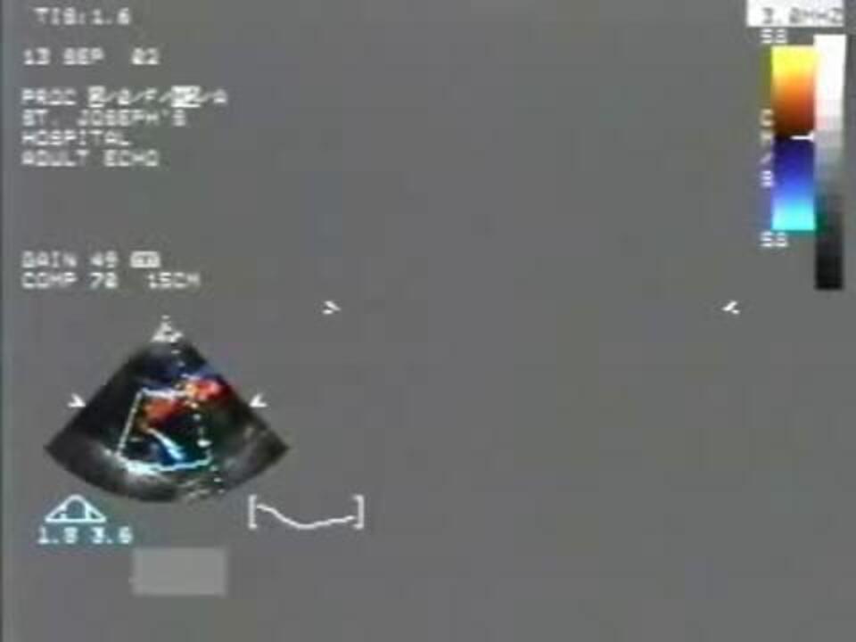
Diagnosis of mitral regurgitation is suspected clinically and confirmed by echocardiography. Doppler echocardiography is used to detect regurgitant flow and pulmonary hypertension. Two-dimensional or 3-dimensional echocardiography is used to evaluate the leaflet morphology and motion; to determine the cause and severity of MR (see table ), the presence and extent of annular calcification, and the size and function of the LV and LA; and to detect pulmonary hypertension.
When it is acute, severe MR may not be apparent on color Doppler echocardiography, but suspicion is raised when acute heart failure is accompanied by hyperdynamic LV systolic function.
Grading of Mitral Regurgitation
Parameter | Moderate MR | Severe MR |
|---|---|---|
Vena contracta* | 3–7 mm | > 7 mm |
Effective regurgitant orifice area | 0.20–0.40 cm2 | > 0.40 cm2 |
Regurgitant volume | 30–60 mL | > 60 mL |
Regurgitant fraction | 30–50% | > 50% |
* The narrowest diameter of the fluid stream downstream of an abnormal valve orifice; it is slightly smaller than the anatomic valve orifice. | ||
MR = mitral regurgitation. | ||
Data from Otto CM, Nishimura RA, Bonow RO, et al: 2020 ACC/AHA Guideline for the Management of Patients With Valvular Heart Disease: Executive Summary: A Report of the American College of Cardiology/American Heart Association Joint Committee on Clinical Practice Guidelines. Circulation 143(5):e35–e71, 2021. doi: 10.1161/CIR.0000000000000932 | ||
If endocarditis or valvular thrombi are suspected, transesophageal echocardiography (TEE) can provide a more detailed view of the mitral valve and LA. TEE, with 3-dimensional imaging when feasible, is also indicated to evaluate the mechanism of MR in more detail when mitral valve repair instead of replacement is being considered.
Cardiac catheterization is performed before surgery, mainly to determine whether coronary artery disease (CAD) is present. A prominent systolic c-v wave is seen on pulmonary artery occlusion pressure (pulmonary capillary wedge pressure) tracings during ventricular systole. Ventriculography can be used to quantify MR. Cardiac MRI can accurately measure regurgitant fraction and determine the cause of dilated myopathy with MR.
Periodic exercise testing (stress ECG) is often performed to detect any decrease in exercise tolerance, which would prompt consideration of surgical intervention. Periodic echocardiography is performed to detect progression of MR.
Phonocardiographic Characteristics of Heart Murmurs


Treatment of Mitral Regurgitation
Mitral valve repair preferred for primary MR
Medical therapy or mitral valve replacement for secondary MR
Anticoagulants for patients with atrial fibrillation
Angiotensin-converting enzyme (ACE) inhibitors and other vasodilators do not delay LV dilation or MR progression and so have no role in asymptomatic MR with preserved LV function. However, if LV dilation or dysfunction is present, therapy using an angiotensin II receptor blocker, neprilysin inhibitor (eg, sacubitril, combined with the angiotensin II blocker valsartan), aldosterone antagonist, and/or vasodilating beta-blocker (eg, carvedilol) is indicated. In secondary MR, these medications can reduce the severity of MR and may improve prognosis.
If the ECG shows left bundle branch block, then biventricular pacing may be beneficial for secondary MR.
Loop diuretics such as furosemide are helpful in patients with exertional or nocturnal dyspnea. Digoxin may reduce symptoms in patients with atrial fibrillation or those in whom valve surgery is not appropriate.
Antibiotic prophylaxis is not longer except for patients who have had valve replacement or repair utilizing prosthetic materials (see table ).
Anticoagulants are used to prevent thromboemboli in patients with atrial fibrillation.
Indications for intervention
Acute mitral regurgitation requires emergency mitral valve repair or replacement with concomitant coronary revascularization as necessary. Pending surgery, nitroprusside or nitroglycerin infusion and an intra-aortic ballon pump may be used to reduce afterload, thus improving forward stroke volume and reducing ventricular and regurgitant volume.
Chronic primary mitral regurgitation that is severe needs intervention at the onset of symptoms or decompensation (left ventricular ejection fraction [LVEF] ≤ 60% or LV end-systolic diameter ≥ 40 mm). Even in the absence of these triggers, intervention may be beneficial when surgical risk is low and valve morphology suggests a high likelihood of successful repair. Atrial fibrillation, pulmonary hypertension, flail leaflet, or left atrial dilation alone is not an indication for intervention (1). When the EF falls to < 30%, surgical risk is high, necessitating a careful weighing of risk and benefit.
Chronic secondary mitral regurgitation has fewer indications for intervention. Because the initial pathology involves the LV muscle, correction of MR is not as beneficial and should be considered only when significant symptoms persist despite a trial of guideline-directed management and therapy, including biventricular pacing, if indicated. Patients with atrial functional mitral regurgitation may benefit from surgery more than patients with other causes of secondary mitral regurgitation (eg, regional or global ischemic changes) (2). Indications for transcatheter edge-to-edge repair (TEER) are wider (more permissive) than those for other surgical options.
For patients undergoing cardiac surgery for other indications, concomitant mitral valve surgery should be considered for a repairable valve with MR that is moderate. However, for secondary MR, the value of this practice is unclear. When studied, the addition of mitral valve repair compared with coronary artery bypass grafting (CABG) alone did not affect LV remodelling or survival, but an excess of adverse events occurred (3). Therefore, only severe secondary MR should be treated when CABG is being performed to treat ischemia.
Choice of intervention
In primary mitral regurgitation, the closer the mitral valve intervention mimics the native valve, the better for LV preservation and mortality. Hence, the order of preference is:
Repair with leaflet remodelling and chordal replacement
Replacement with chordal preservation
Replacement with removal of chordae
If mitral valve repair with leaflet remodelling and chordal replacement is not feasible, replacement with a mechanical prosthesis is preferred because tissue valves have reduced longevity in the mitral position. A bioprosthesis is an option for older patients.
In secondary mitral regurgitation, mitral valve replacement is preferred over repair with a downsized annuloplasty ring because mitral valve replacement results in less mitral regurgitation and heart failure 2 years after treatment (4). When the mechanism of secondary MR is annular dilation due to atrial fibrillation, valve surgery may be beneficial when symptoms cannot be medically controlled.
Another method of repair is transcatheter edge-to-edge repair (TEER) with a device that approximates the mitral leaflets. TEER is an option for patients with severe primary MR and New York Heart Association (NYHA) class III or class IV heart failure symptoms refractory to medical therapy who cannot undergo surgery. In secondary MR, given the lesser benefit of surgery, TEER is indicated even for patients whose surgical risk is not prohibitive if they have NYHA class II or IV symptoms refractory to medical therapy, ejection fraction 20 to 50%, LV end-systolic diameter ≤ 70 mm, pulmonary artery systolic pressure ≤ 70 mm Hg, and an anatomically suitable valve. In carefully selected patients, TEER can reduce symptoms, induce reverse remodelling, and improve clinical outcomes, even though rates of residual and recurrent MR are higher than with surgical repair (5). Clinical success and outcomes depend on careful patient selection and clinical care, so most centers that offer TEER do so through a specialized multidisciplinary heart team (1, 6, 7).
In some decompensated patients, prosthetic valve implantation markedly depresses ejection fraction because in such patients, ventricular function has become dependent on the afterload reduction of MR.
For patients with atrial fibrillation, concomitant procedures such as ablation therapy and surgically occluding the left atrial appendage to reduce the risk of stroke may be beneficial.
Anticoagulation
Lifelong anticoagulation with warfarin is required in patients with a mechanical valve to prevent thromboembolism. A mitral bioprosthetic valve requires anticoagulation with warfarin for 3 to 6 months postoperatively. Long-term non-vitamin K oral anticoagulants or warfarin can be considered in patients with a bioprosthetic valve and atrial fibrillation (see also Anticoagulation for patients with a prosthetic cardiac valve or native valve disease).
Treatment references
1. Otto CM, Nishimura RA, Bonow RO, et al: 2020 ACC/AHA Guideline for the Management of Patients With Valvular Heart Disease: Executive Summary: A Report of the American College of Cardiology/American Heart Association Joint Committee on Clinical Practice Guidelines. Circulation 143(5):e35–e71, 2021. doi: 10.1161/CIR.0000000000000932
2. Farhan S, Silbiger JJ, Halperin JL, et al: Pathophysiology, Echocardiographic Diagnosis, and Treatment of Atrial Functional Mitral Regurgitation: JACC State-of-the-Art Review [published correction appears in J Am Coll Cardiol 2023 Feb 21;81(7):711]. J Am Coll Cardiol 80(24):2314–2330, 2022. doi:10.1016/j.jacc.2022.09.046
3. Michler RE, Smith PK, Parides MK, et al: Two-year outcomes of surgical treatment of moderate ischemic mitral regurgitation. N Engl J Med 374:1932–1941, 2016. doi: 10.1056/NEJMoa1602003
4. Goldstein D, Moskowitz AJ, Gelijns AC, et al: Two-year outcomes of surgical treatment of severe ischemic mitral regurgitation. N Engl J Med 374:344–353, 2016. doi: 10.1056/NEJMoa1512913
5. Feldman T, Kar S, Elmariah S, et al: Randomized comparison of percutaneous repair and surgery for mitral regurgitation: 5-year results of EVEREST II. J Am Coll Cardiol 66:2844–2854, 2015. doi: 10.1016/j.jacc.2015.10.018
6. Obadia JF, Messika-Zeitoun D, Leurent G, et al: Percutaneous repair or medical treatment for secondary mitral regurgitation. N Engl J Med 379:2297–2306, 2018. doi: 10.1056/NEJMoa1805374
7. Stone GW, Lindenfeld J, Abraham WT, et al: Transcatheter mitral-valve repair in patients with heart failure. N Engl J Med 379:2307–2318, 2018. doi: 10.1056/NEJMoa1806640
Prognosis for Mitral Regurgitation
Prognosis of mitral regurgitation varies by duration, severity, and cause.
Some MR worsens and eventually becomes severe. Once MR becomes severe, approximately 8% of asymptomatic patients become symptomatic each year thereafter.
Approximately 15% of patients with chronic moderate or severe MR require surgical intervention, with surgical intervention being more common in primary versus secondary MR (1).
Prognosis reference
1. Otto CM, Nishimura RA, Bonow RO, et al: 2020 ACC/AHA Guideline for the Management of Patients With Valvular Heart Disease: Executive Summary: A Report of the American College of Cardiology/American Heart Association Joint Committee on Clinical Practice Guidelines. Circulation 143(5):e35–e71, 2021. doi: 10.1161/CIR.0000000000000932
Key Points
Common causes of mitral regurgitation (MR) include mitral valve prolapse, rheumatic fever, and left ventricular dilation or infarction.
Acute MR may cause acute pulmonary edema and cardiogenic shock or sudden cardiac death.
Chronic MR causes slowly progressive symptoms of heart failure and, if atrial fibrillation develops, palpitations.
Typical heart sounds are a holosystolic murmur that is heard best at the apex, radiates toward the left axilla, increases in intensity with handgrip or squatting, and decreases in intensity with standing or the Valsalva maneuver.
Symptomatic patients and those meeting certain echocardiographic criteria benefit from valve replacement or repair.
Drug Information for the Topic


