


Erythema nodosum is a specific form of panniculitis characterized by tender, red or violet, palpable, subcutaneous nodules on the shins and occasionally other locations. It often occurs with an underlying systemic disease, notably streptococcal infections, sarcoidosis, and inflammatory bowel disease. Diagnosis is by clinical evaluation and sometimes biopsy. Treatment depends on the cause.
Erythema nodosum primarily affects people in their 20s and 30s but can occur at any age; women are more often affected.
Etiology of Erythema Nodosum
The etiology of erythema nodosum is poorly characterized. It may primarily represent a type IV (delayed) hypersensitivity reaction to various antigenic stimuli (1). Erythema nodosum is frequently accompanied by other disorders.
The most common accompanying disorders are (2):
Streptococcal infection (especially in children)
Other possible triggers include:
Other bacterial infections (eg, Yersinia, Salmonella, mycoplasma, chlamydia, leprosy, lymphogranuloma venereum, tuberculosis)
Fungal infections (eg, kerion, coccidioidomycosis, blastomycosis, histoplasmosis)
Viral infections (eg, Epstein-Barr, hepatitis B)
Use of medications (eg, sulfonamides, iodides, bromides, oral contraceptives)
Hematologic and solid cancers
Pregnancy
Up to approximately 50% of cases of erythema nodosum are idiopathic (3).
Erythema induratum, a similar disorder, manifests with lesions on the calves and classically affects patients with tuberculosis.
Etiology references
1. Ozbagcivan O, Akarsu S, Avci C, et al. Examination of the Microbial Spectrum in the Etiology of Erythema Nodosum: A Retrospective Descriptive Study. J Immunol Res. 2017;2017:8139591. doi:10.1155/2017/8139591
2. Schwartz RA, Nervi SJ. Erythema nodosum: a sign of systemic disease. Am Fam Physician. 2007;75(5):695-700.
3. Mert A, Kumbasar H, Ozaras R, et al. Erythema nodosum: an evaluation of 100 cases. Clin Exp Rheumatol. 2007;25(4):563-570.
Symptoms and Signs of Erythema Nodosum
Erythema nodosum is a subset of panniculitis that manifests as erythematous to purple, tender nodules or plaques, primarily in the pretibial region, often preceded or accompanied by fever, malaise, and arthralgia. Lesions may be detected more easily by palpation than inspection and can evolve into bruise-like areas over weeks.

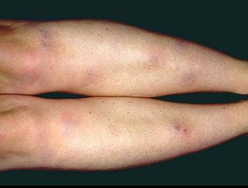
This photo shows characteristic tender, erythematous, subcutaneous nodules on the shins of a patient with erythema nodosum.

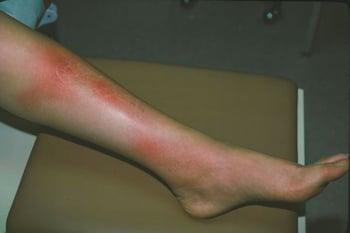
This photo shows characteristic erythematous, tender plaques of erythema nodosum.
Diagnosis of Erythema Nodosum
History and physical examination
Incisional wedge biopsy
The diagnosis of erythema nodosum is usually by clinical appearance of tender pretibial erythematous nodules or plaques and can be confirmed by a deep incisional biopsy of a nodule when necessary (1).
A diagnosis of erythema nodosum should prompt evaluation for an underlying cause (1). Evaluation might include biopsy, tuberculosis testing (using purified protein derivative [PPD] or anergy panel; or using interferon-gamma release assays), and possibly other tests (eg, antinuclear antibodies, complete blood count, chest radiograph, and serial antistreptolysin O titers or a pharyngeal culture). Erythrocyte sedimentation rate is often high.
Diagnosis reference
1. Pérez-Garza DM, Chavez-Alvarez S, Ocampo-Candiani J, et al. Erythema Nodosum: A Practical Approach and Diagnostic Algorithm. Am J Clin Dermatol. 2021;22(3):367-378. doi:10.1007/s40257-021-00592-w
Treatment of Erythema Nodosum
Supportive care
Anti-inflammatory medications (rarely glucocorticoids)
Erythema nodosum almost always resolves spontaneously, typically over approximately 3 to 6 weeks. Treatment of the underlying cause, when identified, is essential. Supportive measures include bed rest, elevation, cool compresses, and nonsteroidal anti-inflammatory drugs (1). Potassium iodide 300 to 900 mg divided orally 3 times a day can be given to decrease inflammation (1). Systemic glucocorticoids are effective but should be used only as a last resort because they can worsen an occult infection.
Treatment reference
1. Pérez-Garza DM, Chavez-Alvarez S, Ocampo-Candiani J, et al. Erythema Nodosum: A Practical Approach and Diagnostic Algorithm. Am J Clin Dermatol. 2021;22(3):367-378. doi:10.1007/s40257-021-00592-w
Key Points
The most common causes of erythema nodosum are streptococcal infections (particularly in children), sarcoidosis, and inflammatory bowel disease.
Diagnose erythema nodosum primarily by clinical appearance but, when necessary, incisional wedge biopsy may be performed for confirmation.
Treat erythema nodosum supportively and use nonsteroidal anti-inflammatory drugs or potassium iodide as needed until the disorder resolves spontaneously.
Drug Information for the Topic







