


Staphylococcal scalded skin syndrome is an acute epidermolysis caused by a staphylococcal toxin. Infants and children are most susceptible. Symptoms include widespread bullae with epidermal sloughing. Diagnosis is by examination and sometimes biopsy. Treatment is antistaphylococcal antibiotics and local care. Prognosis is excellent in children with timely treatment; however, there is high mortality in adults.
(See also Overview of Bacterial Skin Infections.)
Staphylococcal scalded skin syndrome is a severe skin disease associated with blistering and exfoliation that predominantly affects children < 6 years of age (especially infants); it rarely occurs in older adults unless they have kidney failure or are immunocompromised. Occurrence is typically sporadic. For example, epidemics may occur in nurseries, presumably transmitted by the hands of personnel who are in contact with an infected infant or who are nasal carriers of Staphylococcus aureus.
In one cross-sectional study evaluating national inpatient data from the United States, the mean annual incidence of staphylococcal scalded skin syndrome was 7.67 per million children and was 45.1 cases per million for children < 2 years of age (1). Compared to the United States, European data show lower population incidences in France and Germany; however, one study from the Czech republic reported notably higher rates (2–4).There appears to be a slight male predominance, and cases occur more frequently in the summer and fall (1).
General references
1. Staiman A, Hsu DY, Silverberg JI. Epidemiology of staphylococcal scalded skin syndrome in U.S. children. Br J Dermatol. 2018;178(3):704-708. doi:10.1111/bjd.16097
2. Lipový B, Brychta P, Chaloupková Z, Suchánek I. Staphylococcal scalded skin syndrome in the Czech Republic: an epidemiological study. Burns. 2012;38(2):296-300. doi:10.1016/j.burns.2011.08.005
3. Lamand V, Dauwalder O, Tristan A, et al. Epidemiological data of staphylococcal scalded skin syndrome in France from 1997 to 2007 and microbiological characteristics of Staphylococcus aureus associated strains. Clin Microbiol Infect. 2012;18(12):E514-E521. doi:10.1111/1469-0691.12053
4. Mockenhaupt M, Idzko M, Grosber M, Schöpf E, Norgauer J. Epidemiology of staphylococcal scalded skin syndrome in Germany. J Invest Dermatol. 2005;124(4):700-703. doi:10.1111/j.0022-202X.2005.23642.x
Etiology of Staphylococcal Scalded Skin Syndrome
Staphylococcal scalded skin syndrome is caused by Staphylococcus aureus, usually phage group II strains 55 and 71, which elaborate exfoliatin (also called exfoliative toxin [ET] or epidermolysin), a toxin that splits the upper part of the epidermis just beneath the granular cell layer by targeting desmoglein-1 (see Staphylococcal Infections) (1). Methicillin-resistant S. aureus (MRSA) accounts for only 10% of cases (2).
Two major serotypes, exfoliative toxin A (ETA) and exfoliative toxin B (ETB), can cleave desmoglein-1 at a particular peptide bond (3). This cleavage specificity is responsible for the characteristic superficial blister formation at the granular layer of the epidermis. By cleaving desmoglein-1 just below the stratum corneum (see figure ), the toxin allows bacteria to proliferate beneath the epidermal barrier while maintaining a focus that is superficial enough to enable spread by skin contact. Toxin produced in the infected areas of skin may also enter the circulation and affect the entire skin.
Etiology references
1. Handler MZ, Schwartz RA. Staphylococcal scalded skin syndrome: diagnosis and management in children and adults. J Eur Acad Dermatol Venereol. 2014;28(11):1418-1423. doi:10.1111/jdv.12541
2. Azarian T, Cella E, Baines SL, et al. Genomic Epidemiology and Global Population Structure of Exfoliative Toxin A-Producing Staphylococcus aureus Strains Associated With Staphylococcal Scalded Skin Syndrome. Front Microbiol. 2021;12:663831. Published 2021 Aug 18. doi:10.3389/fmicb.2021.663831
3. Hanakawa Y, Schechter NM, Lin C, et al. Molecular mechanisms of blister formation in bullous impetigo and staphylococcal scalded skin syndrome. J Clin Invest. 2002;110(1):53-60. doi:10.1172/JCI15766
Symptoms and Signs of Staphylococcal Scalded Skin Syndrome
In infants, the primary infection often begins in the diaper area or umbilical stump during the first few days of life. In older children, the face is the typical site of initial infection that can then spread systemically.
When a localized area of infection can be found (eg, impetigo), the initial lesion is usually superficial and crusted. Within 24 hours, the surrounding skin becomes painful and erythematous, changes that quickly spread to other areas. The skin may be exquisitely tender and have a wrinkled tissue paper–like consistency.
Large, flaccid blisters arise on the erythematous skin and quickly break to produce erosions. Blisters are frequently present in areas of friction, such as intertriginous areas, buttocks, hands, and feet. Intact blisters extend laterally with gentle pressure (Nikolsky sign).
The epidermis may peel easily, often in large sheets. Widespread desquamation occurs within 36 to 72 hours, and patients may become very ill with systemic manifestations (eg, malaise, chills, fever). Desquamated areas appear scalded. Loss of the protective skin barrier can lead to sepsis and to fluid and electrolyte imbalance.


This photo shows erythema with overlying desquamation in sheets, particularly in the intertriginous area of the groin and axillae and around the umbilicus. Often there is also perioral peeling.
This photo shows erythema with overlying desquamation in sheets, particularly in the intertriginous area of the groin a
Image courtesy of Thomas Habif, MD.

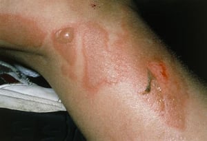
This photo shows the leg of a patient with all 3 stages of the infection: erythema (at center), bullae (left), and desquamation (right).
This photo shows the leg of a patient with all 3 stages of the infection: erythema (at center), bullae (left), and desq
SCIENCE PHOTO LIBRARY

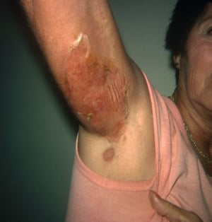
This image shows staphylococcal scalded skin syndrome with superficial skin blistering caused by Staphylococcus aureus infection. This syndrome is rare in adults but can occur in people who are immunocompromised or who have kidney failure or another chronic disease.
This image shows staphylococcal scalded skin syndrome with superficial skin blistering caused by Staphylococcus aureus
DermPics/SCIENCE PHOTO LIBRARY
This photo shows erythema with overlying desquamation in sheets, particularly in the intertriginous area of the groin and axillae and around the umbilicus. Often there is also perioral peeling.
This photo shows erythema with overlying desquamation in sheets, particularly in the intertriginous area of the groin a
Image courtesy of Thomas Habif, MD.
This photo shows the leg of a patient with all 3 stages of the infection: erythema (at center), bullae (left), and desquamation (right).
This photo shows the leg of a patient with all 3 stages of the infection: erythema (at center), bullae (left), and desq
SCIENCE PHOTO LIBRARY
This image shows staphylococcal scalded skin syndrome with superficial skin blistering caused by Staphylococcus aureus infection. This syndrome is rare in adults but can occur in people who are immunocompromised or who have kidney failure or another chronic disease.
This image shows staphylococcal scalded skin syndrome with superficial skin blistering caused by Staphylococcus aureus
DermPics/SCIENCE PHOTO LIBRARY
Diagnosis of Staphylococcal Scalded Skin Syndrome
Primarily history and physical examination
Cultures from areas of suspected primary infection
Rarely toxin detection
The diagnosis of staphylococcal scalded skin syndrome is suspected clinically. Confirmation may rarely require biopsy (frozen section can expedite differentiation from toxic epidermal necrolysis) (1). Specimens show noninflammatory superficial splitting of the epidermis.
Cultures should be taken from the conjunctiva, nasopharynx, blood, urine, and areas of possible primary infection, such as the umbilicus in a neonate or suspect skin lesions. Cultures should not be taken from bullae because they are sterile, unlike in bullous impetigo, where cultures of the blister fluid may yield a pathogen.
Identifying S. aureus in cultures, in the context of suggestive clinical features, lends strong support to a diagnosis of staphylococcal scalded skin syndrome. However, definitive confirmation requires the identification of exfoliative toxins, which can be achieved via polymerase chain reaction (PCR) testing for toxin genes and via Western blot tests or enzyme-linked immunosorbent assays (ELISA) using specific antibodies; assays are not routinely available in hospital laboratories (2).
Differential diagnosis
The differential diagnosis includes drug hypersensitivity, viral exanthemas, scarlet fever, thermal burns, genetic bullous diseases (eg, some types of epidermolysis bullosa), acquired bullous diseases (eg, pemphigus vulgaris, bullous pemphigoid), and toxic epidermal necrolysis (see table and see Stevens-Johnson Syndrome (SJS) and Toxic Epidermal Necrolysis (TEN)).
The mucosal surfaces are spared in staphylococcal scalded skin syndrome; however, they are affected in SJS and TEN.
Differentiating Staphylococcal Scalded Skin Syndrome (SSSS) and Stevens-Johnson Syndrome (SJS)/Toxic Epidermal Necrolysis (TEN)
Feature | SSSS | SJS/TEN |
|---|---|---|
Typical patient population | Infants, young children, and adults with kidney failure or immunocompromise | Older adults |
Usual trigger | Recent staphylococcal infection (toxin-mediated) | Medication exposure (eg, antibiotics, antiseizure medications, NSAIDs) |
Mucous membrane involvement | Absent | Present and prominent |
Systemic illness | Mild to moderate | Severe, multisystem involvement |
Level of epidermal cleavage* | Intraepidermal, within the granular cell (outermost) layer of the epidermis | Subepidermal, at or below the basal cell |
Pathophysiology | Exfoliative toxins cause keratinocyte detachment | Immune-mediated keratinocyte apoptosis |
* Level of epidermal cleavage is determined by frozen section of a fresh skin specimen. | ||
NSAIDs = nonsteroidal anti-inflammatory drugs. | ||
Diagnosis references
1. Handler MZ, Schwartz RA. Staphylococcal scalded skin syndrome: diagnosis and management in children and adults. J Eur Acad Dermatol Venereol. 2014;28(11):1418-1423. doi:10.1111/jdv.12541
2. Ladhani S, Robbie S, Garratt RC, Chapple DS, Joannou CL, Evans RW. Development and evaluation of detection systems for staphylococcal exfoliative toxin A responsible for scalded-skin syndrome. J Clin Microbiol. 2001;39(6):2050-2054. doi:10.1128/JCM.39.6.2050-2054.2001
Treatment of Staphylococcal Scalded Skin Syndrome
Antistaphylococcal antibiotics
Emollients
Gel dressings for weeping lesions
Empiric treatment with IV antistaphylococcal antibiotics should be initiated immediately (1). Typically nafcillin or oxacillin is given until improvement is noted, followed by oral dicloxacillin (2). First-generation cephalosporins (eg, cefazolin, cephalexin) are alternatives. Clindamycin may also be an alternative, but its use is limited by potential resistance. Vancomycin, linezolid, or other antibiotics effective against MRSA should be considered in areas with a high prevalence of MRSA or in patients whose initial therapy failed (3). Glucocorticoids are contraindicated.
Emollients (eg, white petrolatum) are sometimes used to prevent further insensible water loss from ulcerated skin. However, topical therapy (including topical antibiotics) and patient handling must be minimized (2).
If disease is widespread and lesions are weeping, the skin should be treated as for burns. Hydrolyzed polymer gel dressings may be very useful; however, the number of dressing changes should be minimized because they can be painful. Patients should be monitored and treated for complications similar to those that occur with burns (eg, fluid and electrolyte imbalance, sepsis).
Steps to detect carriers and prevent or treat nursery epidemics are discussed elsewhere (see prevention of neonatal hospital-acquired infection).
Treatment references
1. Leung AKC, Barankin B, Leong KF. Staphylococcal-scalded skin syndrome: evaluation, diagnosis, and management. World J Pediatr. 2018;14(2):116-120. doi:10.1007/s12519-018-0150-x
2. Gray L, Hansen AM, Cipriano SD. Pediatric Staphylococcal Scalded Skin Syndrome: A Systematic Review of the Literature to Inform Work-Up and Management. Pediatr Dermatol. 2025;42(5):978-984. doi:10.1111/pde.16029
3. Wang Z, Feig JL, Mannschreck DB, Cohen BA. Antibiotic sensitivity and clinical outcomes in staphylococcal scalded skin syndrome. Pediatr Dermatol. 2020;37(1):222-223. doi:10.1111/pde.14014
Prognosis for Staphylococcal Scalded Skin Syndrome
With prompt diagnosis and therapy, the stratum corneum is quickly replaced, and healing usually occurs within 5 to 7 days after start of treatment.
Mortality is generally low (< 10%) in treated children, despite the higher incidence in this population (1). Mortality is typically higher in adults (up to approximately 60% in some studies), despite initiation of treatment, largely because of underlying comorbidities.
Prognosis reference
1. Handler MZ, Schwartz RA. Staphylococcal scalded skin syndrome: diagnosis and management in children and adults. J Eur Acad Dermatol Venereol. 2014;28(11):1418-1423. doi:10.1111/jdv.12541
Key Points
Generalized desquamation and systemic illness most often indicate staphylococcal scalded skin syndrome (SSSS) in infants and young children (and occasionally in immunocompromised adults), whereas in older adults, they most often indicate toxic epidermal necrolysis.
Culture the conjunctiva, nasopharynx, blood, urine, and areas of possible primary infection, such as the umbilicus and suspect skin lesions.
Treat patients with antistaphylococcal antibiotics and, if disease is widespread, in a burn unit if possible.
Monitor and treat for complications similar to those that occur with burns (eg, fluid and electrolyte imbalance, sepsis).
Drug Information for the Topic


