


Globally, thoracic trauma comprises approximately 25% of deaths caused by trauma (1). Many thoracic injuries cause immediate death (eg, traumatic aortic disruption) while other injuries (eg, tension pneumothorax, cardiac tamponade) result in death during the first minutes or hours after trauma. Early diagnosis of thoracic injuries is paramount because life-saving interventions can frequently be performed at the bedside with definitive or temporizing measures, often by clinicians without advanced surgical training.
General reference
1. Coccolini F, Cremonini C, Moore EE, et al. Thoracic trauma WSES-AAST guidelines. World J Emerg Surg. 2025;20(1):78. Published 2025 Oct 15. doi:10.1186/s13017-025-00651-1
Etiology of Thoracic Trauma
Thoracic injuries result from blunt or penetrating trauma. Blunt thoracic trauma is more common than penetrating trauma. The mechanism of injury determines the injury pattern (1). For example, in a motor vehicle crash, impact to the steering wheel may cause blunt trauma to the chest, resulting in rib fractures or pulmonary contusions. A gunshot wound or stab wound to the chest causes penetrating trauma resulting in hemothorax or cardiac laceration.
The most common serious thoracic injuries include the following:
Pneumothorax (simple pneumothorax, open pneumothorax, or tension pneumothorax)
Additionally, the esophagus and diaphragm (see Overview of Abdominal Trauma) can also be damaged by chest trauma. Because the diaphragm can be as high as the mamillary (nipple) line during exhalation, penetrating trauma to the chest at or below nipple level can also cause intra-abdominal injuries.
Patients may have multiple concurrent types of intrathoracic injuries.
Etiology reference
1. Coccolini F, Cremonini C, Moore EE, et al. Thoracic trauma WSES-AAST guidelines. World J Emerg Surg. 2025;20(1):78. Published 2025 Oct 15. doi:10.1186/s13017-025-00651-1
Pathophysiology of Thoracic Trauma
Most morbidity and mortality due to chest trauma occurs because injuries compromise respiration and/or circulation (1).
Respiration can be compromised by:
Direct damage to the lungs or airways
Altered mechanics of breathing from muscular or bony disruption
Injuries that directly damage the lung or airways include pulmonary contusion and tracheobronchial disruption. Injuries that alter the mechanics of breathing include hemothorax, pneumothorax, and flail chest (paradoxical chest wall movement associated with 3 or more ribs broken in 2 or more places). Injury to the lung, tracheobronchial tree, or rarely esophagus may allow air to enter the soft tissues of the chest and/or neck (subcutaneous emphysema) or mediastinum (pneumomediastinum). This air itself rarely has significant physiologic consequence; the underlying injury is the problem. Tension pneumothorax impairs respiration as well as circulation. Pulmonary contusion creates a ventilation and perfusion mismatch because of lung hemorrhage and edema.
Circulation can be impaired by:
Bleeding
Decreased venous return
Direct cardiac injury
Bleeding, as occurs in hemothorax, can be massive, causing hemorrhagic shock. Respiration can also be impaired if hemothorax is large enough to restrict expansion of the lung during inspiration. Decreased venous return impairs cardiac filling, causing hypotension. Decreased venous return can occur due to increased intrathoracic pressure in tension pneumothorax or to increased intrapericardial pressure in cardiac tamponade. Heart failure and/or conduction abnormalities can result from blunt cardiac injury that damages the myocardium or the heart valves.
Because chest wall injuries typically make breathing very painful, patients often limit inspiration (splinting). A common complication of splinting is atelectasis, which can lead to hypoxemia, pneumonia, or both.
Patients treated with tube thoracostomy may develop empyema, particularly if a hemothorax is incompletely drained.
Significant pulmonary contusion can require similar treatment strategies as needed in patients with acute respiratory distress syndrome (ARDS).
Pathophysiology reference
1. Helsloot D, Fitzgerald MC, Lefering R, et al; and the TraumaRegister DGU®. The first hour of trauma reception is critical for patients with major thoracic trauma: A retrospective analysis from the TraumaRegister DGU. Eur J Anaesthesiol. 2023;40(11):865-873. doi:10.1097/EJA.0000000000001834
Symptoms and Signs of Thoracic Trauma
Symptoms include pain, which usually worsens with breathing if the chest wall is injured, and sometimes shortness of breath.
Common physical examination findings in thoracic trauma include respiratory distress, chest tenderness, and chest wall ecchymoses; hypotension or shock may be present.
Neck vein distention can occur in tension pneumothorax or cardiac tamponade if patients have sufficient intravascular volume.
The trachea can deviate away from the side of a tension pneumothorax.
In flail chest, a segment of the chest wall moves paradoxically—that is, in the opposite direction from the rest of the chest wall (outward during expiration and inward during inspiration); the flail chest is often palpable.
Subcutaneous emphysema causes a crackling or crunch when palpated. Findings may be localized to a small area or involve a large portion of the chest wall and/or extend to the neck. Most often, pneumothorax is the cause; when subcutaneous emphysema is extensive, injury to the tracheobronchial tree or upper airway should be considered. Air in the mediastinum may produce a characteristic crunching sound synchronous with the heartbeat (Hamman sign or Hamman crunch). Hamman sign suggests pneumomediastinum and often tracheobronchial tree injury or, rarely, esophageal injury.
Decreased breath sounds can result from pneumothorax or hemothorax; percussion over the affected areas is dull with hemothorax and hyperresonant with pneumothorax.
Diagnosis of Thoracic Trauma
History and physical examination
Chest radiograph
Sometimes other imaging studies (eg, CT, CT angiography, ultrasound)
Initial evaluation
Five conditions are immediately life threatening and rapidly correctable:
Diagnosis and treatment begin during the primary trauma survey (see Approach to the Trauma Patient) and are based first on physical examination. Depth and symmetry of chest wall excursion are assessed, the lungs are auscultated, and the entire chest wall and neck are inspected and palpated. Patients in respiratory distress should be monitored with serial assessments of clinical status and of oxygenation plus ventilation (eg, with pulse oximetry, arterial blood gas tests, capnometry if intubated).
Penetrating chest wounds should not be probed. However, the location of wounds helps predict which organs may be injured. High-risk wounds are those medial to the nipples or scapulae and those that traverse the chest from side to side (ie, entering one hemithorax and exiting the other). Such wounds may injure the hilar or great vessels, heart, tracheobronchial tree, or rarely the esophagus.
Patients with symptoms of partial or complete airway obstruction following blunt trauma should be immediately intubated to control the airway.
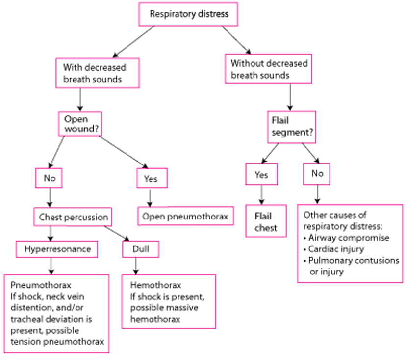
In patients with difficulty breathing, severe injuries to consider during the primary trauma survey include the following:
A simplified, rapid approach to help differentiate these injuries is shown in the figure (see figure ).
A Simplified, Rapid Assessment of Patients With Thoracic Trauma and Respiratory Distress During the Primary Survey

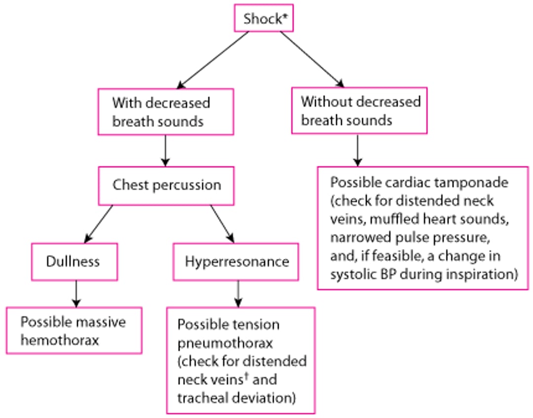
In patients with thoracic trauma and impaired circulation (signs of shock), severe injuries to consider during the primary trauma survey include the following:
Other chest injuries (eg, blunt cardiac injury, aortic disruption) may cause shock but are not treated during the primary trauma survey. Simplified, rapid approaches can help differentiate among rapidly correctable causes of shock due to chest injuries (see figure A Simplified, Rapid Assessment for Chest Injuries in Patients With Shock...). However, hemorrhage should be excluded in all patients who have shock after major trauma, regardless of whether a chest injury that could cause shock is identified.
A Simplified, Rapid Assessment for Chest Injuries in Patients With Shock During the Primary Trauma Survey
* Hemorrhage should be excluded in all patients who are in shock after major trauma, regardless of whether a chest injury that could cause shock is identified. † Neck vein distention may be absent in patients with hypovolemic shock. |

Treatment of injuries affecting the airway, breathing, or circulation begins during the primary trauma survey. After the primary trauma survey, patients are clinically assessed in more detail for other severe chest injuries as well as less severe manifestations of the injuries considered during the primary trauma survey.
Imaging
Imaging studies are typically required in patients with significant chest trauma. Chest radiographs are almost always obtained. Results are usually diagnostic of certain injuries (eg, pneumothorax, hemothorax, moderate or severe pulmonary contusion, clavicle fracture, some rib fractures) and suggestive for others (eg, aortic disruption, diaphragmatic rupture). However, findings may evolve over hours (eg, in pulmonary contusion and diaphragmatic injury). Plain radiographs of the scapula or sternum are sometimes performed when there is tenderness over those structures.
During the E-FAST (Extended Focused Assessment With Sonography in Trauma) examination, ultrasound of the heart is typically performed during the resuscitation phase to look for cardiac tamponade; some pneumothoraces can also be seen.
CT of the chest is often performed when aortic injury is suspected and to diagnose small pneumothoraces, sternal and rib fractures, mediastinal (eg, heart, esophageal, bronchial) injuries, and thoracic spine injuries.
Other tests for aortic injury include aortography and transesophageal echocardiography.
Laboratory and other testing
Complete blood count is often performed but is mainly valuable as a baseline for detecting ongoing hemorrhage. Arterial and venous blood gas results, or end tidal capnography, help monitor patients with hypoxia or respiratory distress. Normal cardiac biomarkers (eg, troponin) can help exclude blunt cardiac injury in patients with chest trauma.
ECG is typically performed for chest trauma that is severe or compatible with cardiac injury. Cardiac injury may cause arrhythmia, conduction abnormalities, ST segment abnormalities, or a combination.
Treatment of Thoracic Trauma
Supportive care
Treatment of specific injuries
Immediately life-threatening injuries are treated at the bedside at the time of diagnosis:
Respiratory distress with suspected tension pneumothorax: Needle thoracostomy or finger thoracostomy.
Respiratory distress with suspected open pneumothorax: Partially occlusive dressing followed by tube thoracostomy
Respiratory distress with suspected flail chest: Mechanical ventilation
Respiratory distress or shock with decreased breath sounds and suspected hemothorax: Tube thoracostomy
Shock with suspected cardiac tamponade: Pericardiocentesis
Suspected hypovolemic shock: Fluid resuscitation and blood products when available
Immediate resuscitative thoracotomy can be considered for trauma victims if the clinician is proficient in the procedure and the patient has one of the following indications (1):
Penetrating thoracic injury with a need for cardiopulmonary resuscitation (CPR) of < 15 minutes
Penetrating trauma to the neck or extremity with a need for CPR of < 5 minutes
Blunt trauma with a need for CPR of < 10 minutes
Persistent systolic blood pressure of < 60 mm Hg due to suspected cardiac tamponade, hemorrhage, or air embolism
Pearls & Pitfalls
|
In the absence of any of these criteria, resuscitative thoracotomy is contraindicated because the procedure has significant risks (eg, transmission of bloodborne diseases, injury to clinician).
Specific treatment is directed at the injury. Whole blood transfusion when immediately available should be used for volume resuscitation but blood component transfusion (eg, red blood cells, platelets and plasma) can also be used (2, 3, 4). Supportive therapy typically includes analgesics, supplemental oxygen, and sometimes mechanical ventilation.
Treatment references
1. Burlew CC, Moore EE, Moore FA, et al. Western Trauma Association critical decisions in trauma: resuscitative thoracotomy. J Trauma Acute Care Surg. 2012;73(6):1359-1363. doi:10.1097/TA.0b013e318270d2df
2. Torres CM, Kent A, Scantling D, et al. Association of whole blood with survival among patients presenting with severe hemorrhage in US and Canadian adult civilian trauma centers. JAMA Surg 2023;158(5):532-540. doi: 10.1001/jamasurg.2022.6978
3. Holcomb JB, Tilley BC, Baraniuk S, et al: Transfusion of plasma, platelets, and red blood cells in a 1:1:1 vs a 1:1:2 ratio and mortality in patients with severe trauma: the PROPPR randomized clinical trial. JAMA 2015;313(5):471-482. doi: 10.1001/jama.2015.12
4. LaGrone LN, Stein D, Cribari C, et al. American Association for the Surgery of Trauma/American College of Surgeons Committee on Trauma: Clinical protocol for damage-control resuscitation for the adult trauma patient. J Trauma Acute Care Surg. 2024;96(3):510-520. doi:10.1097/TA.0000000000004088


