


Congenital eye anomalies include eyes that are absent, deformed, or incompletely developed, often in conjunction with other congenital anomalies and syndromes.
The prevalence of congenital eye anomalies is approximately 1 in 2400 (1). (See also Overview of Congenital Craniofacial Anomalies.)
A clinical geneticist should evaluate affected patients even in cases of apparent isolated congenital anomaly. Anophthalmia and microphthalmia frequently co-occur with craniofacial and brain anomalies (2).
Chromosomal microarray analysis, specific gene tests, or broader gene panel tests should be considered in the evaluation of patients with congenital craniofacial anomalies. If the results of these tests are nondiagnostic, clinical exome sequencing analysis or clinical genome sequencing may be recommended.
Some structural birth defects of the eye, or their complications, are treated surgically using various surgical techniques. Consultation with a pediatric ophthalmologist is important for children with these conditions.
General references
1. Maillet C, Guilbaud L, Monier I, et al. Prevalence and prenatal diagnosis of congenital eye anomalies: A population-based study. BJOG. 2024;131(10):1385-1391. doi:10.1111/1471-0528.17817
2. Schraw JM, Benjamin RH, Scott DA, et al. A Comprehensive Assessment of Co-occurring Birth Defects among Infants with Non-Syndromic Anophthalmia or Microphthalmia. Ophthalmic Epidemiol. 2021;28(5):428-435. doi:10.1080/09286586.2020.1862244
Hypertelorism
Hypertelorism is widely spaced eyes, as determined by increased interpupillary distance, and can occur in numerous congenital syndromes, including frontonasal dysplasia (with midline facial cleft, and brain anomalies), craniofrontonasal dysplasia (with craniosynostosis), and Aarskog syndrome (with limb and genital anomalies).

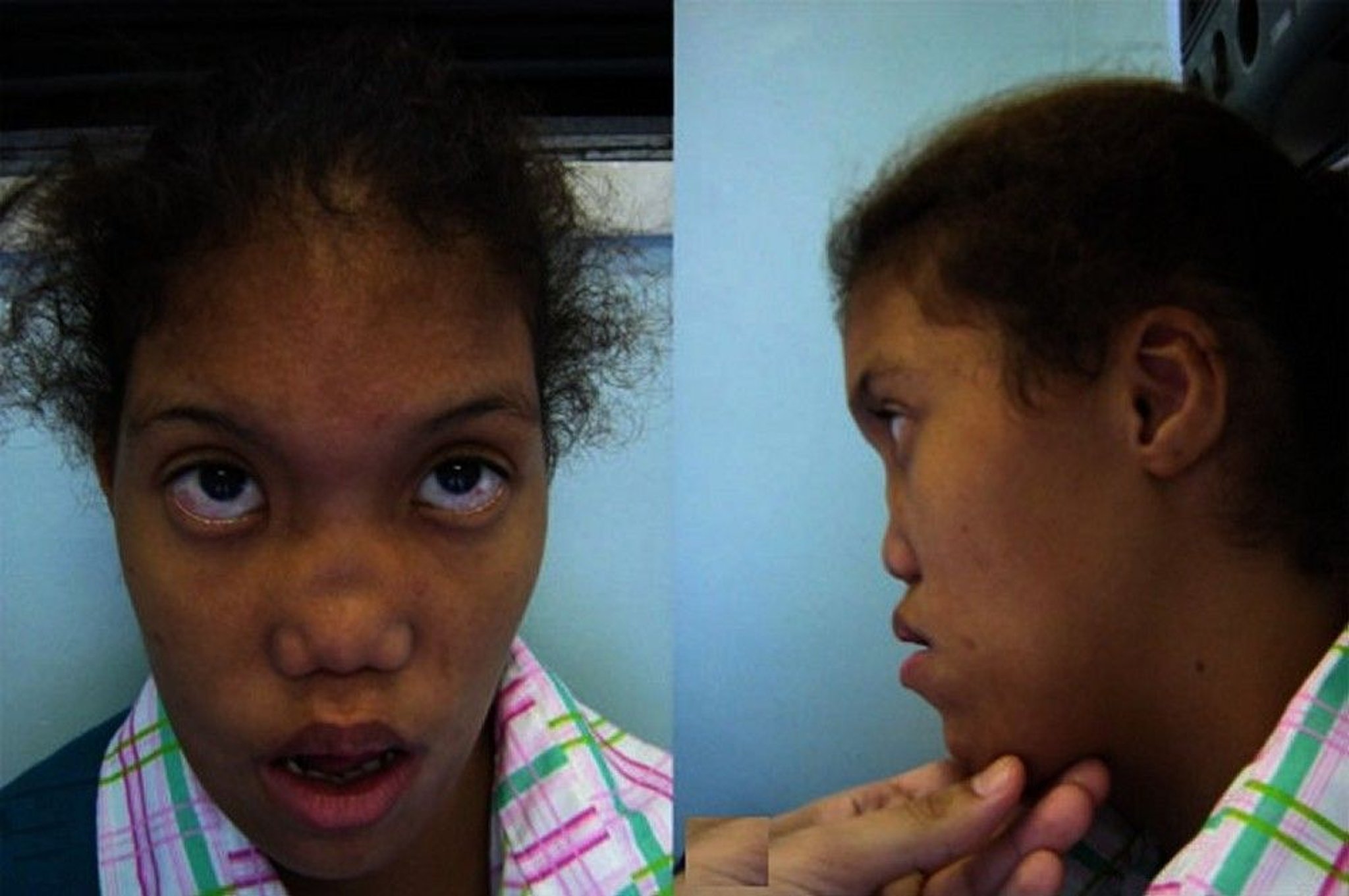
This patient has ocular hypertelorism (widely spaced eyes) and maxillary hypoplasia (small upper mandible). She also has other facial deformities including flat nasal bridge, upslanting palpebral fissures, epicanthal folds, low-set ears, and retrognathism.
© Springer Science+Business Media
Hypotelorism
Hypotelorism is closely spaced eyes, as determined by decreased interpupillary distance. This anomaly should raise suspicion of holoprosencephaly (a midline brain abnormality).
Coloboma
Coloboma is a gap in the structure of the eye that may affect the eyelid, iris, retina, or optic nerve of 1 or both eyes.
Coloboma involving the macula or optic disc can cause vision loss (1).
Coloboma of the eyelid is frequently associated with epibulbar dermoid cysts and is common in Treacher Collins syndrome, Nager syndrome, and Goldenhar syndrome, among many others (1).
Coloboma of the iris raises the possibility of CHARGE association (coloboma, heart defects, choanal atresia, retarded growth and development, genital hypoplasia, and ear abnormalities), Schmid-Fraccaro syndrome (ie, cat eye syndrome), Kabuki syndrome, or Aicardi syndrome.


This photo shows bilateral coloboma with enlarged pupils extending to the lower edge of the iris of each eye.
Coloboma reference
1. Lingam G, Sen AC, Lingam V, Bhende M, Padhi TR, Xinyi S. Ocular coloboma-a comprehensive review for the clinician. Eye (Lond). 2021;35(8):2086-2109. doi:10.1038/s41433-021-01501-5
Microphthalmia
Microphthalmia is a small eye globe, which may be unilateral or bilateral. Even when unilateral, mild anomalies (eg, microcornea, colobomas, congenital cataract) of the other eye are frequently present (1).
Microphthalmia causes sight-threatening complications such as angle-closure glaucoma, chorioretinal pathology (eg, uveal effusion), strabismus, and amblyopia.


This photo shows a patient with bilateral microphthalmia (left greater than right).
Causes of microphthalmia include prenatal exposure to teratogens, alcohol, and infections (eg, TORCH [toxoplasmosis, other pathogens, rubella, cytomegalovirus, and herpes simplex] and Zika virus) (2), and numerous chromosomal or genetic disorders, some of which are suggested by other clinical features (1, 3). Growth and developmental delays are frequently present in microphthalmia that is caused by a chromosomal disorder. Facial asymmetry suggests Goldenhar syndrome or Treacher Collins syndrome, hand anomalies suggest trisomy 13, oculo-dental-digital syndrome, or fetal alcohol syndrome, and genital anomalies may suggest chromosomal defects, Fraser syndrome, or CHARGE association (coloboma, heart defects, choanal atresia, retarded growth and development, genital hypoplasia, and ear abnormalities).
Microphthalmia references
1. Skalicky SE, White AJ, Grigg JR, et al. Microphthalmia, anophthalmia, and coloboma and associated ocular and systemic features: understanding the spectrum. JAMA Ophthalmol. 2013;131(12):1517-1524. doi:10.1001/jamaophthalmol.2013.5305
2. Devakumar D, Bamford A, Ferreira MU, et al. Infectious causes of microcephaly: epidemiology, pathogenesis, diagnosis, and management. Lancet Infect Dis. 2018;18(1):e1-e13. doi:10.1016/S1473-3099(17)30398-5
3. Verma AS, Fitzpatrick DR. Anophthalmia and microphthalmia. Orphanet J Rare Dis. 2007;2:47. Published 2007 Nov 26. doi:10.1186/1750-1172-2-47
Anophthalmia
Anophthalmia is complete absence of the eye globe. It occurs in > 50 genetic syndromes caused by chromosomal abnormalities or pathogenic variants in one of several genes (eg, SOX2, OTX2, BMP4) (1, 2).
When skin covers the orbit, the anomaly is called cryptophthalmos, which suggests Fraser syndrome, Nager syndrome, or ocular disorders associated with intellectual disabilities.


This patient has multiple congenital anomalies including right-sided anophthalmia, deformed pinna, and hemifacial microsomia.
Anophthalmia references
1. Verma AS, Fitzpatrick DR. Anophthalmia and microphthalmia. Orphanet J Rare Dis. 2007;2:47. Published 2007 Nov 26. doi:10.1186/1750-1172-2-47
2. Skalicky SE, White AJ, Grigg JR, et al. Microphthalmia, anophthalmia, and coloboma and associated ocular and systemic features: understanding the spectrum. JAMA Ophthalmol. 2013;131(12):1517-1524. doi:10.1001/jamaophthalmol.2013.5305


