


Obstructive sleep apnea (OSA) consists of recurrent episodes of partial or complete closure of the upper airway that occur during sleep and lead to pauses in breathing (defined as a period of apnea or hypopnea > 10 seconds) with hypoxemia followed by arousals. Symptoms can include excessive daytime sleepiness, restlessness, snoring, recurrent awakening, and morning headache. Diagnosis is based on sleep history and diagnostic testing. Treatment includes continuous positive airway pressure, oral appliances, and, in refractory cases, surgery. In patients with overweight or obesity, weight loss can reduce OSA severity. Prognosis is good with treatment. Untreated patients are at risk for hypertension, atrial fibrillation and other arrhythmias, heart failure, and injury or death due to motor vehicle crashes and other accidents resulting from hypersomnolence.


Obstructive sleep apnea (OSA) is common, and the prevalence is increasing with the increased prevalence of obesity. An estimated 1 billion people are affected worldwide (1), most of whom are undiagnosed and untreated. Some degree of OSA with symptoms is present in 8 to 16% of adults (2). OSA is up to 4 times more common among males (3) and 7 times more common among people with obesity (ie, body mass index [BMI] ≥ 30), although one-third of patients with OSA do not have overweight or obesity (1). (See also Obstructive Sleep Apnea in Children.)
An apneic episode in adults is defined as a cessation of airflow lasting at least 10 seconds. OSA occurs when the cessation of airflow is accompanied by ongoing respiratory effort.
A hypopneic episode in adults can be defined in different ways, typically as a 30% or more reduction in airflow lasting ≥ 10 seconds that results in either oxygen desaturation or an arousal from sleep. A hypopneic episode also typically includes 3% or 4% drop in oxygen saturation levels or an arousal from sleep.
The manifestations, treatment, and prognosis of OSA vary with sex and age (3, 4, 5).
OSA may coexist with central sleep apnea (CSA) in the same individual.
General references
1. Gottlieb DJ, Punjabi NM. Diagnosis and management of obstructive sleep apnea: A review. JAMA. 2020;323(14):1389-1400. doi:10.1001/jama.2020.3514
2. Peppard PE, Young T, Barnet JH, Palta M, Hagen EW, Hla KM. Increased prevalence of sleep-disordered breathing in adults. Am J Epidemiol. 2013;177(9):1006-1014. doi:10.1093/aje/kws342
3. Bonsignore MR, Saaresranta T, Riha RL. Sex differences in obstructive sleep apnoea. Eur Respir Rev. 2019;28(154):190030. doi: 10.1183/16000617.0030-2019
4. Braley TJ, Dunietz GL, Chervin RD, et al. Recognition and diagnosis of obstructive sleep apnea in older Americans. J Am Geriatr Soc. 2018;66(7):1296-1302. doi:10.1111/jgs.15372
5. Jordan AS, McSharry DG, Malhotra A. Adult obstructive sleep apnoea. Lancet. 2014;383(9918):736-747. doi:10.1016/S0140-6736(13)60734-5
Pathophysiology of Obstructive Sleep Apnea (OSA)
Obstructive sleep apnea occurs due to repetitive collapse of the upper airway during sleep. In contrast to CSA, apnea that occurs in OSA is characterized by cessation of both airflow and respiratory effort.
Sleep is associated with decreased muscle tone of the upper airway, leading to partial or complete obstruction of the nasopharynx, oropharynx, or both. Airway patency tends to oscillate, causing recurrent periods of apnea and recovery. Dynamic factors, including redistribution of dependent edema to the neck during the night while the patient is recumbent, may contribute to narrowing of the upper airway (1). Other factors thought to be important include upper airway muscle responsiveness, sleep stability, and ventilatory control (2).
Ventilatory control is often quantified using the engineering term "loop gain". The loop gain of the respiratory system quantifies how the systems responds to disturbances in ventilation, (eg, apneas, hypopneas). In a system with high loop gain, any respiratory disturbance that results in hypoventilation will lead to even bigger response or overcompensation with hyperventilation. Subsequently, periods of hyperventilation can perpetuate unstable breathing and lead to further apneas.
Obstruction causes multiple episodes of apnea or hypopnea, which lead to hypoxia and hypercapnia, all of which disrupt normal sleep, with partial or complete arousals from nonrapid eye movement (NREM) and rapid eye movement (REM) sleep. Inspiratory efforts against a closed upper airway cause swings in intrathoracic pressure that affect cardiac performance. Endothelial and neurotransmitter dysfunction occur. All of these factors interact to increase the risk of significant morbidity and mortality.
Related disorders
Less severe forms of OSA may not produce oxygen desaturation but can interrupt sleep.
Upper airway resistance syndrome can cause symptoms similar to OSA, but breathing reductions do not meet strict criteria for obstructive apneas and hypopneas. Instead, there are hypopneas or snoring that lead to respiratory effort-related arousals (RERAs) from sleep. Patients with upper airway resistance syndrome are typically younger and less likely to have obesity than those with OSA, so drops in oxygen desaturation are less likely to be observed. Symptoms, diagnostic evaluation, and treatment of snoring and upper airway resistance syndrome are similar to those of OSA.
Obesity-hypoventilation syndrome refers to a related disorder in patients with obesity, usually severe OSA, and hypoventilation without any other cause. Treatment of OSA in OHS with positive airway pressure (PAP) therapy can improve hypoventilation (3).
Complications
Obstructive sleep apnea has significant neurocognitive, cardiovascular, and metabolic consequences.
Although insufficient sleep is the most common overall cause of excessive daytime sleepiness, OSA is the leading medical cause. The excessive sleepiness actively increases the risk of automobile crashes, difficulties at work, and sexual dysfunction. There is often some degree of cognitive impairment and also an increased risk of injury (eg, when operating heavy machinery or engaging in other activities during which unintentional sleep episodes would be hazardous).
Relationships with bed partners, roommates, and/or housemates may also be adversely affected because such people may have difficulty sleeping because of the patient's noisy, restless sleep.
Hypertension is strongly associated with OSA (4). Patients with untreated OSA who are normotensive are more likely to develop hypertension within 5 years of diagnosis. Repetitive nocturnal hypoxia and sleep disruption are associated with increased risk of medical disorders, including heart failure, coronary artery disease, atrial fibrillation (including recurrence after catheter ablation) and other arrhythmias, metabolic dysfunction–associated steatotic liver disease (MASLD), and stroke (5). The risk of stroke and all-cause mortality is increased even when controlling for other risk factors (eg, hypertension, diabetes) (6, 7). However, the contribution of OSA to these common disorders is often underappreciated (8).
Perioperative complications can occur with unrecognized OSA because moderate or general anesthesia is a risk for airway obstruction. Patients with diagnosed OSA should inform an anesthesiologist of the diagnosis before undergoing any surgery and should receive continuous positive airway pressure (CPAP) when they receive preoperative medications and during recovery.
Pathophysiology references
1. White LH, Bradley TD. Role of nocturnal rostral fluid shift in the pathogenesis of obstructive and central sleep apnoea. J Physiol. 2013;591(5):1179-1193. doi:10.1113/jphysiol.2012.245159
2. Edwards BA, Redline S, Sands SA, Owens RL. More Than the Sum of the Respiratory Events: Personalized Medicine Approaches for Obstructive Sleep Apnea. Am J Respir Crit Care Med. 2019;200(6):691-703. doi:10.1164/rccm.201901-0014TR
3. Masa JF, Pépin JL, Borel JC, Mokhlesi B, Murphy PB, Sánchez-Quiroga MÁ. Obesity hypoventilation syndrome. Eur Respir Rev. 2019;28(151):180097. doi:10.1183/16000617.0097-2018
4. Van Ryswyk E, Mukherjee S, Chai-Coetzer CL, et al. Sleep disorders, including sleep apnea and hypertension. Am J Hypertens. 2018;31(8):857-864. doi: 10.1093/ajh/hpy082
5. Zinchuk AV, Jeon S, Koo BB, et al. Polysomnographic phenotypes and their cardiovascular implications in obstructive sleep apnoea. Thorax. 2018;73(5):472-480. doi: 10.1136/thoraxjnl-2017-210431
6. Punjabi NM, Caffo BS, Goodwin JL, et al. Sleep-disordered breathing and mortality: a prospective cohort study. PLoS Med. 2009;6(8):e1000132. doi:10.1371/journal.pmed.1000132
7. Yaggi HK, Concato J, Kernan WN, et al. Obstructive sleep apnea as a risk factor for stroke and death. N Engl J Med. 2005;353(19):2034-2041. doi:10.1056/NEJMoa043104
8. Borsoi L, Armeni P, Donin G, et al. The invisible costs of obstructive sleep apnea (OSA): Systematic review and cost-of-illness analysis. PLoS One. 2022;17(5):e0268677. doi: 10.1371/journal.pone.0268677
Etiology of Obstructive Sleep Apnea (OSA)
Anatomic risk factors for obstructive sleep apnea include:
An oropharynx “crowded” by a short or retracted mandible
A prominent tongue base or tonsils
A rounded head shape and a short neck
A neck circumference > 43 cm (> 17 in) in males and > 41cm (> 16 inches) in females
Thick lateral pharyngeal walls and parapharyngeal fat pads


This illustration shows normal breathing (top panel), snoring with partial obstruction of the airway (middle panel), and OSA with complete obstruction of the airway (bottom panel). The top panel also shows normal placement of the tongue, soft palate, and uvula during sleep.
Alila Medical Media/stock.adobe.com
Such risk factors may not predict severity.
Other identified risk factors include postmenopausal status, aging, overweight or obesity, and alcohol or sedative use (1). Medical disorders that have been thought to cause or contribute to OSA include nocturnal gastroesophageal reflux, acromegaly, hypothyroidism, and prior stroke. OSA and obesity-hypoventilation syndrome frequently coexist.
In a twin heritability study, OSA was found to be 73% heritable (2). Factors contributing to the severity of OSA such as apnea-hypopnea index (AHI) also appear to be highly heritable. Genetic factors may explain nearly 40% of the variation in AHI, and genetic risks may vary by ethnicity (3). Likelihood of OSA in a given family member is proportional to the number of other affected family members.
Etiology references
1. Patel SR. Obstructive sleep apnea. Ann Intern Med. 2019;171(11):ITC81-ITC96. doi: 10.7326/AITC201912030
2. Szily M, Tarnoki AD, Tarnoki DL, et al. Genetic influences on the onset of obstructive sleep apnoea and daytime sleepiness: a twin study. Respir Res. 2019;20(1):125. doi:10.1186/s12931-019-1095-x
3. Yi M, Tan Y, Pi Y, et al. Variants of candidate genes associated with the risk of obstructive sleep apnea. Eur J Clin Invest. 2022;52(1):e13673. doi: 10.1111/eci.13673
Symptoms and Signs of Obstructive Sleep Apnea (OSA)
In patients with OSA, habitual snoring is present in 50 to 60%, and witnessed apneas are reported in 10 to 15% (1). However,most people who snore do not have OSA. Other symptoms of OSA may include (2, 3, 4):
Choking, gasping, or snorting during sleep
Restless and unrefreshing sleep
Difficulty staying asleep
Some patients may be unaware of nocturnal symptoms until informed by bed partners, roommates, or housemates. Obtaining a history from a bed partner can be useful in the evaluation. In the morning, some patients have a sore throat, dry mouth, or headache.
During daily activities, patients may experience intrusive sleepiness, fatigue, and impaired concentration. The frequency of sleep complaints and the degree of wake-time sleepiness do not correlate well with the number of sleep events or arousals. Some patients may be relatively asymptomatic, experiencing neither sleep nor wake symptoms despite polysomnographic data indicating moderate or severe disease.
Modified Mallampati Scoring
Modified Mallampati scoring is as follows:
|

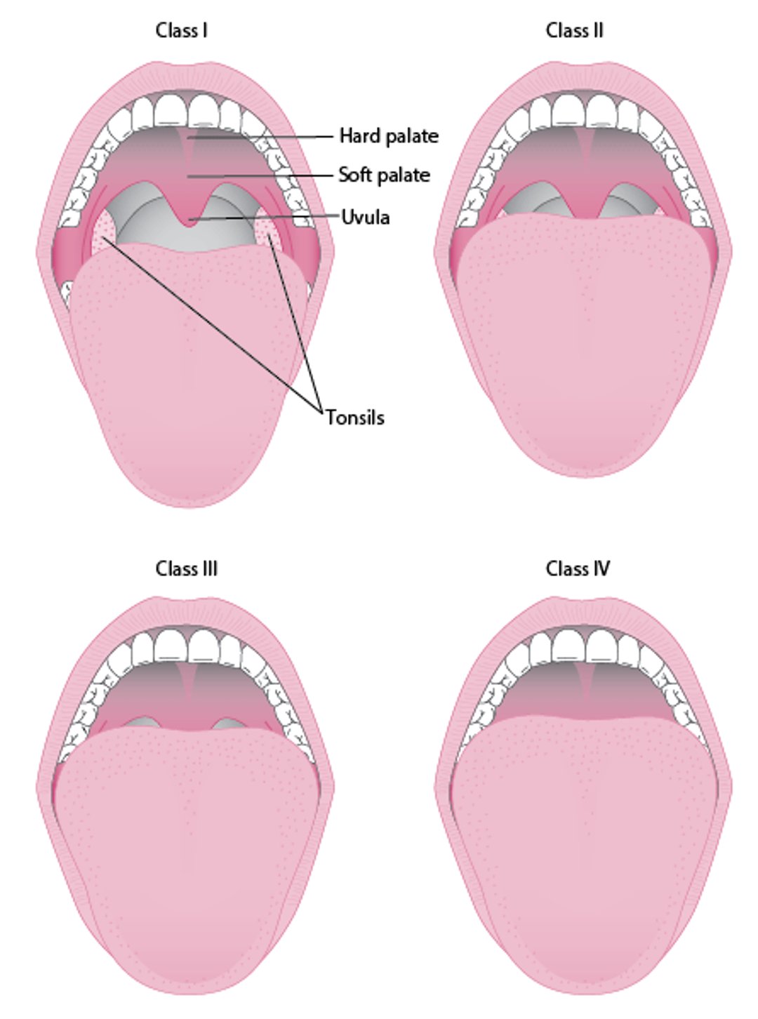
Physical examination may show signs of nasal obstruction, tonsillar hypertrophy, and abnormalities of pharyngeal structure. Anatomic risk factors for OSA should be noted and are typically assessed using the modified Mallampati score (5).
Symptoms and signs references
1. Gottlieb DJ, Punjabi NM. Diagnosis and management of obstructive sleep apnea: A review. JAMA. 2020;323(14):1389-1400. doi:10.1001/jama.2020.3514
2. Epstein LJ, Kristo D, Strollo PJ Jr, et al. Clinical guideline for the evaluation, management and long-term care of obstructive sleep apnea in adults. J Clin Sleep Med. 2009;5(3):263-276.
3. Lee JJ, Sundar KM. Evaluation and management of adults with obstructive sleep apnea syndrome. Lung. 2021;199(2):87-101. doi: 10.1007/s00408-021-00426-w
4. Strohl KP, Redline S. Recognition of obstructive sleep apnea. Am J Respir Crit Care Med. 1996;154(2 Pt 1):279-289. doi: 10.1164/ajrccm.154.2.8756795. PMID: 8756795.
5. Friedman M, Tanyeri H, La Rosa M, et al. Clinical predictors of obstructive sleep apnea. Laryngoscope. 1999;109(12):1901-1907. doi:10.1097/00005537-199912000-00002
Diagnosis of Obstructive Sleep Apnea (OSA)
History and physical examination
Confirmation by sleep studies
The diagnosis of obstructive sleep apnea is initially clinically suspected in patients with identifiable risk factors, symptoms, or both; and confirmed by sleep studies.
In addition to the patient, bed partners, roommates, and/or housemates are all sources for risk assessment information.
At-risk patients who might need more detailed evaluation include those who:
Are age 65 years or older
Report wake-time fatigue, sleepiness, or difficulty staying asleep
Have overweight or obesity
Have poorly controlled hypertension (which may be caused or exacerbated by OSA [1]), atrial fibrillation or other arrhythmias, heart failure (which may cause or coexist with OSA [2]), stroke, or diabetes
Screening questionnaires, such as STOP-BANG (see table ) (3), Berlin Questionnaire (BQ) (4), and Epworth Sleepiness Scale (ESS) (5), can be used by nonspecialists to assess risk and need for more definitive sleep testing. The questionnaires are imperfect and should not be used to make a diagnosis or to direct therapy. The multimodal STOP-BANG and the BQ are more sensitive than the ESS for risk of OSA and have good negative predictive value (6).
Patients whose symptoms, screening questionnaires, and/or risk factors suggest a higher likelihood of OSA should typically have sleep monitoring to determine the AHI or another indicator of sleep-disordered breathing such as the Respiratory Disturbance Index (RDI). The additional information is needed to confirm OSA and grade severity. Patients who report only snoring without other symptoms or cardiovascular risks do not need an extensive evaluation for OSA.
The apnea-hypopnea index (AHI) represents the total number of episodes of apnea and hypopnea occurring during sleep divided by the hours of sleep time; it is expressed as the number of episodes occurring per hour. The more events that occur, the more severe the OSA and generally the greater the likelihood of adverse effects. AHI values can be computed for different sleep stages and body positions (side or back).
Criteria for diagnosis of OSA include wake-time symptoms, sleep-time symptoms, and sleep monitoring results that show an AHI ≥ 5 per hour in patients with symptoms, or ≥ 15 per hour in the absence of symptoms (7). Symptoms should include ≥ 1 of the following:
Unrefreshing sleep
Daytime sleepiness, fatigue
Unintentional sleep episodes
Difficulty staying asleep
Awakening with breath holding, gasping, or choking
Reports by a bed partner of loud snoring, breathing interruptions, or both
Sleepiness that intrudes into daily activities or results in errors or motor vehicle crashes is particularly significant.
The differential diagnosis includes many other conditions and factors that reduce the quantity or quality of sleep or cause wake-time sedation or sleepiness. These include:
Other sleep disorders: Poor sleep hygiene, insufficient total sleep time, narcolepsy and other hypersomnolence disorders, restless legs syndrome, periodic limb movement disorder
Medications and other substances: Alcohol, sedatives, and other medications (eg, opioids)
Medical disorders: Cardiovascular, respiratory, and metabolic disorders (eg, hypothyroidism)
Mood disorders such as depression (which often accompanies as well as contributes to disordered sleep)
History and physical examination (including sleep history) should seek evidence of these conditions, including identification of clinical features of hypothyroidism and acromegaly.
Measurement of thyroid-stimulating hormone can be useful in patients with sleepiness in whom hypothyroidism is clinically suspected but should not be done routinely as it has not been shown to be helpful in diagnosing OSA.
No other adjunctive testing (eg, upper airway imaging, facial photographs,) has sufficient specificity to be recommended routinely.
Sleep studies
Sleep studies include:
Traditional polysomnography conducted in a sleep laboratory
Portable diagnostic tools that can be used by patients at home in their own bed
Polysomnography records and helps classify stages of sleep and the occurrence and duration of apneic and hypopneic periods. It is considered the gold standard for making the diagnosis of OSA and quantifying its severity (8). However, it requires an overnight stay in a sleep laboratory and may thus present logistical challenges. Polysomnography typically includes
Continuous measurement of sleep architecture by EEG (electroencephalography)
Chin electromyography to detect hypotonia
Electro-oculography to assess the occurrence of rapid eye movements
Airflow sensors at the nose and mouth to detect apneas and hypopneas
Chest and/or abdominal sensors to detect respiratory effort
Oxygen saturation by pulse oximetry
ECG monitoring to detect arrhythmias associated with apneic episodes
The patient is also observed by video.
Other variables evaluated include limb muscle activity (to assess nonrespiratory causes of sleep arousal, such as restless legs syndrome and periodic limb movement disorder) and body position, because apnea may occur predominantly in the supine position and is sometimes treated with avoidance of supine sleep.
Alternatively, patients may undergo a "split night" sleep study in which, after a diagnosis of OSA is established with polysomnography, CPAP is then administered and the pressure level is titrated to effect (ie, identifying the airway pressure at which apneas and hypopneas are eliminated). Titrating CPAP levels shortly after diagnosing OSA allows determination of appropriate therapy during the same overnight monitoring period. A whole night CPAP titration can also be performed, if needed, to assess the effectiveness of CPAP treatment after an already established diagnosis of OSA.
Home sleep testing using portable diagnostic tools evaluates a limited subset of polysomnographic measures, typically just heart rate, pulse oximetry, respiratory effort, body position, and nasal airflow to detect apnea and estimate its severity. The role of home sleep testing is expanding because of the convenience, decreased cost, and ability to provide a reasonably accurate estimate of respiratory disturbances during sleep (9).
However, portable tools have some limitations. They do not actually detect the presence of sleep and instead depend on patients to self-report sleeping and total estimated sleeping time, which can be inaccurate due to the potential for recall bias. For example, if patients were not sleeping during part of the study and they did not report this, sleep-disordered breathing may be underestimated. Thus, a negative home sleep test result in a patient with symptoms should be followed by polysomnography. Also, coexisting sleep disorders (eg, restless legs syndrome, seizures, REM behavior disorder, confusional arousals) are not detected. Follow-up polysomnography may still be needed to characterize these disorders as well as to accurately provide AHI and RDI values in the different stages of sleep and with changes in position, especially when surgery or therapy other than positive airway pressure is being considered.
Classification of severity
The AHI is the total number of episodes of apnea and hypopnea occurring during sleep divided by the hours of sleep time. It is a commonly used measure of respiratory disturbance during sleep and is used to classify the severity of OSA. OSA is graded as:
No OSA: AHI < 5 per hour
Mild: AHI ≥ 5 and < 15 per hour
Moderate: AHI ≥ 15 and ≤ 30 per hour
Severe: AHI > 30 per hour
The respiratory disturbance index (RDI) is a related measure that includes the number of arousals related to respiratory effort (called respiratory effort-related arousals or RERAs) plus the number of apnea and hypopnea episodes per hour of sleep.
The arousal index, which is the number of arousals per hour of sleep, can be computed if EEG monitoring is used. The arousal index is loosely correlated with AHI and RDI; 20 to 30% of apneas and desaturation episodes are not accompanied by arousals (10). Other causes of arousals must also be considered.
The oxygen desaturation index (ODI) is the mean number of episodes of desaturation occurring per hour (11). Desaturation episodes are defined as a decrease in the mean oxygen saturation of ≥ 3% (over the last 2 minutes) that lasts for at least 10 seconds.
However, these indices may only be moderately associated with the severity of symptoms. Some patients with a high or extremely high AHI (eg, > 60 per hour) may have few or no symptoms. Additional metrics and combinations of metrics may prove useful in diagnosis (12). Beyond AHI, a composite of clinical and polysomnographic data have been linked to clinical outcomes, including cardiovascular risk and mortality (13). For example, sleepiness even in the absence of known sleep disorders may be linked to an increased risk of cardiovascular disease (14).
See also Approach to the Patient With a Sleep or Wakefulness Disorder.
Diagnosis references
1. Walia HK, Li H, Rueschman M, et al. Association of severe obstructive sleep apnea and elevated blood pressure despite antihypertensive medication use. J Clin Sleep Med. 2014;10(8):835-843. doi:10.5664/jcsm.39461.
2. Gupta A, Quan SF, Oldenburg O, et al. Sleep-disordered breathing in hospitalized patients with congestive heart failure: a concise review and proposed algorithm. Heart Fail Rev. 2018;23(5):701-709. doi:10.1007/s10741-018-9715-y
3. Chung F, Abdullah HR, Liao P. STOP-Bang Questionnaire: A Practical Approach to Screen for Obstructive Sleep Apnea. Chest. 2016;149(3):631-638. doi:10.1378/chest.15-0903
4. Netzer NC, Stoohs RA, Netzer CM, Clark K, Strohl KP. Using the Berlin Questionnaire to identify patients at risk for the sleep apnea syndrome. Ann Intern Med. 1999;131(7):485-491. doi:10.7326/0003-4819-131-7-199910050-00002
5. Johns MW. A new method for measuring daytime sleepiness: the Epworth sleepiness scale. Sleep. 1991;14(6):540-545. doi:10.1093/sleep/14.6.540
6. Gamaldo C, Buenaver L, Chernyshev O, et al. Evaluation of clinical tools to screen and assess for obstructive sleep apnea. J Clin Sleep Med. 2018;14(7):1239-1244. doi:10.5664/jcsm.7232
7. The American Association of Sleep Medicine. The AASM International Classification of Sleep Disorders – Third Edition, Text Revision (ICSD-3-TR). AASM Darien, IL. 2023.
8. Kapur VK, Auckley DH, Chowdhuri S, et al. Clinical Practice Guideline for Diagnostic Testing for Adult Obstructive Sleep Apnea: An American Academy of Sleep Medicine Clinical Practice Guideline. J Clin Sleep Med. 2017;13(3):479-504. doi:10.5664/jcsm.6506
9. Malhotra A, Ayappa I, Ayas N, et al. Metrics of sleep apnea severity: beyond the apnea-hypopnea index. Sleep. 2021;44(7):zsab030. doi: 10.1093/sleep/zsab030
10. Jordan AS, Eckert DJ, Wellman A, Trinder JA, Malhotra A, White DP. Termination of respiratory events with and without cortical arousal in obstructive sleep apnea. Am J Respir Crit Care Med. 2011;184(10):1183-1191. doi:10.1164/rccm.201106-0975OC
11. Varghese L, Rebekah G, N P, Oliver A, Kurien R. Oxygen desaturation index as alternative parameter in screening patients with severe obstructive sleep apnea. Sleep Sci. 2022;15(Spec 1):224-228. doi:10.5935/1984-0063.20200119
12. Collop NA, Anderson WM, Boehlecke B, et al. Clinical guidelines for the use of unattended portable monitors in the diagnosis of obstructive sleep apnea in adult patients. J Clin Sleep Med. 2007;3(7):737-47.
13. Hajipour M, Baumann B, Azarbarzin A, et al. Association of alternative polysomnographic features with patient outcomes in obstructive sleep apnea: a systematic review. J Clin Sleep Med. 2023;19(2):225-242. doi:10.5664/jcsm.10298
14. Bock J, Covassin N, Somers V. Excessive daytime sleepiness: an emerging marker of cardiovascular risk. Heart. 2022;108(22):1761-1766. doi:10.1136/heartjnl-2021-319596
Treatment of Obstructive Sleep Apnea (OSA)
Control of risk factors such as obesity, alcohol use, and sedative use
Continuous positive airway pressure (CPAP) or oral appliances
Sometimes surgery, or nerve stimulation
Obstructive sleep apnea is managed through a stepwise, patient-centered approach that includes risk factor modification, device therapy (eg, CPAP, oral appliances), and selected surgical or neurostimulation options. Treatment decisions are generally guided by symptom burden, disease severity (eg, using validated measures such as AHI), and comorbidities.
The aims of treatment (1) are to:
Reduce symptoms
Reduce episodes of hypoxia and sleep fragmentation
Restore sleep continuity and architecture
Avoid episodic asphyxia
There are many available approaches to treatment. Patient and clinician should engage in shared decision making to align the severity of disease, symptoms, and other relevant comorbidities with feasible interventions and the outcomes most important to the patient.
Patients without symptoms and an apnea-hypopnea index (AHI) between 5 and 15 per hour may not require specific treatment. While an AHI ≥ 5 per hour can be useful to establish a diagnosis of OSA, treatment is provided only to patients who have symptoms (sleepiness with fatigue, nonrestorative sleep, and snoring/gasping/choking). Patients with a low AHI but with symptoms may choose continuous positive airway pressure (CPAP). An AHI value ≥ 15 indicates at least a moderate level of sleep apnea and is considered a threshold to treat even in the absence of symptoms. Patients who report only snoring without other symptoms or cardiovascular risks can respond by increasing physical fitness, losing weight, improving sleep hygiene, and treating nasal allergies (eg, allergic rhinitis, chronic rhinosinusitis with nasal polyposis).
Pending treatment, patients with excessive wake-time sleepiness should be warned of the risks of driving, operating heavy machinery, or engaging in other activities during which unintentional sleep would be hazardous.
Treatment is directed at both risk factors and OSA itself. First-line direct therapy includes use of a CPAP device or an oral appliance. For anatomic encroachment or for disease that does not respond to these devices, surgery or nerve stimulation is considered. Success is defined as a resolution of symptoms with AHI reduction, usually to < 10 per hour and ideally to < 5 per hour.
Treatment of OSA is associated with modest 2 to 3 mm Hg decreases in blood pressure, but data are mixed regarding the effectiveness of treatment of OSA in the prevention of primary or secondary cardiovascular events, atrial fibrillation (2, 3, 4, 5, 6), and other quantifiable outcomes such as improved glycemic control. American Heart Association (AHA) guidelines recommend screening for OSA in patients with atrial fibrillation but recognize the limited data suggesting that treatment of sleep apnea facilitates maintenance of sinus rhythm (7).
Also see Treatment of Snoring.
Control of risk factors
Initial treatment aims to control risk factors for OSA such as obesity, hypertension, alcohol use, and sedative use. Exercise decreases the AHI and increases alertness independent of any effect on body mass index (BMI).
Wake-time sleepiness can be reduced by good sleep hygiene measures, including sleeping longer and discontinuing sedative medications, particularly antihistamines or antidepressants. Avoiding supine sleep can be useful and feasible for some patients who have primarily positional (eg, supine position-related) OSA.
Modest weight loss (≥ 15%) may result in clinically meaningful improvement (8, 9) but should not be considered curative for OSA. However, weight loss may be challenging for many people, especially those who are fatigued or sleepy. Weight loss as a result of bariatric surgery, can reduce the AHI and reduce symptoms (10, 11). Medications such as GLP-1 receptor antagonists used for weight loss have been shown to decrease OSA severity. For example, tirzepatide pharmacotherapy for the treatment of OSA,has shown that 1 year of treatment was associated with substantial improvements in both weight and OSA severity (12).
Continuous positive airway pressure (CPAP)
CPAP is highly efficacious for OSA and can ameliorate symptoms (13, 14, 15). Treatment of OSA with continuous positive airway pressure (CPAP) has been consistently shown to reduce sleepiness and snoring and improve bed partner sleep (16). Racial and socioeconomic disparities in the usage of CPAP have been reported (17, 18).
There are many different CPAP interfaces (masks) available, including those that are inserted into the nose (nasal pillows) and those that cover the nose (nasal masks), nose and mouth (full face masks), or the entire face including the eyes (total face mask). All have cushions to provide an air seal, which is essential for maintaining a pressure gradient. Cushions may be inflatable or made of silicone, foam, or gel. Proper fit and comfort vary widely among patients but must be optimized for both efficacy and adherence.
Types of Masks for CPAP
Yumagema/stock.adobe.com |


The level of CPAP necessary for airway patency is determined either by CPAP titration in the sleep laboratory or increasingly through the use of automatic positive airway pressure (APAP). With APAP, a range of pressures is prescribed (eg, 5 to 15 cm H2O), and the device uses internal algorithms to titrate the pressure up and down throughout the night as needed. CPAP improves upper airway patency by applying positive pressure to the collapsible upper airway segment. Pressure requirements do not correlate with disease severity. Effective pressures typically range from 5 to 15 cm H2O. If necessary, polysomnographic monitoring can be used to guide manual titration of pressure. CPAP can be augmented with inspiratory assistance (bilevel positive airway pressure) to increase tidal volume in patients with comorbid obesity-hypoventilation syndrome and sometimes, for comfort.
Beyond reducing AHI as a part of treatment goals, CPAP may additionally reduce tiredness and improve quality of life. CPAP may also reduce blood pressure, although the impact is usually modest. If CPAP is withdrawn, symptoms recur over several days, although short interruptions of therapy are usually well tolerated. The therapy is usually administered indefinitely (ie, lifelong) unless other lifestyle modifications or treatments are sufficient to resolve OSA.
If clinical improvement is not apparent, CPAP adherence should be reviewed and patients reassessed for comorbid disorders. If patients have septal deviation or nasal polyps, nasal surgery may make CPAP treatment more successful; however, surgery rarely cures OSA by itself.
Adherence is difficult for many people and is lower in patients who do not experience sleepiness. Overall, estimates of regular long-term CPAP use range widely, from 29% to 85%, depending on a number of factors, including tolerability and the method of ascertainment (19). Approximately 65% to 80% of patients continue using CPAP after 4 years (20). Adherence can be improved by efforts to foster a positive attitude toward device use combined with early attention to any problems, particularly mask fit, and close follow-up by a committed caretaker, with reinforcement by the clinician. With many machines, adherence, pressure levels, leak, and residual respiratory events are tracked daily by the devices and available to patients and clinicians.
The treatment efficacy of CPAP may decrease if patient factors change (eg, weight gain occurs, nasal obstruction develops), necessitating a reassessment of the therapeutic approach.
Adverse effects of nasal CPAP include discomfort resulting from a poorly fitting mask, and dryness and nasal irritation, which can be alleviated in some cases with the use of warm, humidified air. However, newer masks are designed to offer improved comfort and ease of use.
Oral appliances


The 2 pieces of the appliance fit over the upper and lower teeth and are held together by a metal fitting that is adjusted to pull the lower jaw forward enough to reduce sleep apnea.
Images courtesy of Robert S. Porter, MD.
Oral appliances are designed to advance the mandible forward or, at the very least, prevent retrusion and tongue prolapse during sleep (21, 22, 23). Some appliances are designed to pull the tongue forward. These appliances are considered mainstream treatments for both snoring and mild to moderate OSA. Comparisons of appliances to CPAP show equivalent effectiveness in mild to moderate OSA, but cost-effectiveness studies are focused on fixed initial costs of fabrication rather than on replacement and follow-up costs.
Surgery
Surgical procedures to correct anatomic factors, such as enlarged tonsils and nasal polyps that contribute to upper airway obstruction, should be considered (23, 24). Surgery is a first-line treatment if specific anatomic encroachment is identified. However, in the absence of encroachment, evidence to support surgery as a first-line treatment is lacking. Surgery for anatomic risk factors (eg, macroglossia, micrognathia) is also an option (see figure Modified Mallampati Scoring).
Uvulopalatopharyngoplasty (UPPP) involves resection of pharyngeal tissue. UPPP has been largely replaced by less aggressive approaches that attempt to stabilize the lateral walls of the pharynx and/or enlarge the velopharyngeal area without risk of altering speech or swallowing. CPAP and UPPP have not been directly compared in rigorous studies, but UPPP may be efficacious in well-selected individuals. Results are less predictable in patients who have severe obesity or anatomic narrowing of the airway (25). The procedure may reduce intrusive snoring, although apneic episodes may remain as severe (although silent) as before surgical intervention.
Other surgical procedures include midline glossectomy, hyoid advancement, and mandibulomaxillary advancement (26). Mandibulomaxillary advancement is sometimes offered as a second-stage procedure if soft-tissue approaches are not curative. The optimal multistage approach is not known.
Tracheostomy is the most effective therapeutic maneuver for OSA but is done as a last resort. It bypasses the site of obstruction and is indicated for patients most severely affected (eg, those with cor pulmonale) who cannot tolerate CPAP.
Upper airway stimulation
Upper airway stimulation using an implanted device to stimulate a branch of the hypoglossal nerve can activate muscles that cause tongue protrusion and other muscles that help maintain airway patency (27, 28). This therapy is successful in selected patients with moderate to severe disease. It is used mainly in those who are unable to tolerate CPAP therapy and in whom oral appliances have proven ineffective. The procedure may also be tried in those in whom mandibulomaxillary advancement is contemplated. In one prospective cohort study, approximately 70% of patients who underwent upper airway stimulation reported reductions in both AHI and ODI to < 10 per hour (29).
An intraoral, removable, tongue muscle stimulator may also be used. Such devices use daytime neuromuscular electrical stimulation to improve tongue muscle tone in patients with snoring or mild OSA (30, 31).
Adjunctive treatments
Various adjunctive treatments are sometimes used but have no proven benefit for OSA.
Supplemental oxygen improves blood oxygenation and may reduce AHI and arousal index among patients who did not respond to upper airway surgery (32). However, a beneficial clinical effect occurs mainly in those with high loop gain (tendency to have repeated apnea or hypopnea after an initial episode), and effects are hard to predict. Also, oxygen may provoke respiratory acidosis and morning headache. For these reasons, supplemental oxygen is not recommended for the treatment of OSA.
Wake-promoting agents (also called wakefulness-promoting agents; eg, modafinil, solriamfetol) may be used to adjunctively treat excessive sleepiness that occurs with OSA or in the 20% of treated patients who have residual sleepiness despite adequate treatment with CPAP (33, 34, 35, 36).
A number of other medications have been tried, including tricyclic antidepressants, theophylline, dronabinol, combined atomoxetine plus oxybutynin (37). However, these agents cannot be routinely recommended because of factors including limited clinical experience and evidence supporting their use, a low therapeutic index, and a lack of consistent results. Improved methods for recognizing sleep apnea subtypes may allow for a better selection of patients for pharmacotherapy.
Exercises for upper airway muscles (myofunctional therapy) have been proposed on the theory that improved muscle strength and tone might help improve airway patency during sleep (38). There are a number of exercises that seem to reduce AHI and symptoms, making this approach interesting, particularly because it is noninvasive and with no adverse effects. However, this approach is not a mainstream recommendation because of the wide variety of techniques proposed, uncertainty about their mechanisms of action and efficacy, and practical difficulties with adherence. At least one device based on daytime electrical stimulation of the upper airway dilator muscles is available for the treatment of snoring and mild OSA (39).
Nasal dilatory devices and throat sprays sold over-the-counter for snoring are not conclusively proven to be efficacious in the treatment of OSA (40, 41).
Laser-assisted uvuloplasty, uvular splints, and radiofrequency tissue ablation have been used to treat snoring in patients without OSA. Although they may transiently decrease snoring loudness, their efficacy in treating OSA is neither predictable nor durable.
Patient education and support
An informed patient and family are better able to cope with an OSA treatment strategy, including tracheostomy. Patient support groups provide helpful information and effectively support timely treatment and follow-up. The role of patient support groups and digital support tools for management continues to be under investigation (42, 43).
Prognosis for Obstructive Sleep Apnea (OSA)
The prognosis for obstructive sleep apnea is excellent provided treatment is instituted, accepted, and effective. Evidence from multiple longitudinal studies indicates that although mild OSA may progress over time, such progression (ie, moderate or severe obstructive sleep apnea) is not clinically significant (1). Untreated or unrecognized OSA is accompanied by a lower quality of life and increased risk of hypertension and injuries related to falling asleep while engaging in potentially hazardous activities. Predictors of OSA progression in untreated adults include a higher baseline BMI, weight gain, more severe baseline hypoxemia, and longer sleep latency (2).
Prognosis references
1. Fong PY, Tay ZY, Soh RY, Fook SMC, Li HH, Phua CQ. Should mild obstructive sleep apnoea be treated? A systematic review from the standpoint of disease progression. J Laryngol Otol. 2023;137(8):828-839. doi:10.1017/S0022215122002419
2. Lavie P. Predicting sleep apnea worsening and cardiometabolic risk without treatment. Sleep Breath. 2026;30(2):131. doi:10.1007/s11325-026-03617-y
Key Points
Obesity, anatomic abnormalities in the upper airway passages, family history, certain disorders (eg, hypothyroidism, stroke), and use of alcohol or sedatives increase the risk of obstructive sleep apnea (OSA).
Patients typically snore, have restless and unrefreshing sleep, and often feel daytime sleepiness and fatigue; however, some have few or no symptoms.
Most people who snore do not have OSA.
Disorders that occur more commonly in patients with OSA include hypertension, stroke, diabetes, gastroesophageal reflux disease, metabolic dysfunction–associated steatotic liver disease (MASLD), nocturnal angina, heart failure, and atrial fibrillation or other arrhythmias.
The diagnosis is usually confirmed by sleep testing, including using portable devices used in the home.
Modifiable risk factors such as obesity should be controlled and most patients treated with continuous positive airway pressure and/or oral appliances designed to maintain airway patency.
Surgery may be considered for abnormalities causing airway encroachment or if the disorder is intractable.
Drug Information for the Topic







