

Renal artery stenosis is a decrease in blood flow through one or both of the main renal arteries or their branches. Renal artery occlusion is a complete blockage of blood flow through one or both of the main renal arteries or its branches. Stenosis and occlusion are usually due to thromboemboli, atherosclerosis, or fibromuscular dysplasia. Symptoms of acute occlusion include steady, aching flank pain, abdominal pain, fever, nausea, vomiting, and hematuria. Acute kidney injury may develop. Chronic, progressive stenosis causes refractory hypertension and may lead to chronic kidney disease. Diagnosis is by imaging tests (eg, CT angiography, magnetic resonance angiography). Treatment of acute occlusion is with anticoagulation and sometimes fibrinolytics and surgical or catheter-based embolectomy, or a combination. Treatment of chronic, progressive stenosis includes angioplasty with stent placement or surgical bypass.
Renal hypoperfusion results in renovascular hypertension, kidney failure and, if complete occlusion occurs, renal infarction and necrosis.
Etiology of Renal Artery Stenosis and Occlusion
Occlusion may be acute or chronic. Acute occlusion is usually unilateral. Chronic occlusion may be unilateral or bilateral.
Acute renal artery occlusion
The most common cause is thromboembolism. Emboli may originate in the heart (due to atrial fibrillation, after myocardial infarction, or from vegetations due to bacterial endocarditis) or the aorta (as atheroemboli); less often, fat or tumor emboli are the cause. Thrombosis may occur in a renal artery spontaneously or after trauma, surgery, angiography, or angioplasty. Other causes of acute occlusion include aortic dissection and rupture of a renal artery aneurysm.
Rapid, total occlusion of large renal arteries for 30 to 60 minutes results in infarction. The infarct is typically wedge-shaped, radiating outward from the affected vessel.
Chronic progressive renal artery stenosis
About 90% of cases are due to atherosclerosis, which is usually bilateral. Almost 10% of cases are due to fibromuscular dysplasia, which is commonly unilateral. Less than 1% of cases result from Takayasu arteritis, Kawasaki disease, neurofibromatosis type 1, aortic wall hematoma, or aortic dissection (1).
Atherosclerosis develops primarily in patients > 45 years (more often men) and usually affects the aortic orifice or proximal segment of the renal artery. Chronic progressive stenosis tends to become clinically evident after about 10 years of atherosclerosis, causing renal atrophy and chronic kidney disease.
Fibromuscular dysplasia is pathologic thickening of the arterial wall, most often of the distal main renal artery or the intrarenal branches. The thickening tends to be irregular and can involve any layer (but most often the media). This disorder develops primarily in younger adults, particularly in women aged 20 to 50 years. It is more common among first-degree relatives of patients with fibromuscular dysplasia and among people with the ACE1 gene.
Etiology reference
1. Whelton PK, Carey RM, Aronow WS, et al. 2017 ACC/AHA/AAPA/ABC/ACPM/AGS/APhA/ASH/ASPC/NMA/PCNA Guideline for the Prevention, Detection, Evaluation, and Management of High Blood Pressure in Adults: A Report of the American College of Cardiology/American Heart Association Task Force on Clinical Practice Guidelines. Circulation 2018;138(17):e484-e594. doi:10.1161/CIR.0000000000000596
Symptoms and Signs of Renal Artery Stenosis and Occlusion
Manifestations depend on rapidity of onset, extent, whether occlusion is unilateral or bilateral, and duration of renal hypoperfusion. Stenosis of one renal artery is often asymptomatic for a considerable time.
Acute complete occlusion of one or both renal arteries causes steady and aching flank pain, abdominal pain, fever, nausea, and vomiting. Gross hematuria, oliguria, or anuria may occur; hypertension is rare. After 24 hours, symptoms and signs of acute kidney injury may develop. If the cause was thromboembolic, features of arterial thromboembolism at other sites (eg, "blue toe" syndrome, livedo reticularis, retinal lesions on fundoscopic examination) also may be present.
Chronic progressive stenosis causes hypertension, which may begin at an atypical age (eg, before age 30 years or after age 50 years) and which may be refractory to control despite use of multiple antihypertensives. Physical examination may detect an abdominal bruit or signs of atherosclerosis. Symptoms and signs of chronic kidney disease develop slowly.
Diagnosis of Renal Artery Stenosis and Occlusion
Clinical suspicion
Imaging
Renal artery occlusion is suspected in patients with acute kidney injury who have
Symptoms of acute renal artery occlusion
Symptoms or signs of thromboembolism
Renal artery stenosis is suspected in patients who have
Hypertension that begins before age 30 in a patient with no family history of hypertension
Severe or resistant hypertension
Unexplained creatinine elevation and/or acute rise of creatinine of at least 50% after administration of an angiotensin-converting enzyme (ACE) inhibitor or angiotensin II receptor blocker (ARB)

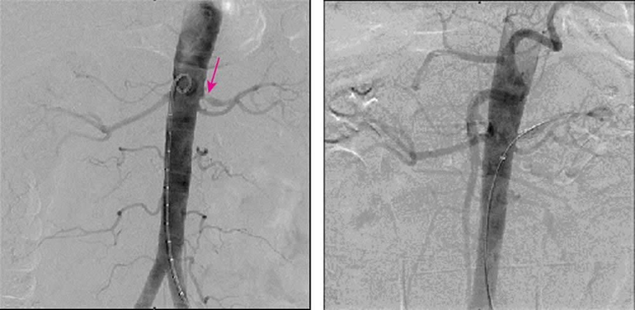
The left image shows 90% stenosis at the origin of the left main renal artery on digital subtraction angiography. The right image shows complete resolution of stenosis after stent placement.
Image provided by Jan N. Basile, MD.
Blood and urine tests are performed to confirm kidney failure. Diagnosis is confirmed by imaging (see table ). Which tests are done depends on the patient’s renal function and other characteristics and on test availability.
Some tests (CT angiography, arteriography, digital subtraction angiography) require an IV ionic radiocontrast agent, which may be nephrotoxic; the risk of nephrotoxicity is lower with the nonionic hypo-osmolar or iso-osmolar contrast agents that are in widespread use (see Radiographic Contrast Agents and Contrast Reactions).
Magnetic resonance angiography (MRA) with gadolinium contrast carries the risk of nephrogenic systemic fibrosis (a condition that closely resembles systemic sclerosis and has no satisfactory method of treatment) in patients with a decreased glomerular filtration rate (GFR), but group II gadolinium contrast agents can be used to minimize this potential risk when medically necessary.
When results of other tests are inconclusive or negative but clinical suspicion is strong, arteriography is necessary for definitive diagnosis. Arteriography may also be needed before invasive interventions.
Imaging Tests for Diagnosis of Renal Artery Stenosis or Occlusion
Test | Advantages | Disadvantages |
|---|---|---|
Noninvasive Fast Generally available | Requires IV iodinated contrast, which may be nephrotoxic | |
Highly accurate Noninvasive Safe in patients with GFR > 60 mL/minute and possibly GFR 30–60 mL/minute | Requires gadolinium contrast, which increases risk of nephrogenic systemic fibrosis | |
Noninvasive Provides information about kidney function | Operator-dependent, time-consuming, and not always available; limited accuracy in patients with obesity | |
Radionuclide renography | Noninvasive Images renal blood flow | More accurate in unilateral than in bilateral stenosis; more accurate when captopril is used; at least 10% false-positive and false-negative rates, even when captopril is usedMore accurate in unilateral than in bilateral stenosis; more accurate when captopril is used; at least 10% false-positive and false-negative rates, even when captopril is used Usually not used as the initial test |
Arteriography | Diagnostic gold standard Provides anatomic detail for surgical and invasive radiologic procedures | Invasive Risk of atheroembolism (due to arterial catheterization) and contrast-induced nephropathy |
Uses less iodinated contrast than arteriography | Invasive Requires iodinated contrast, but in smaller amounts than arteriography | |
Carbon dioxide angiography | No need for contrast agent | Relatively unavailable |
GFR = glomerular filtration rate. | ||
When a thromboembolic disorder is suspected, electrocardiography or rhythm monitoring (to detect atrial fibrillation) and hypercoagulability studies may be needed to identify treatable embolic sources. Transesophageal echocardiography is performed to detect atheromatous lesions in the ascending and thoracic aorta and cardiac sources of thrombi or valvular vegetations.
Blood and urine tests are nondiagnostic but are performed to confirm kidney failure, indicated by elevated creatinine, blood urea nitrogen, and potassium. Leukocytosis, gross or microscopic hematuria, and proteinuria may also be present.
Treatment of Renal Artery Stenosis and Occlusion
Restoration of vascular patency in acute occlusions and, if patients have refractory hypertension or potential for kidney failure, in chronic stenosis
Treatment depends on the cause.
Acute renal artery occlusion
A renal thromboembolic disorder may be treated with a combination of catheter directed thrombolysis, systemic anticoagulation, and surgical or catheter-based embolectomy. Treatment within 3 hours of symptom onset is likely to improve kidney function. However, complete recovery is unusual, and early and late mortality rates are high because of extrarenal embolization or underlying atherosclerotic heart disease.
Patients presenting within 3 hours may benefit from fibrinolytic (thrombolytic) therapy (eg, streptokinase, alteplase) given IV or by local intra-arterial infusion. However, such rapid diagnosis and treatment are relatively rare.(eg, streptokinase, alteplase) given IV or by local intra-arterial infusion. However, such rapid diagnosis and treatment are relatively rare.
All patients with a thromboembolic disorder require anticoagulation with IV heparin, unless contraindicated. Long-term anticoagulation with oral warfarin can be initiated simultaneously with with IV heparin, unless contraindicated. Long-term anticoagulation with oral warfarin can be initiated simultaneously withheparin if no invasive intervention is planned. Non–vitamin K (direct) oral anticoagulants (eg, dabigatran, apixaban, rivaroxaban) can be considered in appropriate patients, although there is no clear consensus on their use. Anticoagulation should be continued for at least 6 to 12 months—indefinitely for patients with a recurrent thromboembolic disorder or a hypercoagulability disorder.if no invasive intervention is planned. Non–vitamin K (direct) oral anticoagulants (eg, dabigatran, apixaban, rivaroxaban) can be considered in appropriate patients, although there is no clear consensus on their use. Anticoagulation should be continued for at least 6 to 12 months—indefinitely for patients with a recurrent thromboembolic disorder or a hypercoagulability disorder.
Surgery to restore vascular patency has a higher mortality rate than fibrinolytic therapy and has no advantage in recovery of kidney function. However, surgery, particularly if done within the first few hours, is preferred for patients with traumatic renal artery thrombosis. If patients with nontraumatic, severe kidney failure do not recover function after 4 to 6 weeks of medical therapy, surgical revascularization (embolectomy) can be considered, but it helps only a few.
If the cause is thromboemboli, the source should be identified and treated appropriately. Statins are generally offered to stabilize existing plaques and limit further atherosclerotic disease.
Chronic progressive renal artery stenosis
Revascularization may be considered for patients who meet one or more of the following criteria (1):
Failure or intolerance of medical therapy for optimal blood pressure
A short duration of elevated blood pressure prior to the diagnosis of renovascular disease
Recurrent flash pulmonary edema
Unexplained rapid progression of chronic kidney disease
Revascularization is usually achieved by percutaneous transluminal angioplasty (PTA) with stent placement or with surgical bypass of the stenotic segment. Surgery is usually more effective than PTA for atherosclerotic occlusion; it cures or attenuates hypertension in 60 to 70% of patients. However, surgery is considered only if patients have complex anatomic lesions or if PTA is unsuccessful, particularly with repeated in-stent restenosis. PTA is preferred for patients with fibromuscular dysplasia; risk is minimal, success rate is high, and restenosis rate is low.
Renovascular hypertension
For patients who do not meet one of the criteria above, medical therapy is recommended based on data from the randomized trials, which showed that renal-artery stenting plus medical therapy had no significant benefit over medical therapy alone for preventing adverse cardiovascular or renal events (2). Angiotensin-converting enzyme (ACE) inhibitors or angiotensin II receptor blockers (ARBs) can be used in unilateral and, if glomerular filtration rate (GFR) is monitored closely, in bilateral renal artery stenosis. These medications can potentially reduce GFR and increase serum blood urea nitrogen (BUN) and creatinine levels. Additional antihypertensive medications are frequently required.
Treatment references
1. Whelton PK, Carey RM, Aronow WS, et al: 2017 ACC/AHA/AAPA/ABC/ACPM/AGS/APhA/ASH/ASPC/NMA/PCNA Guideline for the Prevention, Detection, Evaluation, and Management of High Blood Pressure in Adults: A Report of the American College of Cardiology/American Heart Association Task Force on Clinical Practice Guidelines [published correction appears in Hypertension 2018 Jun;71(6):e140-e144. doi: 10.1161/HYP.0000000000000076]. Hypertension 71(6):e13-e115, 2018. doi:10.1161/HYP.0000000000000065
2. Cooper CJ, Murphy TP, Cutlip DE, et al: Stenting and medical therapy for atherosclerotic renal-artery stenosis. N Engl J Med 370(1):13-22, 2014. doi: 10.1056/NEJMoa1310753
Key Points
Renal artery stenosis or occlusion may be acute (usually due to thromboembolism) or chronic (usually due to atherosclerosis or fibromuscular dysplasia).
Suspect acute occlusion if patients have steady, aching flank or abdominal pain, and sometimes fever, nausea and vomiting, and/or gross hematuria.
Suspect chronic stenosis in patients who develop unexplained severe or early-onset hypertension.
Confirm the diagnosis with vascular imaging.
Restore vascular patency for patients who have acute occlusion and for selected patients (eg, with severe complications or refractory disease) who have chronic occlusion.
Angiotensin-converting enzyme (ACE) inhibitors and angiotensin II receptor blockers (ARBs) are effective for hypertension control in patients with renovascular hypertension, but additional antihypertensive medications are frequently required.
Drug Information for the Topic


