


Cardiomyopathy refers to progressive impairment of the structure and function of the muscular walls of the heart chambers.
There are 3 main types of cardiomyopathy:
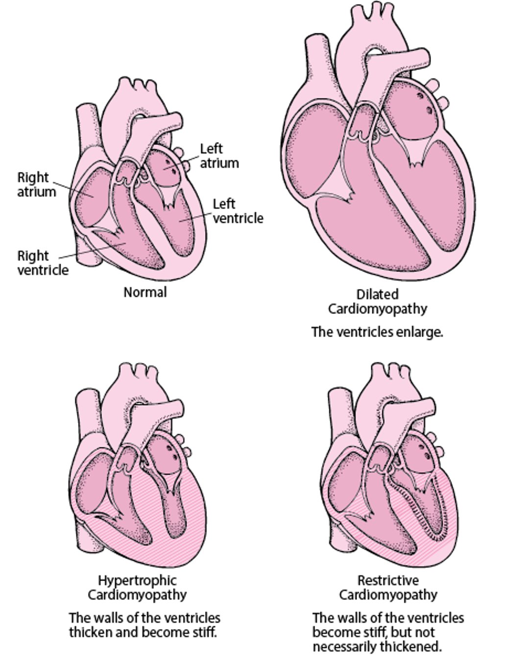
Dilated cardiomyopathy, in which the ventricles (the 2 lower heart chambers) enlarge (dilate)
Hypertrophic cardiomyopathy, in which the walls of the ventricles thicken and become stiff
Restrictive cardiomyopathy, in which the walls of the ventricles become stiff but not necessarily thickened

The main types of cardiomyopathy may overlap, that is, people may have features of more than one type.
The term cardiomyopathy is used only when a disorder directly affects the heart muscle. Other heart disorders such as coronary artery disease and heart valve disorders, as well as high blood pressure, also can eventually cause the ventricles to enlarge or thicken.
Types of Cardiomyopathy
There are 3 main types of cardiomyopathy—dilated, hypertrophic, and restrictive. |

Cardiomyopathy can be caused by many disorders, or it may have no identifiable cause.
Cardiomyopathies often result in the heart not pumping blood adequately. Inadequate blood pumping can cause symptoms of heart failure, including shortness of breath and fatigue. Some cardiomyopathies may also cause chest pain, fainting, abnormal heart rhythms, or sudden death.
To diagnose a cardiomyopathy, doctors ask whether the person has a family history of cardiomyopathy and then do blood tests, electrocardiography, chest x-ray, echocardiography, radionuclide imaging of the heart (sometimes), and magnetic resonance imaging of the heart. In some people, doctors take a sample of tissue from the inner wall of the heart to examine under a microscope (endomyocardial biopsy). Other tests are done as needed to determine the cause.
Treatment depends on the specific type and cause of cardiomyopathy.
More Information
The following English-language resources may be useful. Please note that THE MANUAL is not responsible for the content of these resources.
American Heart Association: Cardiomyopathy in adults: Provides comprehensive information on symptoms, diagnosis, and treatment of cardiomyopathies
American Heart Association: Pediatric cardiomyopathies: Provides comprehensive information on diagnosis, including genetic diagnosis and treatment of cardiomyopathy in children


