


Dilated cardiomyopathy (DCM) is a primary myocardial disorder, defined as the presence of left ventricular or biventricular dilatation and systolic dysfunction in the absence of congenital heart disease, abnormal loading conditions (eg, primary valve disease), or coronary artery disease sufficient to account for the observed ventricular remodelling. Patients typically present with clinical heart failure: exertional dyspnea, fatigue, and signs of congestion, including peripheral edema. The diagnosis is established through clinical evaluation and supported by elevated natriuretic peptide levels and cardiac imaging, including chest radiography, echocardiography, and cardiac MRI. Management is directed toward identification and treatment of the underlying etiology, when possible, in conjunction with guideline-directed medical therapy for heart failure with reduced ejection fraction (HFrEF). Advanced disease may require implantation of a durable left ventricular assist device or cardiac transplantation.
DCM has an estimated lifetime prevalence of approximately 1 in 250 individuals. It can occur at any age but is most commonly diagnosed in adults younger than 50 years, and it represents the most common cause of childhood cardiomyopathy (1, 2). DCM exhibits a male predominance (2, 3) and, in the United States, it is more frequently reported among Black than White patients (4), likely reflecting a combination of genetic susceptibility, comorbid conditions, and social determinants of health (1).

General references
1. Bozkurt B, Colvin M, Cook J, et al. Current Diagnostic and Treatment Strategies for Specific Dilated Cardiomyopathies: A Scientific Statement From the American Heart Association. Circulation. 2016;134(23):e579-e646. doi:10.1161/CIR.0000000000000455
2. Heymans S, Lakdawala NK, Tschöpe C, Klingel K. Dilated cardiomyopathy: causes, mechanisms, and current and future treatment approaches. Lancet. 2023;402(10406):998-1011. doi:10.1016/S0140-6736(23)01241-2
3. Bergan N, Prachee I, Curran L, et al. Systematic Review, Meta-Analysis, and Population Study to Determine the Biologic Sex Ratio in Dilated Cardiomyopathy. Circulation. 2025;151(7):442-459. doi:10.1161/CIRCULATIONAHA.124.070872
4. Ntusi NAB, Sliwa K. Impact of Racial and Ethnic Disparities on Patients With Dilated Cardiomyopathy: JACC Focus Seminar 7/9. J Am Coll Cardiol. 2021;78(25):2580-2588. doi:10.1016/j.jacc.2021.10.021
Pathophysiology of Dilated Cardiomyopathy
Phenotypically, dilated cardiomyopathy (DCM) is characterized by ventricular dilatation and systolic dysfunction, frequently accompanied by compensatory eccentric hypertrophy, atrial dilation, and functional mitral regurgitation and/or tricuspid regurgitation. Both ventricles are frequently involved (1). Ventricular dilation and reduced contractility predispose to intracardiac thrombus formation due to stasis of blood. Atrial fibrillation frequently develops in the setting of left atrial enlargement (2), and ventricular arrhythmias may occur, particularly in the context of acute myocarditis or in the presence of myocardial fibrosis (3, 4, 5, 6).
Pathophysiology references
1. Manca P, Nuzzi V, Cannatà A, et al. The right ventricular involvement in dilated cardiomyopathy: prevalence and prognostic implications of the often-neglected child. Heart Fail Rev. 2022;27(5):1795-1805. doi:10.1007/s10741-022-10229-7
2. Newman JD, O'Meara E, Böhm M, et al. Implications of Atrial Fibrillation for Guideline-Directed Therapy in Patients With Heart Failure: JACC State-of-the-Art Review. J Am Coll Cardiol. 2024;83(9):932-950. doi:10.1016/j.jacc.2023.12.033
3. Abbate A, Lazzerini PE. Myocarditis and Arrhythmias: A Dangerous Intersection. JACC Clin Electrophysiol. 2024;10(6):1175-1177. doi:10.1016/j.jacep.2024.03.020
4. Disertori M, Masè M, Ravelli F. Myocardial fibrosis predicts ventricular tachyarrhythmias. Trends Cardiovasc Med. 2017;27(5):363-372. doi:10.1016/j.tcm.2017.01.011
5. Rav-Acha M, Shah K, Hasin T, et al. Incidence and Predictors for Recurrence of Ventricular Arrhythmia Presenting During Acute Myocarditis: A Multicenter Study. JACC Clin Electrophysiol. 2024;10(6):1161-1174. doi:10.1016/j.jacep.2024.02.027
6. Spezzacatene A, Sinagra G, Merlo M, et al. Arrhythmogenic Phenotype in Dilated Cardiomyopathy: Natural History and Predictors of Life-Threatening Arrhythmias. J Am Heart Assoc. 2015;4(10):e002149. doi:10.1161/JAHA.115.002149
Etiology of Dilated Cardiomyopathy
Etiologies of dilated cardiomyopathy (DCM) include both genetic (primary) and acquired (secondary) causes (see Table ), and DCM is best regarded as a phenotypic response to diverse myocardial insults rather than a single disease entity.
Familial disease is present in approximately 30 to 40% of cases, with monogenic variants identified in a substantial proportion (25 to 40%) of these patients (1). TTN and LMNA are the most commonly implicated genes in DCM.
Acquired etiologies include infections (eg, coxsackievirus B, Trypanosoma cruzi in endemic regions), toxins (eg, alcohol, cardiotoxic chemotherapeutic agents), tachycardia-mediated cardiomyopathy, and systemic disorders, including autoimmune, endocrine (eg, thyrotoxicosis), nutritional (eg, thiamine deficiency), and neuromuscular diseases (see table ). Persistent tachyarrhythmias, frequent ventricular ectopy, or chronic right ventricular pacing may result in potentially reversible left ventricular systolic dysfunction. Similarly, hyperadrenergic states can precipitate acute LV systolic dysfunction (eg, stress [Takotsubo] cardiomyopathy) that is typically reversible.
Some conditions, including pregnancy, can also act as modifiers, aggravating cardiomyopathy without being directly causal, and leading to the clinical manifestation of previously asymptomatic DCM. (Note that the exacerbation of a pre-existing clinical or subclinical cardiomyopathy by pregnancy is distinct from peripartum cardiomyopathy.)
Causes of Dilated Cardiomyopathy
Cause | Examples |
|---|---|
Chronic tachycardia | Frequent ventricular ectopy Uncontrolled atrial fibrillation or other persistent tachyarrhythmias |
Eosinophilic myocarditis | |
Genetic abnormality | Familial disease in 30–40% of patients: autosomal dominant, X-linked, autosomal recessive, mitochondrial inheritance, or polygenic |
Granulomatous disorders | Granulomatosis with polyangiitis Granulomatous or giant cell myocarditis |
Hereditary neuromuscular and neurologic disorders | Dystrophinopathies (Duchenne/Becker muscular dystrophy/X-linked DCM) Emery-Dreifuss muscular dystrophy Fascioscapulohumeral muscular dystrophy Limb-girdle muscle dystrophies Myotonic dystrophy |
Infections (acute or chronic) | Bacterial (eg, brucellosis, diphtheria, psittacosis, typhoid fever) Fungal Helminthic Protozoal (eg, Chagas disease, schistosomiasis, toxoplasmosis) Rickettsial Spirochetal (eg, borreliosis, leptospirosis) Viral (eg, adenovirus, coronavirus [including COVID-19], coxsackievirus A, coxsackievirus B, cytomegalovirus, Epstein-Barr virus, human herpes virus 6, hepatitis B virus, hepatitis C virus, HIV, influenza A, influenza B, parvovirus B19, varicella) |
Medications, drugs, and toxins | Amphetamines Anthracyclines Carbon monoxide Catecholamines Cocaine Cyclophosphamide Doxorubicin Heavy metals (cobalt, lead, mercury) Organic solvents Psychotherapeutic medications (tricyclic and quadricyclic antidepressants, phenothiazine) Radiation Trastuzumab |
Metabolic disorders/endocrinopathies | Electrolyte disturbances (hypocalcemia, hypophosphatemia, uremia) Endocrine abnormalities (Cushing disease, acromegaly, hypothyroidism, hyperthyroidism) Nutritional deficiencies (thiamine, carnitine, selenium, kwashiorkor) Iron overload (hereditary hemochromatosis and secondary iron overload) |
Pregnancy (peripartum period) | — |
Systemic rheumatic diseases | |
Tumors | Certain endocrinologically active tumors (eg, pheochromocytoma, paraganglioma, adrenal tumors, thyroid tumors) |
Heymans S, Lakdawala NK, Tschöpe C, Klingel K. Dilated cardiomyopathy: causes, mechanisms, and current and future treatment approaches. Lancet. 2023;402(10406):998-1011. doi:10.1016/S0140-6736(23)01241-2; Japp AG, Gulati A, Cook SA, Cowie MR, Prasad SK. The Diagnosis and Evaluation of Dilated Cardiomyopathy. J Am Coll Cardiol. 2016;67(25):2996-3010. doi:10.1016/j.jacc.2016.03.590; Schultheiss HP, Fairweather D, Caforio ALP, et al. Dilated cardiomyopathy. Nat Rev Dis Primers. 2019;5(1):32. doi:10.1038/s41572-019-0084-1; Tayal U, Ware JS, Lakdawala NK, Heymans S, Prasad SK. Understanding the genetics of adult-onset dilated cardiomyopathy: what a clinician needs to know. Eur Heart J. 2021;42(24):2384-2396. doi:10.1093/eurheartj/ehab286 | |
Etiology reference
1. Heymans S, Lakdawala NK, Tschöpe C, Klingel K. Dilated cardiomyopathy: causes, mechanisms, and current and future treatment approaches. Lancet. 2023;402(10406):998-1011. doi:10.1016/S0140-6736(23)01241-2
Symptoms and Signs of Dilated Cardiomyopathy
The clinical onset of dilated cardiomyopathy (DCM) is typically insidious, with gradual progression of symptoms; however, more abrupt presentations may occur in patients with acute myocarditis, stress (Takotsubo) cardiomyopathy, or tachycardia-mediated cardiomyopathy.
Clinical manifestations reflect the predominant ventricular involvement. Left ventricular systolic dysfunction most commonly leads to exertional dyspnea, orthopnea, and fatigue, which is attributable to elevated left-sided filling pressures and/or reduced cardiac output. Right ventricular involvement results in systemic venous congestion, manifesting as peripheral edema, hepatomegaly, abdominal distension, and elevated jugular venous pressure.
Other cardiac findings on examination may include an S3 or S4 gallop, a mitral or tricuspid regurgitation murmur, a displaced point of maximal impulse, and, in severe cases, pulsus alternans.
Patients may also present with atrial and ventricular arrhythmias or sudden cardiac death due to malignant ventricular tachyarrhythmias. Acute fulminant myocarditis may present with cardiogenic shock and multiorgan failure.
Diagnosis of Dilated Cardiomyopathy
History and physical examination, including family history
ECG
Chest radiography
Echocardiography
Cardiac MRI
Endomyocardial biopsy (select cases)
Testing for cause as indicated
The diagnosis of dilated cardiomyopathy (DCM) is established through comprehensive clinical evaluation and the exclusion of other common causes of LV systolic dysfunction (eg, systemic hypertension, primary valvular disorders, significant coronary artery disease—see table ). A detailed family history should be taken to identify family members with possible early-onset heart disease, heart failure, or sudden death and to guide consideration of genetic testing (1). Initial evaluation includes electrocardiography, chest radiography, transthoracic echocardiography, and cardiac MRI.
Laboratory testing includes natriuretic peptides, which are typically elevated and provide both diagnostic and prognostic information (2, 3). Additional testing should be guided by clinical context and may include assessment for reversible or contributory conditions (eg, iron studies, thyroid function tests, viral serology, nucleic acid testing). Troponin concentrations may be elevated, particularly in patients with myocarditis or ongoing myocardial injury; however, in the absence of the characteristic dynamic rise and fall, troponin elevation is not specific for acute myocardial ischemia. Other laboratory testing should be performed to evaluate for end-organ dysfunction (eg, liver, kidney).
Given the high prevalence of coronary artery disease, an ischemia evaluation should be performed to exclude obstructive coronary artery disease as the cause of LV systolic dysfunction.
ECG or ambulatory rhythm monitoring may be normal or show abnormalities, including T-wave changes, left bundle branch block, conduction abnormalities including prolongation of atrioventricular conduction (4), or atrial fibrillation. Atrial and ventricular ectopy, including nonsustained ventricular tachycardia, is common.
Chest radiograph may reveal an enlarged cardiac silhouette, splaying of the carina, interstitial edema, and pleural effusions.

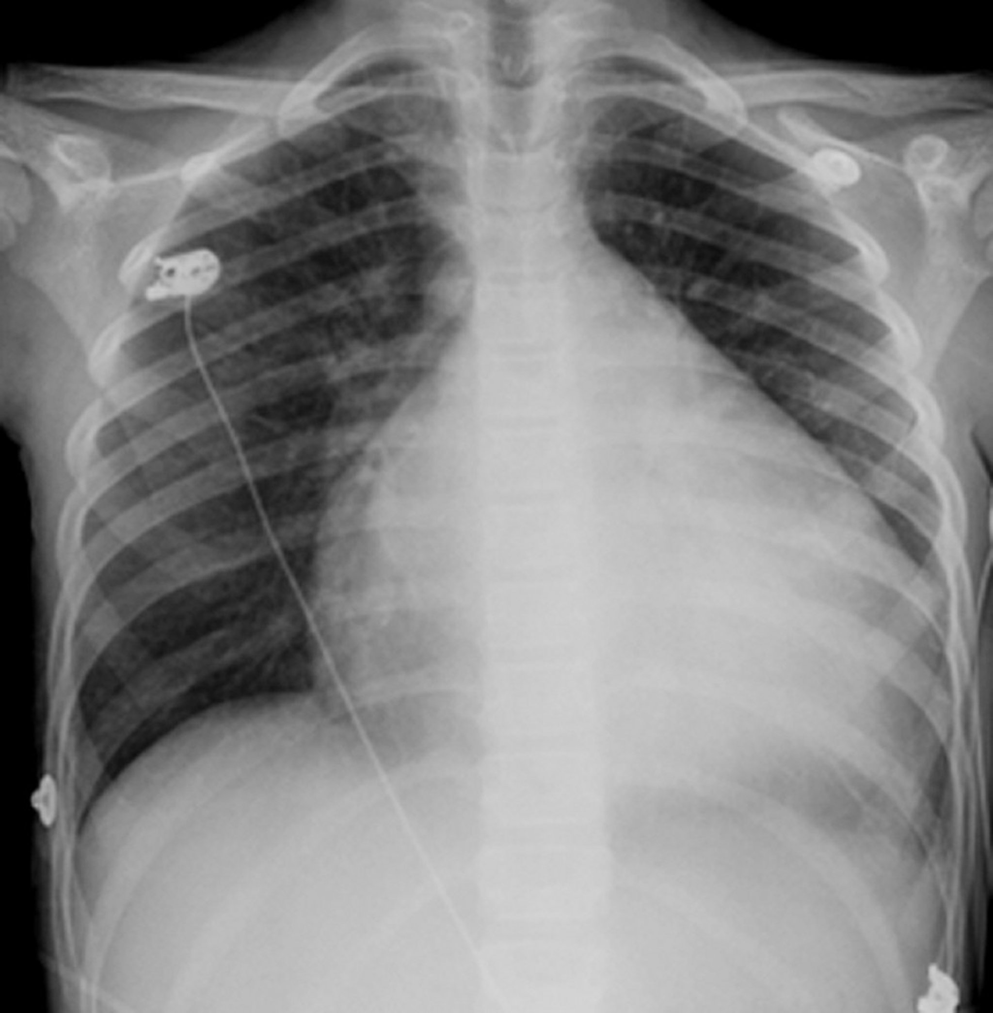
This radiograph shows severe cardiomegaly in a patient with dilated cardiomyopathy.
© Springer Science+Business Media
Transthoracic echocardiography characteristically demonstrates left ventricular dilation with global hypokinesis and atrial enlargement and excludes primary valvular disorders. Functional mitral and tricuspid regurgitation may result from annular dilation and papillary muscle displacement (4). Although the typical pattern is one of diffuse hypokinesis, regional wall motion abnormalities may occur, reflecting the heterogeneous or patchy nature of myocardial involvement in DCM. In selected cases, use of contrast echocardiography may be required to improve endocardial border definition and to exclude the presence of a left ventricular thrombus.
Cardiac MRI provides detailed structural and tissue characterization. Patterns of inflammation and late gadolinium enhancement assist in identifying specific etiologies (eg, myocarditis or infiltrative cardiomyopathies) and contribute to risk stratification, particularly with respect to future risk of ventricular arrhythmias and lack of reverse (ie, favorable) remodeling (4, 5, 6, 7, 8).The presence of late gadolinium enhancement in a ring-like pattern ("ring sign") is one such unfavorable prognostic marker.

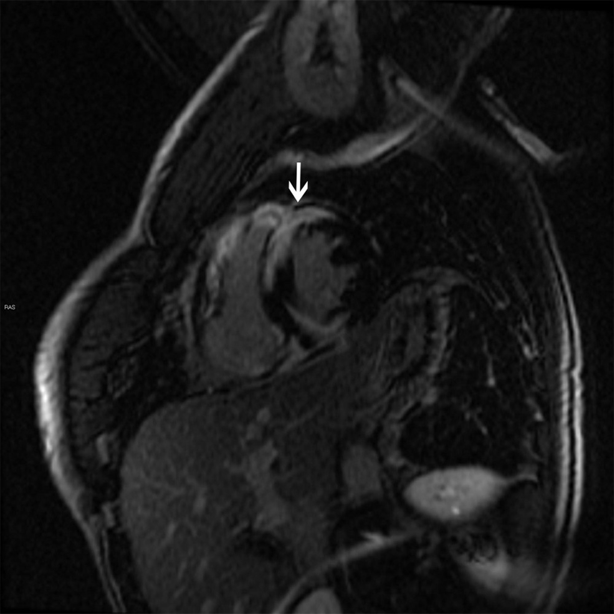
This cardiac MRI shows a late gadolinium enhancement in a patchy pattern (arrow) characteristic of sarcoidosis.
© 2017 Elliot K. Fishman, MD.
Positron-emission tomography (PET) is a valuable tool for the diagnosis of cardiac sarcoidosis and monitoring disease activity (6).
Endomyocardial biopsy should be considered in selected patients with rapidly progressive heart failure despite appropriate medical therapy when there is a probability of a specific and potentially treatable diagnosis (eg, giant cell myocarditis, eosinophilic myocarditis) that requires histopathologic confirmation (2, 3).
Diagnosis references
1. Tayal U, Ware JS, Lakdawala NK, Heymans S, Prasad SK. Understanding the genetics of adult-onset dilated cardiomyopathy: what a clinician needs to know. Eur Heart J. 2021;42(24):2384-2396. doi:10.1093/eurheartj/ehab286
2. Heidenreich PA, Bozkurt B, Aguilar D, et al. 2022 AHA/ACC/HFSA Guideline for the Management of Heart Failure: A Report of the American College of Cardiology/American Heart Association Joint Committee on Clinical Practice Guidelines. Circulation. 2022;145(18):e895-e1032. doi:10.1161/CIR.0000000000001063
3. Authors/Task Force Members:, McDonagh TA, Metra M, et al. 2021 ESC Guidelines for the diagnosis and treatment of acute and chronic heart failure: Developed by the Task Force for the diagnosis and treatment of acute and chronic heart failure of the European Society of Cardiology (ESC). With the special contribution of the Heart Failure Association (HFA) of the ESC. Eur J Heart Fail. 2022;24(1):4-131. doi:10.1002/ejhf.2333
4. Schultheiss HP, Fairweather D, Caforio ALP, et al. Dilated cardiomyopathy. Nat Rev Dis Primers. 2019;5(1):32. doi:10.1038/s41572-019-0084-1
5. Ganesan AN, Gunton J, Nucifora G, McGavigan AD, Selvanayagam JB. Impact of Late Gadolinium Enhancement on mortality, sudden death and major adverse cardiovascular events in ischemic and nonischemic cardiomyopathy: A systematic review and meta-analysis. Int J Cardiol. 2018;254:230-237. doi:10.1016/j.ijcard.2017.10.094
6. Heymans S, Lakdawala NK, Tschöpe C, Klingel K. Dilated cardiomyopathy: causes, mechanisms, and current and future treatment approaches. Lancet. 2023;402(10406):998-1011. doi:10.1016/S0140-6736(23)01241-2
7. McNally EM, Mestroni L. Dilated Cardiomyopathy: Genetic Determinants and Mechanisms. Circ Res. 2017;121(7):731-748. doi:10.1161/CIRCRESAHA.116.309396
8. Teraoka Y, Kato S, Yasuda N, Sawamura S, Horita N, Utsunomiya D. Late Gadolinium Enhancement Magnetic Resonance Imaging (MRI) for Predicting Left Ventricular Reverse Remodeling in Non-Ischemic Cardiomyopathy: A Systematic Review and Meta-Analysis. J Clin Med. 2025;14(3):895. doi:10.3390/jcm14030895
Treatment of Dilated Cardiomyopathy
Treat the cause (if a reversible etiology has been identified)
Guideline-directed medical therapy for heart failure with reduced ejection fraction (HFrEF)
Implantable cardioverter-defibrillator or cardiac resynchronization therapy (CRT) for symptomatic patients with left ventricular ejection fraction (LVEF) ≤ 35% despite optimal medical therapy
Consideration of durable left ventricular assist device or cardiac transplantation for patients with advanced disease
Reversible causes (eg, tachyarrhythmias, infections, metabolic or nutritional disorders) should be identified and treated (1). Immunosuppressive therapy should be reserved for patients with inflammatory causes (eg, giant cell myocarditis, eosinophilic myocarditis, sarcoidosis) and certain patients with autoimmune disorders (eg, systemic lupus erythematosus).
Patients with dilated cardiomyopathy (DCM) should be treated with guideline-directed medical therapy for HFrEF because these agents reduce morbidity and mortality (2). The cornerstones of treatment include beta blockers, angiotensin receptor/neprilysin inhibitor (ARNi) (or angiotensin-converting enzyme [ACE] inhibitor or angiotensin II receptor blocker (ARB) if ARNi not feasible), mineralocorticoid receptor antagonists (MRA), and sodium glucose cotransporter-2 protein inhibitors (SGLT2i), with diuretics for volume management (3, 4).
Anticoagulation may be considered in patients with documented intracardiac thrombus, and atrial fibrillation (3, 5, 6).
Special considerations apply in peripartum cardiomyopathy. Many standard heart failure treatments are potentially teratogenic (eg, ACE inhibitors, ARB, ARNi, MRA, SGLT2i, some anticoagulants). Anticoagulation may be considered in patients with severe left ventricular dysfunction due to heightened thrombotic risk (7, 8).
Implantable cardioverter-defibrillator therapy is recommended for primary prevention in patients with symptoms and persistent LVEF ≤ 35% despite chronic guideline-directed medical therapy, and cardiac resynchronization therapy (CRT) is indicated in patients with symptoms with LVEF ≤ 35% and QRS prolongation, particularly in the presence of a left bundle branch block (3, 4). Percutaneous edge-to-edge mitral valve repair can be considered in carefully selected patients with severe secondary mitral regurgitation despite optimal medical therapy and/or CRT. Advanced heart failure refractory to medical therapy may require durable left ventricular assist device support or cardiac transplantation in appropriate candidates (3, 4).
The management of acute heart failure and chronic heart failure is discussed in more detail elsewhere.
Treatment references
1. Bozkurt B, Colvin M, Cook J, et al: Current Diagnostic and Treatment Strategies for Specific Dilated Cardiomyopathies: A Scientific Statement From the American Heart Association. Circulation. 2016;134(23):e579-e646. doi:10.1161/CIR.0000000000000455
2. Vaduganathan M, Claggett BL, Jhund PS, et al. Estimating lifetime benefits of comprehensive disease-modifying pharmacological therapies in patients with heart failure with reduced ejection fraction: a comparative analysis of three randomised controlled trials. Lancet. 2020;396(10244):121-128. doi:10.1016/S0140-6736(20)30748-0
3. Heidenreich PA, Bozkurt B, Aguilar D, et al. 2022 AHA/ACC/HFSA Guideline for the Management of Heart Failure: A Report of the American College of Cardiology/American Heart Association Joint Committee on Clinical Practice Guidelines. Circulation. 2022;145(18):e895-e1032. doi:10.1161/CIR.0000000000001063
4. Authors/Task Force Members:, McDonagh TA, Metra M, et al. 2021 ESC Guidelines for the diagnosis and treatment of acute and chronic heart failure: Developed by the Task Force for the diagnosis and treatment of acute and chronic heart failure of the European Society of Cardiology (ESC). With the special contribution of the Heart Failure Association (HFA) of the ESC. Eur J Heart Fail. 2022;24(1):4-131. doi:10.1002/ejhf.2333
5. Gopinathannair R, Chen LY, Chung MK, et al. Managing Atrial Fibrillation in Patients With Heart Failure and Reduced Ejection Fraction: A Scientific Statement From the American Heart Association. Circ Arrhythm Electrophysiol. 2021;14(6):HAE0000000000000078. doi:10.1161/HAE.0000000000000078
6. Levine GN, McEvoy JW, Fang JC, et al. Management of Patients at Risk for and With Left Ventricular Thrombus: A Scientific Statement From the American Heart Association. Circulation. 2022;146(15):e205-e223. doi:10.1161/CIR.0000000000001092
7. Halpern, D, Weinberg, C, Pinnelas, R. et al. Use of Medication for Cardiovascular Disease During Pregnancy: JACC State-of-the-Art Review. JACC. 2019;73(4):457-476. doi:10.1016/j.jacc.2018.10.075
8. Regitz-Zagrosek V, Roos-Hesselink JW, Bauersachs J, et al. 2018 ESC Guidelines for the management of cardiovascular diseases during pregnancy: The Task Force for the Management of Cardiovascular Diseases during Pregnancy of the European Society of Cardiology (ESC). Eur Heart J. 2018;39(34): 3165-3241. doi: 10.1093/eurheartj/ehy340
Prognosis for Dilated Cardiomyopathy
Outcomes in dilated cardiomyopathy (DCM) have significantly improved over recent decades, although they remain variable, depending on etiology, disease severity, and response to treatment (1, 2, 3, 4). Contemporary 5-year mortality rates range from 20 to 30%, compared with historical rates of approximately 50% (5, 6, 7, 8). The principal causes of death are pump failure and sudden cardiac death (9, 10). Survival differs by demographic factors, with generally worse outcomes observed in men compared with women. Population-based studies also demonstrate higher age-adjusted mortality rates among Black patients compared with White patients (1, 9).
Prognosis references
1. Abdul Jabbar AB, Javed MA, Mohammed SF. Dilated Cardiomyopathy-Related Mortality in the United States: Demographic and Regional Trends Over the Past 2 Decades. Circ Heart Fail. 2026;19(1):e012987. doi:10.1161/CIRCHEARTFAILURE.125.012987
2. Felker GM, Thompson RE, Hare JM, et al. Underlying causes and long-term survival in patients with initially unexplained cardiomyopathy. N Engl J Med. 2000;342(15):1077-1084. doi:10.1056/NEJM200004133421502
3. Merlo M, Cannatà A, Pio Loco C, et al. Contemporary survival trends and aetiological characterization in non-ischaemic dilated cardiomyopathy. Eur J Heart Fail. 2020;22(7):1111-1121. doi:10.1002/ejhf.1914
4. Sjöland H, Silverdal J, Bollano E, Pivodic A, Dahlström U, Fu M. Temporal trends in outcome and patient characteristics in dilated cardiomyopathy, data from the Swedish Heart Failure Registry 2003-2015. BMC Cardiovasc Disord. 2021;21(1):307. doi:10.1186/s12872-021-02124-0
5. Bardy GH, Lee KL, Mark DB, et al. Amiodarone or an implantable cardioverter-defibrillator for congestive heart failure. N Engl J Med. 2005;352(3):225-237. doi:10.1056/NEJMoa043399
6. Køber L, Thune JJ, Nielsen JC, et al. Defibrillator Implantation in Patients with Nonischemic Systolic Heart Failure. N Engl J Med. 2016;375(13):1221-1230. doi:10.1056/NEJMoa1608029
7. Weintraub RG, Semsarian C, Macdonald P. Dilated cardiomyopathy. Lancet. 2017;390(10092):400-414. doi:10.1016/S0140-6736(16)31713-5
8. Zhou D, Zhu L, Li S, et al. Prognosis and Risk Stratification in Dilated Cardiomyopathy With LVEF≤35%: Cardiac MRI Insights for Better Outcomes. Circ Cardiovasc Imaging. 2025;18(3):e017246. doi:10.1161/CIRCIMAGING.124.017246
9. Bozkurt B, Colvin M, Cook J, et al: Current Diagnostic and Treatment Strategies for Specific Dilated Cardiomyopathies: A Scientific Statement From the American Heart Association. Circulation. 2016;134(23):e579-e646. doi:10.1161/CIR.0000000000000455
10. Schultheiss HP, Fairweather D, Caforio ALP, et al. Dilated cardiomyopathy. Nat Rev Dis Primers. 2019;5(1):32. doi:10.1038/s41572-019-0084-1
Key Points
Dilated cardiomyopathy is characterized by left ventricular dilation and systolic dysfunction; wall thickness is typically normal or reduced relative to chamber size.
Causes include infection (commonly viral), toxins, and metabolic, genetic, systemic, or autoimmune disorders.
Evaluation includes ECG, chest radiograph, and echocardiography; cardiac MRI provides additional tissue characterization.
Management includes treatment of reversible causes and guideline-directed medical therapy for heart failure with reduced ejection fraction.
Device therapy (implantable cardioverter-defibrillator, cardiac resynchronization therapy) is indicated in appropriately selected patients; mechanical support or transplantation is necessary for some.
Anticoagulation is indicated for atrial fibrillation or intracardiac thrombus; immunosuppressive therapy is reserved for selected inflammatory etiologies.







